Skip to main content Accessibility help
×
Hostname: page-component-78c5997874-xbtfd Total loading time: 0 Render date: 2024-11-14T06:23:41.017Z Has data issue: false hasContentIssue false

Chapter 22 - Assessment of fetal wellbeing

from Section 6 - Fetal growth and wellbeing

Published online by Cambridge University Press:  05 April 2016

Bidyut Kumar
Affiliation:
Wrexham Maelor Hospital
Zarko Alfirevic
Affiliation:
University of Liverpool
Get access

Summary

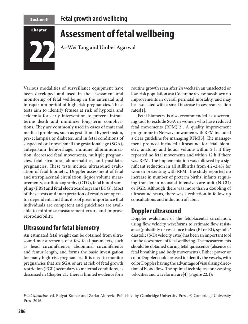
Image of the first page of this content. For PDF version, please use the ‘Save PDF’ preceeding this image.'
Type
Chapter
Information
Fetal Medicine , pp. 286 - 296
Publisher: Cambridge University Press
Print publication year: 2016

Access options

Get access to the full version of this content by using one of the access options below. (Log in options will check for institutional or personal access. Content may require purchase if you do not have access.)

References

Bricker, L, Neilson, JP, Dowswell, T. Routine ultrasound in late pregnancy (after 24 weeks’ gestation). Cochrane Database Syst Rev 2008; CD001451.Google Scholar
Royal College of Obstetricians and Gynaecologists (RCOG). Reduced fetal movements. RCOG Green-Top Guidelines No.57. London: RCOG Press, February 2011.Google Scholar
Tveit, JV, Saastad, E, Stray-Pedersen, B, et al. Reduction of late stillbirth with the introduction of fetal movement information and guidelines – a clinical quality improvement. BMC Pregnancy Childbirth 2009; 9: 32.Google Scholar
Bhide, A, Acharya, G, Bilardo, CM, et al. ISUOG Practice Guidelines: use of Doppler ultrasonography in obstetrics. Ultrasound Obstet Gynecol 2013; 41: 233–9.Google ScholarPubMed
Gómez, O, Figueras, F, Fernández, S, et al. Reference ranges for uterine artery mean pulsatility index at 11–41 weeks of gestation. Ultrasound Obstet Gynecol 2008; 32(2): 128–32.Google Scholar
Velauthar, L, Plana, MN, Kalidindi, M, et al. First-trimester uterine artery Doppler and adverse pregnancy outcome: a meta-analysis involving 55,974 women. Ultrasound Obstet Gynecol 2014; 43(5): 500–7.CrossRefGoogle ScholarPubMed
Gallo, DM, Poon, LC, Akolekar, R, et al. Prediction of preeclampsia by uterine artery Doppler at 20–24 weeks’ gestation. Fetal Diagn Ther 2013; 34(4): 241–7.CrossRefGoogle ScholarPubMed
Alfirevic, Z, Stampalija, T, Gyte, GML. Fetal and umbilical Doppler ultrasound in high-risk pregnancies. Cochrane Database Syst Rev 2013; (11): CD007529.Google Scholar
Acharya, G, Wilsgaard, T, Berntsen, GK, et al. Reference ranges for serial measurements of blood velocity and pulsatility index at the intra-abdominal portion, and fetal and placental ends of the umbilical artery. Ultrasound Obstet Gynecol 2005; 26(2): 162–9.CrossRefGoogle ScholarPubMed
Mari, G, Deter, RL, Carpenter, RL, et al. Noninvasive diagnosis by Doppler ultrasonography of fetal anemia due to maternal red-cell alloimmunization. Collaborative Group for Doppler Assessment of the Blood Velocity in Anemic Fetuses. N Engl J Med 2000; 342(1): 914.CrossRefGoogle Scholar
Stirnemann, JJ, Mougeot, M, Proulx, F, et al. Profiling fetal cardiac function in twin-twin transfusion syndrome. Ultrasound Obstet Gynecol 2010; 35(1): 1927.Google Scholar
Nabhan, AF, Abdelmoula, YA. Amniotic fluid index versus single deepest vertical pocket as a screening test for preventing adverse pregnancy outcome. Cochrane Database Syst Rev 2008; 3: CD006593.Google ScholarPubMed
Grivell, RM, Alfirevic, Z, Gyte, GML, et al. Antenatal cardiotocography for fetal assessment. Cochrane Database Syst Rev 2012; 2: CD007863.Google ScholarPubMed
Devane, D, Lalor, JG, Daly, S, et al. Cardiotocography versus intermittent auscultation of fetal heart on admission to labour ward for assessment of fetal wellbeing. Cochrane Database Syst Rev 2012; 2: CD005122.Google ScholarPubMed
Serra, V, Moulden, M, Bellver, J, et al. The value of the short-term fetal heart rate variation for timing the delivery of growth-retarded fetuses. BJOG 2008; 115(9): 1101–7.CrossRefGoogle ScholarPubMed
Huntleigh Healthcare Ltd. Sonicaid Team Operator’s Manual. Cardiff: Huntleigh Healthcare Diagnostic Products Division, 2005.Google Scholar
Manning, FA. Fetal biophysical profile. Obstet Gynecol Clin North Am 1999; 26(4): 557–77.Google Scholar
Lalor, JG, Fawole, B, Alfirevic, Z, et al. Biophysical profile for fetal assessment in high risk pregnancies. Cochrane Database Syst Rev 2008; 1: CD000038.Google Scholar
Turan, S, Turan, OM, Berg, C, et al. Computerized fetal heart rate analysis, Doppler ultrasound and biophysical profile score in the prediction of acid-base status of growth-restricted fetuses. Ultrasound Obstet Gynecol 2007; 30(5): 750–6.CrossRefGoogle ScholarPubMed
Hill, JB, Chauhan, SP, Magann, EF, et al. Intrapartum fetal surveillance: review of three national guidelines. Am J Perinatol 2012; 29(7): 539–50.Google ScholarPubMed
National Institute for Health and Care Excellence (NICE). Intrapartum care: care of healthy women and their babies during childbirth. CG190. London: NICE, December 2014.Google Scholar
Alfirevic, Z, Devane, D, Gyte, GML. Continuous cardiotocography (CTG) as a form of electronic fetal monitoring (EFM) for fetal assessment during labour. Cochrane Database Syst Rev 2013; 5: CD006066.Google Scholar
Skupski, DW, Rosenberg, CR, Eglinton, GS. Intrapartum fetal stimulation tests: a meta‐analysis. Obstet Gynecol 2002; 99(1): 129–34.Google Scholar
East, CE, Smyth, RMD, Leader, LR, et al. Vibroacoustic stimulation for fetal assessment in labour in the presence of a nonreassuring fetal heart rate trace. Cochrane Database Syst Rev 2013; 1: CD004664.Google Scholar
East, CE, Chan, FY, Colditz, PB, et al. Fetal pulse oximetry for fetal assessment in labour. Cochrane Database Syst Rev 2007; 2: CD004075.Google ScholarPubMed
East, CE, Leader, LR, Sheehan, P, et al. Intrapartum fetal scalp lactate sampling for fetal assessment in the presence of a non-reassuring fetal heart rate trace. Cochrane Database Syst Rev 2010; 3: CD006174.Google Scholar
MacLennan, A. A template for defining a causal relation between acute intrapartum events and cerebral palsy: international consensus statement. BMJ 1999; 319(7216): 1054–9.Google Scholar
Kruger, K, Hallberg, B, Blennow, M, et al. Predictive value of fetal scalp blood lactate concentration and pH as markers of neurologic disability. Am J Obstet Gynecol 1999; 181(5): 1072–8.Google Scholar
Neilson, JP. Fetal electrocardiogram (ECG) for fetal monitoring during labour. Cochrane Database Syst Rev 2013; 5: CD000116.Google Scholar
Becker, JH, Bax, L, Amer-Wåhlin, I, et al. ST analysis of the fetal electrocardiogram in intrapartum fetal monitoring: a meta-analysis. Obstet Gynecol 2012; 119(1): 145–54.CrossRefGoogle ScholarPubMed

Save book to Kindle

To save this book to your Kindle, first ensure coreplatform@cambridge.org is added to your Approved Personal Document E-mail List under your Personal Document Settings on the Manage Your Content and Devices page of your Amazon account. Then enter the ‘name’ part of your Kindle email address below. Find out more about saving to your Kindle.

Note you can select to save to either the @free.kindle.com or @kindle.com variations. ‘@free.kindle.com’ emails are free but can only be saved to your device when it is connected to wi-fi. ‘@kindle.com’ emails can be delivered even when you are not connected to wi-fi, but note that service fees apply.

Find out more about the Kindle Personal Document Service.

Available formats
×

Save book to Dropbox

To save content items to your account, please confirm that you agree to abide by our usage policies. If this is the first time you use this feature, you will be asked to authorise Cambridge Core to connect with your account. Find out more about saving content to Dropbox.

Available formats
×

Save book to Google Drive

To save content items to your account, please confirm that you agree to abide by our usage policies. If this is the first time you use this feature, you will be asked to authorise Cambridge Core to connect with your account. Find out more about saving content to Google Drive.

Available formats
×