Anterior spinal artery syndrome is a rare clinical syndrome, occurring in 5–8% of acute spinal cord injury (SCI).Reference Nedeltchev, Loher and Stepper1 Nontraumatic vascular causes are usually due to severe systemic hypotensionReference Kumral, Polat, Gulluoglu, Uzunkopru, Tuncel and Alpaydin2 or aortic aneurysm repair surgery.Reference Mawad, Rivera, Crawford, Ramirez and Breitbach3 Acute ischemic spinal stroke is rare, responsible for 5–14% of acute myelopathies.Reference Nedeltchev, Loher and Stepper1,Reference de Seze, Stojkovic and Breteau4 In a review from Kumral et al.Reference Kumral, Polat, Gulluoglu, Uzunkopru, Tuncel and Alpaydin2 identifying the pathogenetic mechanisms for ischemic spinal stroke, cardioembolic sources were not reported despite extensive work-up. Cardioembolism was also rarely (3.5–6%) the cause of medullary infarct among other studies.Reference Nedeltchev, Loher and Stepper1
An 86-year-old male presented with complaints of acute paraplegia over two hours. Past medical history included mild cognitive impairment, hypertension, and a new-onset atrial fibrillation, which was not anticoagulated. The patient was previously ambulating independently. Upon presentation, the patient sustained an acute painless flaccid paraplegia, with sparing of right ankle dorsiflexion (MRC graded at 3/5, Medical Research Council Scale 3/5: active movement against gravity). Deep tendon reflexes were absent. A pinprick sensory level was demonstrated at D3. Dorsal column sensory modalities were preserved. The rest of the neurological examination was unremarkable. Furthermore, a painless right limb ischemia was also noted with pulselessness and pallor. The patient developed urinary retention and constipation over 4 days and subsequent dysautonomia, including orthostatic hypotension, was noted.
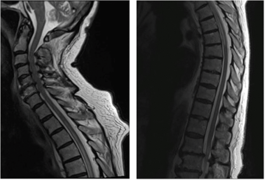
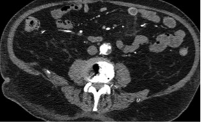
An MRI performed 2 days after initial presentation revealed a T2-hyperintense signal on the thoracic spinal cord from D2 to D7. This lesion abnormality was predominantly centro-anterior, sparing the posterior columns (Figure 1). A contrast-injected thoraco-abdomino-pelvic scan revealed a 3.6 cm descending aortic aneurysm containing an extensive thrombus with irregular borders on the left posterolateral wall extending from D5 to D10 (Figure 2). Furthermore, a 28 × 20 mm thrombus in the left appendix atrium was found and considered the probable embolic etiology. Multiple other thrombi were discovered in the infra-renal abdominal aorta, both lungs, the right popliteal artery, and both posterior inferior cerebellar arteries. Extensive workup, including a lumbar puncture, showed no evidence of inflammatory or neoplastic causes to the medullary infarct. Upon discharge, 1.5 months after admission, no motor or sensory recovery was noted.

Figure 1: Cervical and thoracic spine MRI reveals T2 hyperintensity in the spinal cord from D2 to D7.

Figure 2: Contrast-injected thoraco-abdomino-pelvic scan reveals 3.6 cm descending aortic aneurysm containing extensive thrombus with irregular borders on the left posterolateral wall.
Anterior spinal artery syndrome is characterized by bilateral paralysis with spinothalamic deficits, sparing posterior cord function.Reference Kumral, Polat, Gulluoglu, Uzunkopru, Tuncel and Alpaydin2 It is often sudden and involves bowel and bladder dysfunction.Reference Mawad, Rivera, Crawford, Ramirez and Breitbach3 Historical descriptions of ischemic myelopathies included a prominent radicular pain component.Reference Nedeltchev, Loher and Stepper1 In a more recent study, 40% had pain associated with spinothalamic dysfunction.Reference Masson, Pruvo and Meder5
Although the classical findings of anterior spinal artery infarct were not found (infarct limited to the anterior horns and surrounding white matterReference Kumral, Polat, Gulluoglu, Uzunkopru, Tuncel and Alpaydin2), an ischemic etiology was retained because of the important centro-anterior T2-hypersignal intensity and cord enlargement on MRI extending from D2 to D7.Reference Kumral, Polat, Gulluoglu, Uzunkopru, Tuncel and Alpaydin2 In a study on vascular myelopathies, an ischemic lesion matching an arterial territory was only found in 72% of patients.Reference Kumral, Polat, Gulluoglu, Uzunkopru, Tuncel and Alpaydin2 Masson et al. suggested that infarcts were, for the majority of cases, more frequently located in the central territory of the anterior spinal artery.Reference Masson, Pruvo and Meder5 Another typical MRI pattern of focal signal involving the anterior horns of the grey matter, described as the “owl’s eyes” pattern,Reference Mawad, Rivera, Crawford, Ramirez and Breitbach3 was only diagnosed in 25% of their patients.Reference Masson, Pruvo and Meder5
In this particular clinical case, as it is highly unlikely that individual ischemic events arising from penetrating arteries would have caused the extensive medullary infarct, we believe the medullary infarct was in fact provoked by the arterial thrombi-occlusion of the great anterior radicular artery of Adamkiewicz. This artery has been described in 62–75% of cases arising from the 9th to the 12th intercostal arteries. However, its origin is variable, ranging from the levels D5 to D8.Reference Mawad, Rivera, Crawford, Ramirez and Breitbach3 In a 2010 review of 73 thoraco-abdominal CT (multidetector CT angiography) angiographies, the artery arose from the left wall of the aorta in 65–75% of cases.Reference Sukeeyamanon, Siriapisith and Wasinrat6 This is consistent with the findings in our case, where the thrombus was on the left posterolateral wall of the aorta at D5.
Previously reported cases of acute ischemic myelopathies are most frequently associated with atherosclerosis, aortic aneurysm repair, degenerative spine disease, or prolonged systemic hypotension.Reference Nedeltchev, Loher and Stepper1,Reference Kumral, Polat, Gulluoglu, Uzunkopru, Tuncel and Alpaydin2 Atrial fibrillation as the direct cause has been mentioned sporadically as the trigger agent,Reference Nedeltchev, Loher and Stepper1 but only a few cases have been reported in the literature, accounting for approximately 3.5% of all acute anterior spinal artery syndromes in one retrospective study.Reference Nedeltchev, Loher and Stepper1 We feel the patient’s prothrombotic state represents an unusual cause of acute spinal cord ischemia.
Despite standard rehabilitation and treatment with therapeutic low-molecular-weight heparin, the patient remained paraplegic. A 2010 retrospective series of patients with ischemic medullary infarcts reported a general but incomplete recovery with mild gait impairment at discharge.Reference Kumral, Polat, Gulluoglu, Uzunkopru, Tuncel and Alpaydin2 In addition, for spinal cord infarction, a poor prognostic factor was older age, regardless of the degree of completeness of the injury, as evaluated by the American Spinal Injury Association Impairment Scale.Reference Sturt, Holland and New7 A retrospective study stated that a poor or fair outcome was to be expected in 91% of patients who suffered from acute spinal cord ischemia.Reference de Seze, Stojkovic and Breteau4 To our knowledge, only one prediction rule for walking exists for SCI patients, but it has been tested on patients with traumatic injury.Reference van Middendorp, Hosman and Donders8 This rule used the age and the motor and sensory function of different radicular nerves to predict the walking capacity at 1 year. Had we applied the aforementioned clinical prediction rule, this patient would not have been expected to walk again. However, the ambulatory outcome can be quite different in non-traumatic SCI since patients are often older, have more comorbidities, and are more likely to have an incomplete SCI.Reference Sturt, Holland and New7 Optimal medical treatment remains controversial, and the most appropriate therapy (including steroids, anticoagulation, antiplatelets, or thombolytics) is not established.Reference Kumral, Polat, Gulluoglu, Uzunkopru, Tuncel and Alpaydin2
Recognition of unusual causes of acute spinal cord ischemia should include the possibility of multi-system arterial emboli, and prognosis of these myelopathies is generally poor.
Disclosures
The authors have no conflicts of interest to declare.
Statement of authorship
SJ conceived of the manuscript, did the research, and drafted the initial manuscript. BHN, ARD, and AL reviewed the manuscript and provided linguistic revision. AL supervised the overall project.


