Introduction
Post-coronavirus disease 2019 (Covid-19) acute invasive fungal sinusitis is a new clinical entity that has been described by many authors since 2019.Reference El-Kholy, Abd El-Fattah and Khafagy1–Reference Sebastian, Kumar, Gupta and Covid3 There has been a marked increased incidence of acute invasive fungal sinusitis in post-Covid-19 patients.Reference Ismaiel, Abdelazim, Eldsoky, Ibrahim, Alsobky and Zafan4 Increased susceptibility of fungal infections in Covid-19 patients may be due to overexpression of inflammatory cytokines and impairment of cell-mediated immunity with a decrease in T-helper cell count, elevated blood glucose (diabetes, new-onset hyperglycaemia, steroid-induced hyperglycaemia), hypoxia, acidic medium (metabolic acidosis, diabetic ketoacidosis), decreased phagocytic activity of white blood cells, and immunosuppression (Covid-19 mediated, steroid-mediated, or background comorbidities).Reference Singh, Singh, Joshi and Misra5,Reference Song, Liang and Liu6
Acute invasive fungal sinusitis is a life-threatening condition in which the mucous membrane of the nose and sinuses is invaded by a fungus that has a high risk of invasion and destruction of the adjacent structures.Reference Wandell, Miller, Rathor, Wai, Guyer and Schmidt7 Reported mortality rates in the literature are variable (14–80 per cent),Reference Gardner, Hunter, Vickers, King and Kanaan8–Reference Sen, Honavar, Bansal, Sengupta, Rao and Kim12 and ranging up to almost 100 per cent in disseminated infections despite medical and surgical treatments.Reference Jung, Kim, Park, Song, Cho and Lee13
Factors that may affect the prognosis of acute invasive fungal sinusitis remain not fully understood. Some factors include advancing age,Reference Sun, Forrest, Gupta, Aguado, Lortholary and Julia14,Reference Turner, Soudry, Nayak and Hwang15 early aggressive surgical debridement,Reference Turner, Soudry, Nayak and Hwang15,Reference Wandell, Miller, Rathor, Wai, Guyer and Schmidt16 type of fungus,Reference Deutsch, Whittaker and Prasad9,Reference Turner, Soudry, Nayak and Hwang15 presence of haematologic malignancy,Reference Chen, Sheng, Cheng, Chen, Tsay and Tang17 and orbital and intracranial extensions.Reference Jung, Kim, Park, Song, Cho and Lee13 However, there is a paucity of literature regarding precise prognostic factors. The aim of the current study is to identify the prognostic factors that may have an effect on the outcome of acute invasive fungal sinusitis and the survival of this patient population, in order to help optimise diagnosis and management.
Patients and methods
This retrospective study was conducted in a tertiary referral centre; Otorhinolaryngology Department, Mansoura University Hospitals, Egypt, over two years duration (October 2020–October 2022). Informed written consents were obtained from all participants, and the study was approved by the Mansoura Faculty of Medicine Institutional Research Board (MFM-IRB: MS.22.02.1870).
After reviewing patients’ records, all patients with post-Covid-19 acute invasive fungal sinusitis managed at our institute during the study duration (n = 60) were included in the study. All patients in the study had definite diagnosis of previous Covid-19 infection. The interval between the diagnosis of Covid-19 and acute invasive fungal sinusitis was 13–42 days (mean = 23.7 days). Diagnosis and management of Covid-19 followed the national management protocol.Reference Masoud, Elassal, Hakim, Shawky, Zaky and Baki18
Patients’ management included detailed histories and clinical examinations. All patients in the study underwent high-resolution, contrast-enhanced computed tomography (CT) scans. Additionally, patients with intra-orbital or intra-cranial extension of infection underwent magnetic resonance imaging. Diagnosis and management followed the global guideline for the diagnosis and management of acute invasive fungal sinusitis.Reference Cornely, Alastruey-Izquierdo, Arenz, Chen, Dannaoui and Hochhegger19 Diagnosis relied upon endoscopic endonasal findings of mucosal pallor, blackening, gangrene, and/or palatal ischemia, gangrene, and/or orbital findings as proptosis, ophthalmoplegia, or vision loss. Additionally, histopathological examination confirmed the diagnosis by presence of fungal invasion within sinus mucosa, submucosa, or bone.
Antifungal therapy with surgical debridement was the main line of treatment for acute invasive fungal sinusitis. Upon clinical diagnosis, we started treatment immediately without waiting for histology or culture results because delay in treatment is associated with significant morbidity and mortality.
Antifungal drugs included liposomal amphotericin B, voriconazole, and posaconazole. The choice of antifungal agents followed the national management protocolReference Masoud, Elassal, Hakim, Shawky, Zaky and Baki18 under supervision of the infectious disease department. Duration of the antifungal treatment was 19–62 days (median 33 days), until complete resolution of signs and symptoms, with monitoring of renal functions, liver functions, complete blood count, and blood-sugar levels.
We tailored surgical procedures according to the disease extension. We performed endoscopic, open, and combined approaches. Furthermore, we performed serial debridement until complete eradication of infection. After surgical debridement, histopathological examination of tissues was of utmost importance to confirm acute invasive fungal sinusitis as well as to determine the causative fungus based on the morphology of the hyphae using haematoxylin and eosin, periodic acid-Schiff, and Grocott–Gömöri's methenamine silver staining. Moreover, specimens were sent for fungal culture for determination of the type of growth.
After discharge of the patients, we scheduled follow-up visits in the outpatient department on a weekly basis for three months. The follow-up period was 6–30 months (average 21.5 months). The authors of this work considered the outcome as successful when patients were symptom free, and serial examinations revealed adequate healing with no evidence of disease (ischemia or gangrene) in the follow up visits.
We identified and studied several factors that may have had an effect on the prognosis. These factors included patient-related factors (age, gender, medical comorbidities), disease-related factors (type of fungus, extension of infection) and treatment-related factors (history of intensive care unit (ICU) admission, type of antifungal drug, type of surgical treatment).
Results
This study included 60 patients with post-Covid-19 acute invasive fungal sinusitis, whether admitted from the outpatient clinic (n = 24), from the emergency department (n = 19), or referred from other centres (n = 17). Patients mean age was 56.23 years. Medical comorbidities occurred in 59 of 60 patients (98.3 per cent). Diabetes mellitus was the commonest (95 per cent). Other comorbidities included hypertension (55 per cent), hepatic impairment (13.3 per cent), renal impairment (11.7 per cent) and cardiac disease (3.3 per cent).
Diminution of vision was the most frequent patient presentation (38.3 per cent), followed by palatal necrosis (28.3 per cent). Other presentations included facial pain (16.7 per cent), sino-nasal symptoms (nasal obstruction and discharge) (11.7 per cent) and facial swelling (8.3 per cent).
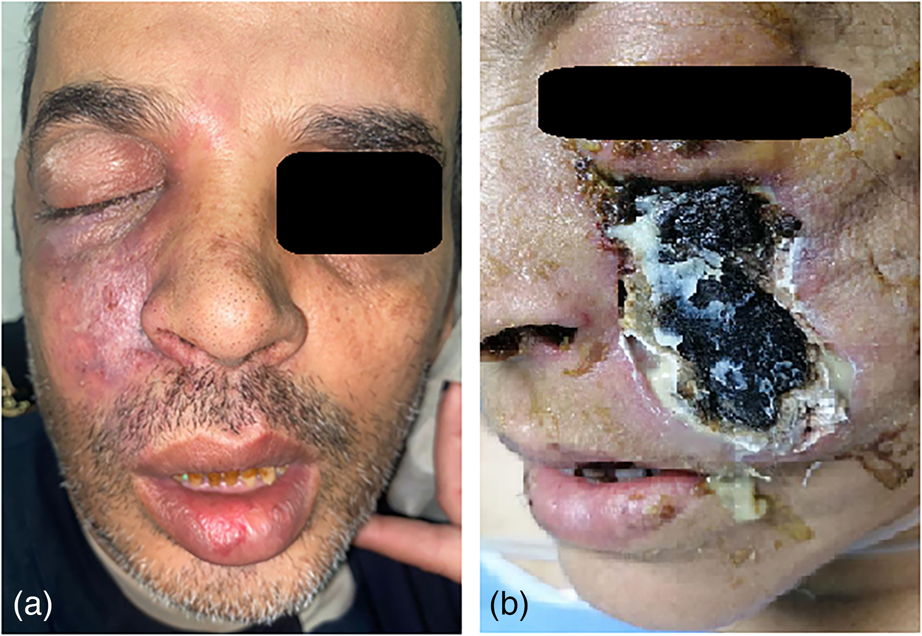
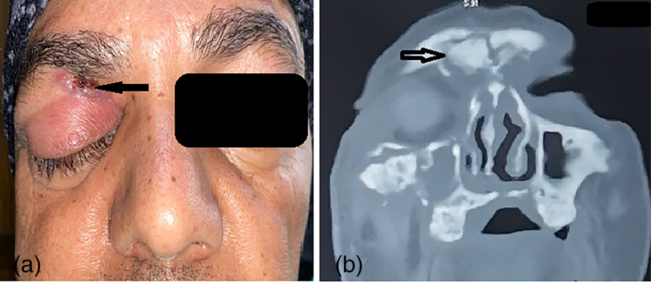
The extension of infection among the studied group was sino-nasal in 58 of 60 (96.7 per cent), orbital in 28 of 60 (46.7 per cent), and palatal in 21 of 60 (35 per cent) (Figure 1). Intracranial involvement occurred in nine patients in the form of cavernous sinus thrombosis (11.7 per cent) and brain abscess (3.3 per cent). Additionally, skin affection in the form of gangrene occurred in two cases (3.3 per cent) (Figure 2), and frontal sinus osteomyelitis occurred in two patients (3.3 per cent) (Figure 3).

Figure 1. A: Osteomyelitis and sequestrum in the right side of the palate (arrow). B, C: Coronal and axial CT scan of the same patient showing osteomyelitis (arrows).

Figure 2. Skin affection in two patients. A: Ischemia and blackening in the right cheek. B: More-severe necrosis and gangrene in the left cheek.

Figure 3. Right frontal osteomyelitis with a fistula formation (arrow). B: CT scan of the same patient showing osteomyelitis and sequestrum formation in the right frontal sinus.
Based on the national protocolReference Masoud, Elassal, Hakim, Shawky, Zaky and Baki18 and recommendations of the infectious diseases specialists, we prescribed empirical IV liposomal amphotericin B (1 mg/kg/day) immediately, before waiting for the results of the culture. Consequently, 58 out of 60 patients received empirical amphotericin. The remaining two cases had a history of liver transplant, and therefore amphotericin B was contra-indicated. After consultation of gastroenterologists, we used posaconazole.
After obtaining culture results, we immediately prescribed voriconazole (200 mg 12 hourly) instead of amphotericin. In two patients the culture revealed mixed infection with both mucor and aspergillus. Therefore, those patients received a combination of liposomal amphotericin B and voriconazole. All patients (n = 60) completed the course of antifungal drugs with no reported major complications of treatment.
Fifty-seven patients (95 per cent) underwent surgical debridement. Three patients received only medical treatment due to poor anaesthetic fitness (n = 2) and patient refusal of surgery (n = 1). We performed endoscopic endonasal debridement in 54 cases. Repeated surgical debridement was necessary in 21 patients until endoscopic examination revealed healthy tissues without evidence of ischemia, necrosis, or gangrene (2 sessions in 17 patients, 3 sessions in 3 patients, and 4 sessions in 1 patient).
We utilised external approaches in 23 patients. After multidisciplinary consultations of ophthalmology, neurosurgery, and maxillofacial surgery, we performed orbital exenteration in 13 patients (21.7 per cent), palatectomy and/or maxillectomy in 11 patients (18.3 per cent), debridement and sequestrectomy for frontal sinus osteomyelitis in 2 cases (3.3 per cent), skin debridement in 2 cases, and craniotomy for evacuation of a brain abscess in 1 patient.
At the end of treatment, the overall success rate was 91.7 per cent. In five patients (8.3 per cent), the prognosis was poor despite medical and/or surgical treatment. All five of these patients died from the disease or its complications and were listed as failed outcomes.
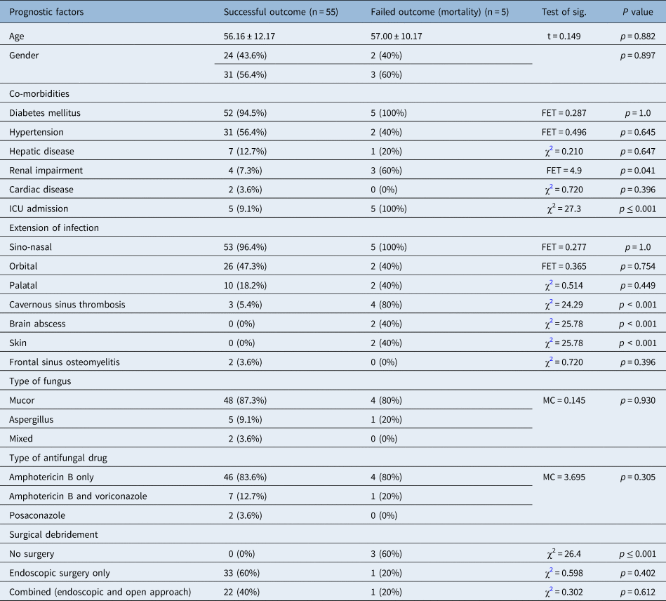
In the current study, we analysed multiple variables that may have affected the success rate (Table 1). Patients’ age and gender did not have an effect on prognosis. Similarly, the type of fungus, whether mucor, aspergillus, or mixed infection, as well as the antifungal agent were not associated with a significant effect on the outcome.
Table 1. The effects of multiple factors on prognosis; ICU = intensive care unit; χReference Mekonnen, Ashraf, Jankowski, Grob, Vagefi and Kersten2 = chi-square test, FET = Fisher's exact test, MC = Monte Carlo test, t = Student's t test

Regarding co-morbidities, renal impairment was detected in 4 of 55 cases (7.3 per cent) in the successful group and in 3 of 5 (60 per cent) in the failed group (p = 0.041). Consequently, renal impairment showed a significant poor effect on the outcome. Other co-morbidities (diabetes mellitus, hypertension, hepatic disease, cardiac disease) did not have a significant effect on the outcome. Additionally, history of ICU admission was associated with a statistically significant poor effect on the prognosis, as it was noted in 5 of 60 cases (9.1 per cent) in the successful group and in 5 of 5 (100 per cent) in the failed group (p ≤ 0.001).
Similarly, extension of infection was one of the significant prognostic factors. Intracranial extension (cavernous sinus thrombosis and brain abscess) had a negative effect on the prognosis (p ≤ 0.001). Additionally, skin affection was associated with a significant poor outcome (p ≤ 0.001). However, orbital and palatal involvement did not have a statistically significant effect on the outcome (p = 0.754 and 0.449, respectively).
Three patients in the current study received only medical antifungal treatment without surgical debridement, and the outcome was poor in those three patients with reported mortality. Consequently, medical treatment alone was associated with a poor effect on the prognosis.
Discussion
Acute invasive fungal sinusitis is a highly morbid disease. It can directly invade intracranial and intra-orbital spaces with a potentially fatal outcome. The reported mortality rates are variable (14–100 per cent).Reference Gardner, Hunter, Vickers, King and Kanaan8–Reference Jung, Kim, Park, Song, Cho and Lee13 In the large meta-analysis conducted by Turner et al.Reference Turner, Soudry, Nayak and Hwang15 on 807 patients with acute invasive fungal sinusitis, the overall survival rate was 49.7 per cent. Few data are available regarding the prognostic factors that may determine the survival rate.
Nearly all patients with acute invasive fungal sinusitis involve immunosuppression, most commonly related to diabetes mellitus, haematologic malignancy, or organ transplantation.Reference Ashraf, Idowu, Hirabayashi, Kalin-Hajdu, Grob and Winn20 In the current study, comorbidities occurred in 98.3 per cent of patients. Diabetes mellitus was the commonest (95 per cent). A diabetic patient is more susceptible to invasive fungal infections due to derangement of granulocyte-phagocytic activity, altered polymorphonuclear leukocyte response, microangiopathy and peripheral vascular diseases, with subsequent local-tissue ischemia and increased susceptibility to infections. Additionally, the acidic environment, low oxygen, and high glucose levels facilitate germination and growth of fungal spores.Reference Kheirkhah, Asoubar, Abdi and Mahmoudi21,Reference Tugsel, Sezer and Akalin22
Comorbidities have been identified as a negative prognostic factor. Previous reports found that underlying haematologic malignancy as well as liver and renal failure are associated with a poor outcome.Reference Turner, Soudry, Nayak and Hwang15,Reference Cho, Jang, Hong, Chung, Kim and Dhong23 In the current study, patients with renal impairment had a statistically significant poor prognosis.
Similarly, some authors have reported that advanced age results in decreased survival, independent of other factors.Reference Sun, Forrest, Gupta, Aguado, Lortholary and Julia14,Reference Turner, Soudry, Nayak and Hwang15 Not surprisingly, such patients can be assumed to have additional comorbidities and are generally more vulnerable to aggressive infections and other medical problems. However, in the present work, age did not have a significant effect on the outcome.
History of ICU admission in the current study had a negative effect on the prognosis. Eldsouky et al.Reference Eldsouky, Shahat, Al-Tabbakh, El Rahman, Marei and Mohammed24 reported that patients with severe Covid-19, who are admitted in the ICU, are especially vulnerable to bacterial and fungal infections.
Disease extension is an additional significant prognostic factor. Progression from limited disease or isolated nasal lateral wall involvement to involvement of the hard palate and orbit has been observed to portend a poorer prognosis.Reference Wandell, Miller, Rathor, Wai, Guyer and Schmidt16,Reference Valera, do Lago, Tamashiro, Yassuda, Silveira and Anselmo-Lima25 In one retrospective series, orbital involvement increased mortality nearly four-fold compared to isolated sinonasal involvement.Reference Trief, Gray, Jakobiec, Durand, Fay and Freitag10 However, other studies have shown that only intracranial and cavernous sinus extension is a negative prognostic factor and that dissemination is a better indicator of morbidity.Reference Turner, Soudry, Nayak and Hwang15,Reference DelGaudio and Clemson26 Orbital and palatal extensions in the current study were not associated with poor outcomes or mortality. Nevertheless, patients with brain abscess, cavernous sinus thrombosis, and skin affection had statistically significant poor prognosis.
Mucor species infection has been associated with higher mortality, given the more aggressive nature of these organisms.Reference Deutsch, Whittaker and Prasad9,Reference Turner, Soudry, Nayak and Hwang15 In our study, no significant difference between mucor and aspergillus infections in terms of survival was reported. This may be due to the relatively small sample size and the small number of patients diagnosed with aspergillus infection (6 out of 60 patients).
According to the global guideline for diagnosis and management of invasive fungal sinusitis, 2019,Reference Cornely, Alastruey-Izquierdo, Arenz, Chen, Dannaoui and Hochhegger19 an early complete surgical treatment is strongly recommended whenever possible, in addition to systemic antifungal treatment. In the current series, relying on medical treatment only without surgical debridement was associated with a statistically significant poor outcome. Systemic antifungal treatment alone is typically insufficient for eliminating the infection because of its angio-invasive nature, which leads to obliteration of local blood supply and rapid tissue necrosis that limits the penetration of systemic therapy.Reference Turner, Soudry, Nayak and Hwang15,Reference Ashraf, Idowu, Hirabayashi, Kalin-Hajdu, Grob and Winn20 Therefore, debridement of necrotic tissue plays a key role in the management of acute invasive fungal sinusitis.
Many authors reported that prompt surgical debridement was associated with better outcomes.Reference Turner, Soudry, Nayak and Hwang15,Reference Wandell, Miller, Rathor, Wai, Guyer and Schmidt16,Reference Malleshappa, Rupa, Varghese and Kurien27 Similarly, in patients with orbital extension, Ashraf et al.Reference Ashraf, Idowu, Hirabayashi, Kalin-Hajdu, Grob and Winn20 reported that orbital exenteration was associated with higher survival rates. Moreover, in patients with fungal osteomyelitis in the maxilla, palate, or frontal bone, debridement and sequestrectomy is the treatment of choice.Reference Srivastava, Mohpatra and Mahapatra28,Reference Ebada, Abd El-Fattah and Tawfik29
It is likely that debriding necrotic areas may lead to improved antimicrobial penetration, and that surgically decreasing the burden of fungal organisms may reduce the risk of regional and systemic spread of disease. In the current work, the survival rate was 91.7 per cent. The relatively low mortality (8.3 per cent) among patients with acute invasive fungal sinusitis in the current study may be attributed to early diagnosis and timely intervention. In the late post-Covid-19 era, increased public awareness of this disease (black fungus disease) has prompted earlier patient referrals from other specialties and facilities, as well as patients seeking their own medical attention upon any disease suspicion. Furthermore, in the late post-Covid era, subsequent variants and mutants of the Covid-19 virus are known to have lower virulence and mortality than the ancestor ones.Reference El-Shabasy, Nayel, Taher, Abdelmonem, Shoueir and Kenawy30
Additionally, improved learning curve and experience at our tertiary referral centre, as well as the multidisciplinary team approach has led to better decision making and patient care. Early aggressive surgical debridement with variable combinations of endoscopic endonasal techniques, orbital exenteration, maxillofacial approaches (frontal, maxillary, and palatal osteomyelitis) and neurosurgical craniotomy approaches according to the disease extension, may have a role in achieving better disease control.
• Although post-coronavirus disease 2019 acute invasive fungal sinusitis recently has been described by many authors, there is a paucity of relevant literature regarding the precise prognostic factors that have an effect on the prognosis of acute invasive fungal sinusitis.
• Researchers have identified some factors that affect acute invasive fungal sinusitis including advancing age, early aggressive surgical debridement, type of fungus, presence of haematologic malignancy and orbital and intracranial extensions
• The current study concluded that comorbidities, especially renal impairment, previous intensive care unit admission, skin involvement, and intracranial spread of infection, are associated with significantly poor outcomes
• Early aggressive surgical debridement was an independent factor associated with better prognosis in this study
Identifying prognostic factors may have a role in predicting prognosis, and tailoring patient-specific treatment protocols. Furthermore, prophylactic antifungal use in high-risk groups such as immunocompromised patients in intensive care units may be warranted to protect against this life-threatening invasive fungal infection.
Conclusion
The current study concludes that comorbidities, especially renal impairment, previous ICU admission, skin involvement, and intracranial spread of infection, are associated with significantly poorer outcomes. Early aggressive surgical debridement is an independent factor associated with better prognosis.
Conflicts of interest and financial support
There are no conflicts of interest or financial disclosures to be made.


