Hostname: page-component-cd9895bd7-dk4vv Total loading time: 0 Render date: 2024-12-28T15:13:49.976Z Has data issue: false hasContentIssue false

Band Excitation Piezoresponse Force Microscopy Adapted for Weak Ferroelectrics: On-the-Fly Tuning of the Central Band Frequency

Published online by Cambridge University Press:  10 March 2021
Maxim Spiridonov
Affiliation:
Moscow Institute of Physics and Technology, 9 Institutskiy Lane, Dolgoprudny, Moscow Region141700, Russia
Anastasia Chouprik*
Affiliation:
Moscow Institute of Physics and Technology, 9 Institutskiy Lane, Dolgoprudny, Moscow Region141700, Russia
Vitalii Mikheev
Affiliation:
Moscow Institute of Physics and Technology, 9 Institutskiy Lane, Dolgoprudny, Moscow Region141700, Russia
Andrey M. Markeev
Affiliation:
Moscow Institute of Physics and Technology, 9 Institutskiy Lane, Dolgoprudny, Moscow Region141700, Russia
Dmitrii Negrov
Affiliation:
Moscow Institute of Physics and Technology, 9 Institutskiy Lane, Dolgoprudny, Moscow Region141700, Russia
*
*Author for correspondence: Anastasia Chouprik, E-mail: chouprik.aa@mipt.ru
Get access

Abstract

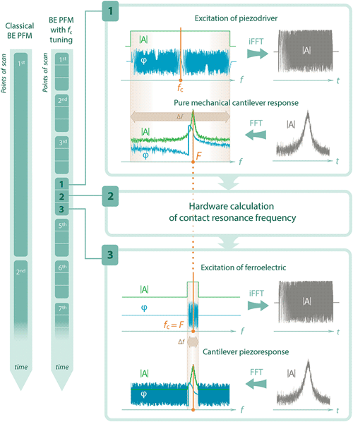
New interest in microscopic studies of ferroelectric materials with low piezoelectric coefficient, $d_{33}^\ast$, has emerged after the discovery of ferroelectric properties in HfO2 thin films, which are the main candidate for the next generation of nonvolatile ferroelectric memory. The study of the microscopic structure of ferroelectric HfO2 capacitors is crucial to get insights into the device behavior and performance. However, a small $d_{33}^\ast$ of ferroelectric HfO2 films leads to a low piezoresponse, especially in band excitation piezoresponse force microscopy (BE-PFM). In this work, we have implemented the BE-PFM technique with an increased scanning rate, thus improving this versatile tool for weak ferroelectrics. The acceleration of measurement was achieved by focusing excitation into a narrow frequency band and tuning the central frequency on-the-fly using an online real-time model estimation by fitting a complex BE response. The tracking of the contact resonance frequency was implemented using a pure mechanical cantilever response acquired in BE atomic force acoustic microscopy. To obtain optimal excitation parameters, we perform statistical analysis by minimizing estimator variance. The measurement precision of several PFM techniques was compared both by the simulation and experimentally using a Hf0.5Zr0.5O2-based ferroelectric capacitor.

Type
Software and Instrumentation
Copyright
Copyright © The Author(s), 2021. Published by Cambridge University Press on behalf of the Microscopy Society of America

Access options

Get access to the full version of this content by using one of the access options below. (Log in options will check for institutional or personal access. Content may require purchase if you do not have access.)

References

Böscke, TS, Müller, J, Brauhaus, D, Schröder, U & Böttger, U (2011). Ferroelectricity in hafnium oxide thin films. Appl Phys Lett 99, 102903.CrossRefGoogle Scholar
Cane, MG (2014). Characterisation of Ferroelectric Bulk Materials and Thin Films. Springer Series in Measurement Science and Technology, Springer Netherlands, eBook ISBN 978-1-4020-9311-1.CrossRefGoogle Scholar
Chouprik, A, Kirtaev, R, Spiridonov, M, Markeev, AM & Negrov, D (2020). Nanoscale tailoring of ferroelectricity in a thin dielectric film. ACS Appl Mater Interfaces. doi:10.1021/acsami.0c16741CrossRefGoogle Scholar
Chouprik, A, Kondratyuk, E, Mikheev, V, Matveyev, Y, Spiridonov, M, Chernikova, A, Kozodaev, MG, Markeev, AM, Zenkevich, A & Negrov, D (2021). Origin of the retention loss in ferroelectric Hf0.5Zr0.5O2-based memory devices. Acta Mater 204, 116515.CrossRefGoogle Scholar
Chouprik, A, Spiridonov, M, Zarubin, S, Kirtaev, R, Mikheev, V, Lebedinskii, Y, Zakharchenko, S & Negrov, D (2019). Wake-up in a Hf0.5Zr0.5O2 film: A cycle-by-cycle emergence of the remnant polarization via the domain depinning and the vanishing of the anomalous polarization switching. ACS Appl Electron Mater 1(3), 275.CrossRefGoogle Scholar
Chouprik, A, Zakharchenko, S, Spiridonov, M, Zarubin, S, Chernikova, A, Kirtaev, R, Buragohain, P, Gruverman, A, Zenkevich, A & Negrov, D (2018). Ferroelectricity in Hf0.5Zr0.5O2 thin films: A microscopic study of the polarization switching phenomenon and field-induced phase transformations. ACS Appl Mater Interfaces 10(10), 8818.CrossRefGoogle ScholarPubMed
Doetsch, G (2011). Introduction to the Theory and Application of the Laplace Transformation. Berlin, Heidelberg: Springer.Google Scholar
Enriquez-Flores, CI, Gervacio-Arciniega, JJ, Cruz-Valeriano, E, de Urquijo-Ventura, P, Gutierrez-Salazar, BJ & Espinoza-Beltran, FJ (2012). Fast frequency sweeping in resonance-tracking SPM for high-resolution AFAM and PFM imaging. Nanotechnology 23, 495705.CrossRefGoogle ScholarPubMed
Franke, K, Besold, J, Haessler, W & Seegebarth, C (1994). Modification and detection of domains on ferroelectric PZT films by scanning force microscopy. Surf Sci Lett 302, L283.CrossRefGoogle Scholar
Gannepalli, A, Yablon, DG, Tsou, AH & Proksch, R (2011). Mapping nanoscale elasticity and dissipation using dual frequency contact resonance AFM. Nanotechnology 22, 355705.CrossRefGoogle ScholarPubMed
Gustavsen, B & Semlyen, A (1999).Rational approximation of frequency domain responses by vector fitting. IEEE Trans Power Deliv 14(3), 1052.CrossRefGoogle Scholar
Güthner, P & Dransfeld, K (1992). Local poling of ferroelectric polymers by scanning force microscopy. Appl Phys Lett 61, 1137.CrossRefGoogle Scholar
Jesse, S, Baddorf, AP & Kalinin, SV (2006). Dynamic behaviour in piezoresponse force microscopy. Nanotechnology 17, 1615.CrossRefGoogle ScholarPubMed
Jesse, S & Kalinin, S (2011). Band excitation in scanning probe microscopy: Sines of change. J. Phys D: Appl Phys 44, 464006.CrossRefGoogle Scholar
Jesse, S & Kalinin, SV (2006). Band excitation method applicable to scanning probe microscopy. US Patent 7775086 B2.Google Scholar
Jesse, S, Kalinin, SV, Proksch, R, Baddorf, AP & Rodriguez, BJ (2007). The band excitation method in scanning probe microscopy for rapid mapping of energy dissipation on the nanoscale. Nanotechnology 18, 435503.CrossRefGoogle Scholar
Jungk, T, Hoffmann, A & Soergel, E (2006). Quantitative analysis of ferroelectric domain imaging with piezoresponse force microscopy. Appl Phys Lett 89, 163507.CrossRefGoogle Scholar
Kalinin, A, Atepalikhin, V, Pakhomov, O, Kholkin, AL & Tselev, A (2018). An atomic force microscopy mode for nondestructive electromechanical studies and its application todiphenylalanine peptide nanotubes. Ultramicroscopy 185, 49.CrossRefGoogle ScholarPubMed
Kos, AB & Hurley, DC (2008). Nanomechanical mapping with resonance tracking scanned probe microscope. Meas Sci Technol 19, 015504.CrossRefGoogle Scholar
Kos, AB, Killgore, JP & Hurley, DC (2014). SPRITE: A modern approach to scanning probe contact resonance imaging. Meas Sci Technol 25, 025405.CrossRefGoogle Scholar
MacDonald, GA, DelRio, FW & Killgore, JP (2018). Higher-eigenmode piezoresponse force microscopy: A path towards increased sensitivity and the elimination of electrostatic artifacts. Nano Futures 2, 015005.CrossRefGoogle Scholar
Rabe, U (2006). Atomic force acoustic microscopy. In Applied Scanning Probe Methods II, Scanning Probe Microscopy Techniques, Bhushan, B & Fuchs, H (Eds.), pp. 3790. Springer-Verlag Berlin Heidelberg.CrossRefGoogle Scholar
Rabe, U & Arnold, W (1994). Acoustic microscopy by atomic force microscopy. Appl Phys Lett 64, 1493.CrossRefGoogle Scholar
Rodriguez, BJ, Callahan, C, Kalinin, SV & Proksch, R (2007). Dual-frequency resonance-tracking atomic force microscopy. Nanotechnology 18, 475504.CrossRefGoogle Scholar
Romanyuk, K, Luchkin, SY, Ivanov, M, Kalinin, A & Kholkin, AL (2015). Single- and multi-frequency detection of surface displacements via scanning probe microscopy. Microsc Microanal 21(1), 154.CrossRefGoogle ScholarPubMed
Sarid, D (1994). Scanning Force Microscopy: With Applications to Electric, Magnetic, and Atomic Forces. Oxford: Oxford University Press.Google Scholar
Shishkin, EI, Ievlev, AV, Nikolaeva, EV, Nebogatikov, MS & Shur, VY (2008). Local study of polarization reversal kinetics in ferroelectric crystals using scanning probe microscopy. Ferroelectrics 374(1), 2632.CrossRefGoogle Scholar
Simonyi, K (2016). Part III in Foundations of Electrical Engineering. Fields-Networks-Waves. Elsevier.Google Scholar
Yamanaka, K (1996). UFM observation of lattice defects in highly oriented pyrolytic graphite. Thin Solid Films 273, 116.CrossRefGoogle Scholar
Supplementary material: PDF

Spiridonov et al. supplementary material

Spiridonov et al. supplementary material

Download Spiridonov et al. supplementary material(PDF)
PDF 489.3 KB