Hostname: page-component-78c5997874-dh8gc Total loading time: 0 Render date: 2024-11-10T06:19:06.397Z Has data issue: false hasContentIssue false

Early Upper Aerodigestive Tract Cancer Detection Using Electron Microscopy to Reveal Chromatin Packing Alterations in Buccal Mucosa Cells

Published online by Cambridge University Press:  10 June 2021
Oisín Bugter
Affiliation:
Department of Otorhinolaryngology and Head and Neck Surgery, Erasmus MC Cancer Institute, Dr. Molewaterplein 40, Postbus 2040, 3000CARotterdam, the Netherlands Center for Optical Diagnostics and Therapy, Erasmus MC Cancer Institute, Dr. Molewaterplein 40, Postbus 2040, 3000CARotterdam, the Netherlands
Yue Li
Affiliation:
Department of Biomedical Engineering, Northwestern University, 2145 Sheridan Road, Evanston, IL60208, USA
Anouk H.G. Wolters
Affiliation:
Department of Biomedical Sciences of Cells and Systems, University of Groningen, University Medical Center Groningen, Antonius Deusinglaan 1, 9713 AVGroningen, the Netherlands
Vasundhara Agrawal
Affiliation:
Department of Biomedical Engineering, Northwestern University, 2145 Sheridan Road, Evanston, IL60208, USA
Amil Dravid
Affiliation:
Department of Electrical Engineering and Computer Science, Northwestern University, 2145 Sheridan Road, Evanston, IL60208, USA
Andrew Chang
Affiliation:
Department of Biomedical Engineering, Northwestern University, 2145 Sheridan Road, Evanston, IL60208, USA
Jose Hardillo
Affiliation:
Department of Otorhinolaryngology and Head and Neck Surgery, Erasmus MC Cancer Institute, Dr. Molewaterplein 40, Postbus 2040, 3000CARotterdam, the Netherlands
Ben N.G. Giepmans
Affiliation:
Department of Biomedical Sciences of Cells and Systems, University of Groningen, University Medical Center Groningen, Antonius Deusinglaan 1, 9713 AVGroningen, the Netherlands
Robert J. Baatenburg de Jong
Affiliation:
Department of Otorhinolaryngology and Head and Neck Surgery, Erasmus MC Cancer Institute, Dr. Molewaterplein 40, Postbus 2040, 3000CARotterdam, the Netherlands
Arjen Amelink
Affiliation:
Department of Optics, the Netherlands Organization for Applied Scientific Research (TNO), Stieltjesweg 1, 2628 CKDelft, the Netherlands Department of Physics and Astronomy, Vrije Universiteit Amsterdam, De Boelelaan 1081, 1081 HVAmsterdam, the Netherlands
Vadim Backman
Affiliation:
Department of Biomedical Engineering, Northwestern University, 2145 Sheridan Road, Evanston, IL60208, USA
Dominic J. Robinson*
Affiliation:
Department of Otorhinolaryngology and Head and Neck Surgery, Erasmus MC Cancer Institute, Dr. Molewaterplein 40, Postbus 2040, 3000CARotterdam, the Netherlands Center for Optical Diagnostics and Therapy, Erasmus MC Cancer Institute, Dr. Molewaterplein 40, Postbus 2040, 3000CARotterdam, the Netherlands
*
*Author for correspondence: Dominic J. Robinson, E-mail: d.robinson@erasmusmc.nl
Get access

Abstract

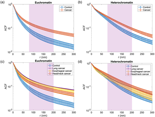
A profound characteristic of field cancerization is alterations in chromatin packing. This study aimed to quantify these alterations using electron microscopy image analysis of buccal mucosa cells of laryngeal, esophageal, and lung cancer patients. Analysis was done on normal-appearing mucosa, believed to be within the cancerization field, and not tumor itself. Large-scale electron microscopy (nanotomy) images were acquired of cancer patients and controls. Within the nuclei, the chromatin packing of euchromatin and heterochromatin was characterized. Furthermore, the chromatin organization was quantified through chromatin packing density scaling. A significant difference was found between the cancer and control groups in the chromatin packing density scaling parameter for length scales below the optical diffraction limit (200 nm) in both the euchromatin (p = 0.002) and the heterochromatin (p = 0.006). The chromatin packing scaling analysis also indicated that the chromatin organization of cancer patients deviated significantly from the control group. They might allow for novel strategies for cancer risk stratification and diagnosis with high sensitivity. This could aid clinicians in personalizing screening strategies for high-risk patients and follow-up strategies for treated cancer patients.

Type
Biological Applications
Copyright
Copyright © The Author(s), 2021. Published by Cambridge University Press on behalf of the Microscopy Society of America

Access options

Get access to the full version of this content by using one of the access options below. (Log in options will check for institutional or personal access. Content may require purchase if you do not have access.)

Footnotes

The first two authors contributed equally to this work.

References

Angadi, PV, Savitha, JK, Rao, SS & Sivaranjini, Y (2012). Oral field cancerization: Current evidence and future perspectives. Oral Maxillofac Surg 16(2), 171180.CrossRefGoogle ScholarPubMed
Bauer, GM, Stypula-Cyrus, Y, Subramanian, H, Cherkezyan, L, Viswanathan, P, Zhang, D, Iyengar, R, Bagalkar, S, Derbas, J, Graff, T, Gladstein, S, Almassalha, LM, Chandler, JE, Roy, HK & Backman, V (2017). The transformation of the nuclear nanoarchitecture in human field carcinogenesis. Future Sci OA 206, 2017–0027.Google Scholar
Bugter, O, Hardillo, JA, Baatenburg de Jong, RJ, Amelink, A & Robinson, DJ (2018 a). Optical pre-screening for laryngeal cancer using reflectance spectroscopy of the buccal mucosa. Biomed Opt Express 9(10), 46654678.CrossRefGoogle ScholarPubMed
Bugter, O, Spaander, MCW, Bruno, MJ, Baatenburg de Jong, RJ, Amelink, A & Robinson, DJ (2018 b). Optical detection of field cancerization in the buccal mucosa of patients with esophageal cancer. Clin Transl Gastroenterol 9(4), 152.CrossRefGoogle ScholarPubMed
Bugter, O, van Brummelen, SE, van der Leest, KH, Aerts, J, Maat, A, Baatenburg de Jong, RJ, Amelink, A & Robinson, DJ (2019). Towards the optical detection of field cancerization in the buccal mucosa of patients with lung cancer. Trans Oncol 12(12), 15331538.CrossRefGoogle ScholarPubMed
Cherkezyan, L, Stypula-Cyrus, Y, Subramanian, H, White, C, Cruz, MD, Wali, RK, Goldberg, MJ, Bianchi, LK, Roy, HK & Backman, V (2014). Nanoscale changes in chromatin organization represent the initial steps of tumorigenesis: A transmission electron microscopy study. BMC Cancer 14(1), 189.CrossRefGoogle ScholarPubMed
de Boer, P, Pirozzi, NM, Wolters, AHG, Kuipers, J, Kusmartseva, I, Atkinson, MA, Campbell-Thompson, M & Giepmans, BNG (2020). Large-scale electron microscopy database for human type 1 diabetes. Nat Commun 11(1), 2475.CrossRefGoogle ScholarPubMed
De Gennes, P-G & Gennes, P-G (1979). Scaling Concepts in Polymer Physics. Ithaca, NY: Cornell University Press.Google Scholar
de Koning, HJ, van der Aalst, CM, de Jong, PA, Scholten, ET, Nackaerts, K, Heuvelmans, MA, Lammers, JJ, Weenink, C, Yousaf-Khan, U, Horeweg, N, van ’t Westeinde, S, Prokop, M, Mali, WP, Mohamed Hoesein, FAA, van Ooijen, PMA, Aerts, J, den Bakker, MA, Thunnissen, E, Verschakelen, J, Vliegenthart, R, Walter, JE, Ten Haaf, K, Groen, HJM & Oudkerk, M (2020). Reduced lung-cancer mortality with volume CT screening in a randomized trial. N Engl J Med 382(6), 503513.CrossRefGoogle ScholarPubMed
Domper Arnal, MJ, Ferrandez Arenas, A & Lanas Arbeloa, A (2015). Esophageal cancer: Risk factors, screening and endoscopic treatment in western and eastern countries. World J Gastroenterol 21(26), 79337943.CrossRefGoogle ScholarPubMed
Dravid, A (2019). Employing deep networks for image processing on small research datasets. Microsc Today 27(1), 1823.CrossRefGoogle Scholar
Evers, D, Hendriks, B, Lucassen, G & Ruers, T (2012). Optical spectroscopy: Current advances and future applications in cancer diagnostics and therapy. Future Oncol 8(3), 307320.CrossRefGoogle ScholarPubMed
Ferlay, J, Soerjomataram, I, Dikshit, R, Eser, S, Mathers, C, Rebelo, M, Parkin, DM, Forman, D & Bray, F (2015). Cancer incidence and mortality worldwide: Sources, methods and major patterns in GLOBOCAN 2012. Int J Cancer 136, E359E386.CrossRefGoogle ScholarPubMed
Flory, PJ (1949). The configuration of real polymer chains. J Chem Phys 17(3), 303310.CrossRefGoogle Scholar
Gerchman, S & Ramakrishnan, V (1987). Chromatin higher-order structure studied by neutron scattering and scanning transmission electron microscopy. Proc Natl Acad Sci USA 84(22), 78027806.CrossRefGoogle ScholarPubMed
Gladstein, S, Damania, D, Almassalha, LM, Smith, LT, Gupta, V, Subramanian, H, Rex, DK, Roy, HK & Backman, V (2018). Correlating colorectal cancer risk with field carcinogenesis progression using partial wave spectroscopic microscopy. Cancer Med 7(5), 21092120.CrossRefGoogle ScholarPubMed
Global Cancer Observatory (GCO). https://gco.iarc.fr/.Google Scholar
Hall, SF, Groome, PA, Rothwell, D & Dixon, PF (1999). Using TNM staging to predict survival in patients with squamous cell carcinoma of head & neck. Head Neck 21(1), 3038.3.0.CO;2-Y>CrossRefGoogle ScholarPubMed
He, K, Zhang, X, Ren, S & Sun, J (2016). Deep residual learning for image recognition. Proceedings of the IEEE Conference on Computer Vision and Pattern Recognition, pp. 770–778.CrossRefGoogle Scholar
Horeweg, N & de Koning, H (2014). The importance of screening for lung cancer. Expert Rev Respir Med 8(5), 597614.CrossRefGoogle ScholarPubMed
Huang, K, Li, Y, Shim, AR, Virk, RKA, Agrawal, V, Eshein, A, Nap, RJ, Almassalha, LM, Backman, V & Szleifer, I (2020). Physical and data structure of 3D genome. Sci Adv 6(2), eaay4055.CrossRefGoogle ScholarPubMed
Kingma, DP & Ba, J (2014). Adam: A method for stochastic optimization. arXiv:1412.6980.Google Scholar
Kollarova, H, Machova, L, Horakova, D, Janoutova, G & Janout, V (2007). Epidemiology of esophageal cancer–An overview article. Biomed Pap Med Fac Univ Palacky Olomouc Czech Repub 151(1), 1720.CrossRefGoogle ScholarPubMed
Kopelovich, L, Henson, DE, Gazdar, AF, Dunn, B, Srivastava, S, Kelloff, GJ & Greenwald, P (1999). Surrogate anatomic/functional sites for evaluating cancer risk: An extension of the field effect. Clin Cancer Res 5(12), 38993905.Google ScholarPubMed
Krishnatreya, M, Rahman, T, Kataki, AC, Das, A, Das, AK & Lahkar, K (2013). Synchronous primary cancers of the head and neck region and upper aero digestive tract: Defining high-risk patients. Indian J Cancer 50(4), 322326.CrossRefGoogle Scholar
Kuipers, J, Kalicharan, RD, Wolters, AH, van Ham, TJ & Giepmans, BN (2016). Large-scale scanning transmission electron microscopy (nanotomy) of healthy and injured zebrafish brain. J Vis Exp 111, 53635.Google Scholar
Le Gros, MA, Clowney, EJ, Magklara, A, Yen, A, Markenscoff-Papadimitriou, E, Colquitt, B, Myllys, M, Kellis, M, Lomvardas, S & Larabell, CA (2016). Soft X-ray tomography reveals gradual chromatin compaction and reorganization during neurogenesis in vivo. Cell Rep 17(8), 21252136.CrossRefGoogle ScholarPubMed
Li, Y, Eshein, A, Virk, R, Eid, A, Wu, W, Frederick, J, VanDerway, D, Gladstein, S, Huang, K & Anthony, N (2020). Nanoscale chromatin imaging and analysis (nano-ChIA) platform bridges 4-D chromatin organization with molecular function. Sci Adv 6(2), eaay4055.Google Scholar
Malhotra, J, Malvezzi, M, Negri, E, La Vecchia, C & Boffetta, P (2016). Risk factors for lung cancer worldwide. Eur Respir J 48(3), 889902.CrossRefGoogle ScholarPubMed
Marur, S & Forastiere, AA (2016). Head and neck squamous cell carcinoma: Update on epidemiology, diagnosis, and treatment. Mayo Clin Proc 91(3), 386396.CrossRefGoogle Scholar
Metzger, R, Wachowiak, R & Kluth, D (2011). Embryology of the early foregut. Semin Pediatr Surg 20(3), 136144.CrossRefGoogle ScholarPubMed
Ou, HD, Phan, S, Deerinck, TJ, Thor, A, Ellisman, MH & O'shea, CC (2017). ChromEMT: Visualizing 3D chromatin structure and compaction in interphase and mitotic cells. Science 357(6349), eaag0025.CrossRefGoogle ScholarPubMed
Pennathur, A, Gibson, MK, Jobe, BA & Luketich, JD (2013). Oesophageal carcinoma. Lancet 381(9864), 400412.CrossRefGoogle ScholarPubMed
Phillips-Cremins, JE, Sauria, ME, Sanyal, A, Gerasimova, TI, Lajoie, BR, Bell, JS, Ong, C-T, Hookway, TA, Guo, C & Sun, Y (2013). Architectural protein subclasses shape 3D organization of genomes during lineage commitment. Cell 153(6), 12811295.CrossRefGoogle ScholarPubMed
Rizvi, MA, Zaki, M, Afzal, M, Mane, M, Kumar, M, Shah, BA, Srivastav, S, Srikrishna, S, Peerzada, GM & Tabassum, SJ (2015). Nuclear blebbing of biologically active organoselenium compound towards human cervical cancer cell (HeLa): In vitro DNA/HSA binding, cleavage and cell imaging studies. Eur J Med Chem 90, 876888.CrossRefGoogle ScholarPubMed
Rogers, JD, Radosevich, AJ, Yi, J & Backman, V (2014). Modeling light scattering in tissue as continuous random media using a versatile refractive index correlation function. Selected topics in quantum electronics. IEEE J Sel Top Quantum Electron 20(2), 173186.CrossRefGoogle Scholar
Ronneberger, O, Fischer, P & Brox, T (2015). U-net: Convolutional networks for biomedical image segmentation. International Conference on Medical Image Computing and Computer-assisted Intervention, pp. 234241. Springer.CrossRefGoogle Scholar
Roy, HK, Hensing, T & Backman, V (2011). Nanocytology for field carcinogenesis detection: Novel paradigm for lung cancer risk stratification. Future Oncol 7(1), 13.CrossRefGoogle ScholarPubMed
Roy, HK, Subramanian, H, Damania, D, Hensing, TA, Rom, WN, Pass, HI, Ray, D, Rogers, JD, Bogojevic, A, Shah, M, Kuzniar, T, Pradhan, P & Backman, V (2010). Optical detection of buccal epithelial nanoarchitectural alterations in patients harboring lung cancer: Implications for screening. Cancer Res 70(20), 77487754.CrossRefGoogle ScholarPubMed
Sidransky, D (2008). The oral cavity as a molecular mirror of lung carcinogenesis. Cancer Prev Res (Phila) 1(1), 1214.CrossRefGoogle ScholarPubMed
Siegel, RL, Miller, KD & Jemal, A (2015). Cancer statistics, 2015. CA Cancer J Clin 65(1), 529.CrossRefGoogle ScholarPubMed
Sokol, E, Kramer, D, Diercks, GFH, Kuipers, J, Jonkman, MF, Pas, HH & Giepmans, BNG (2015). Large-scale electron microscopy maps of patient skin and Mucosa provide insight into pathogenesis of blistering diseases. J Invest Dermatol 135(7), 17631770.CrossRefGoogle ScholarPubMed
Subramanian, H, Pradhan, P, Liu, Y, Capoglu, IR, Rogers, JD, Roy, HK, Brand, RE & Backman, V (2009 a). Partial-wave microscopic spectroscopy detects subwavelength refractive index fluctuations: An application to cancer diagnosis. Opt Lett 34(4), 518520.CrossRefGoogle ScholarPubMed
Subramanian, H, Roy, HK, Pradhan, P, Goldberg, MJ, Muldoon, J, Brand, RE, Sturgis, C, Hensing, T, Ray, D, Bogojevic, A, Mohammed, J, Chang, JS & Backman, V (2009 b). Nanoscale cellular changes in field carcinogenesis detected by partial wave spectroscopy. Cancer Res 69(13), 53575363.CrossRefGoogle ScholarPubMed
Torre, LA, Bray, F, Siegel, RL, Ferlay, J, Lortet-Tieulent, J & Jemal, A (2015). Global cancer statistics, 2012. CA Cancer J Clin 65(2), 87108.CrossRefGoogle ScholarPubMed
van de Ven, S, Bugter, O, Hardillo, JA, Bruno, MJ, Baatenburg de Jong, RJ & Koch, AD (2019). Screening for head and neck second primary tumors in patients with esophageal squamous cell cancer: A systematic review and meta-analysis. United European Gastroenterol J 7(10), 13041311.Google ScholarPubMed
Virk, R, Wu, W, Almassalha, LM, Bauer, GM, Li, Y, VanDerway, D, Frederick, J, Zhang, D, Eshein, A & Szleifer, I (2020 a). Disordered chromatin packing regulates ensemble gene expression and phenotypic plasticity. Biophys J 118(3), 549a550a.CrossRefGoogle Scholar
Virk, RKA, Wu, W, Almassalha, LM, Bauer, GM, Li, Y, VanDerway, D, Frederick, J, Zhang, D, Eshein, A, Roy, HK, Szleifer, I & Backman, V (2020 b). Disordered chromatin packing regulates phenotypic plasticity. Sci Adv 6(2), eaax6232.CrossRefGoogle ScholarPubMed
Zhang, Z, Liu, Q & Wang, Y (2018). Road extraction by deep residual U-net. IEEE Geosci Remote Sens Lett 15(5), 749753.CrossRefGoogle Scholar