Hostname: page-component-78c5997874-ndw9j Total loading time: 0 Render date: 2024-11-10T07:09:14.291Z Has data issue: false hasContentIssue false

Improving Quantitative EDS Chemical Analysis of Alloy Nanoparticles by PCA Denoising: Part I, Reducing Reconstruction Bias

Published online by Cambridge University Press:  03 January 2022
Murilo Moreira
Affiliation:
Instituto de Física “Gleb Wataghin”, Universidade Estadual de Campinas-UNICAMP, Campinas, SP 13083-859, Brazil
Matthias Hillenkamp
Affiliation:
Instituto de Física “Gleb Wataghin”, Universidade Estadual de Campinas-UNICAMP, Campinas, SP 13083-859, Brazil Institute of Light and Matter, University Lyon, Université Claude Bernard Lyon 1, CNRS, UMR5306, Villeurbanne F-69622, France
Giorgio Divitini
Affiliation:
Department of Materials Science and Metallurgy, University of Cambridge, Cambridge CB3 0FS, UK
Luiz H. G. Tizei
Affiliation:
Laboratoire de Physique des Solides, Université Paris-Saclay, CNRS, Orsay 91405, France
Caterina Ducati
Affiliation:
Department of Materials Science and Metallurgy, University of Cambridge, Cambridge CB3 0FS, UK
Monica A. Cotta
Affiliation:
Instituto de Física “Gleb Wataghin”, Universidade Estadual de Campinas-UNICAMP, Campinas, SP 13083-859, Brazil
Varlei Rodrigues
Affiliation:
Instituto de Física “Gleb Wataghin”, Universidade Estadual de Campinas-UNICAMP, Campinas, SP 13083-859, Brazil
Daniel Ugarte*
Affiliation:
Instituto de Física “Gleb Wataghin”, Universidade Estadual de Campinas-UNICAMP, Campinas, SP 13083-859, Brazil
*
*Corresponding author: Daniel Ugarte, E-mail: dmugarte@ifi.unicamp.br
Get access

Abstract

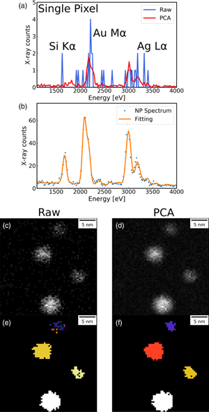
Scanning transmission electron microscopy is a crucial tool for nanoscience, achieving sub-nanometric spatial resolution in both image and spectroscopic studies. This generates large datasets that cannot be analyzed without computational assistance. The so-called machine learning procedures can exploit redundancies and find hidden correlations. Principal component analysis (PCA) is the most popular approach to denoise data by reducing data dimensionality and extracting meaningful information; however, there are many open questions on the accuracy of reconstructions. We have used experiments and simulations to analyze the effect of PCA on quantitative chemical analysis of binary alloy (AuAg) nanoparticles using energy-dispersive X-ray spectroscopy. Our results demonstrate that it is possible to obtain very good fidelity of chemical composition distribution when the signal-to-noise ratio exceeds a certain minimal level. Accurate denoising derives from a complex interplay between redundancy (data matrix size), counting noise, and noiseless data intensity variance (associated with sample chemical composition dispersion). We have suggested several quantitative bias estimators and noise evaluation procedures to help in the analysis and design of experiments. This work demonstrates the high potential of PCA denoising, but it also highlights the limitations and pitfalls that need to be avoided to minimize artifacts and perform reliable quantification.

Type
Software and Instrumentation
Copyright
Copyright © The Author(s), 2022. Published by Cambridge University Press on behalf of the Microscopy Society of America

Access options

Get access to the full version of this content by using one of the access options below. (Log in options will check for institutional or personal access. Content may require purchase if you do not have access.)

References

Alloyeau, D, Mottet, C & Ricolleau, C (2012). Nanoalloys: Synthesis, Structure and Properties. London: Springer-Verlag.CrossRefGoogle Scholar
Belter, M, Sajnóg, A & Barałkiewicz, D (2014). Over a century of detection and quantification capabilities in analytical chemistry – Historical overview and trends. Talanta 129, 606616.CrossRefGoogle Scholar
Binns, C (2014). Nanomagnetism: Fundamentals and Applications, vol. 6. Amsterdam, Netherlands: Elsevier.Google Scholar
Braidy, N, Jakubek, ZJ, Simard, B & Botton, GA (2008). Quantitative energy dispersive X-ray microanalysis of electron beam-sensitive alloyed nanoparticles. Microsc Microanal 14, 166175.CrossRefGoogle ScholarPubMed
Brown, KA, Brittman, S, Maccaferri, N, Jariwala, D & Celano, U (2020). Machine learning in nanoscience: Big data at small scales. Nano Lett 20, 210.CrossRefGoogle ScholarPubMed
Burke, MG, Watanabe, M, Williams, DB & Hyde, JM (2006). Quantitative characterization of nanoprecipitates in irradiated low-alloy steels: Advances in the application of FEG-STEM quantitative microanalysis to real materials. J Mater Sci 41, 45124522.CrossRefGoogle Scholar
Cliff, G & Lorimer, GW (1975). The quantitative analysis of thin specimens. J Microsc 103, 203207.CrossRefGoogle Scholar
Cueva, P, Hovden, R, Mundy, JA, Xin, HL & Muller, DA (2012). Data processing for atomic resolution electron energy loss spectroscopy. Microsc Microanal 18, 667675.CrossRefGoogle ScholarPubMed
Currie, LA (1968). Limits for qualitative detection and quantitative determination. Application to radiochemistry. Anal Chem 40, 586593.CrossRefGoogle Scholar
Currie, LA (1999). Detection and quantification limits: Origins and historical overview. Anal Chim Acta 391, 127134.CrossRefGoogle Scholar
de la Peña, F, Ostasevicius, T, Fauske, VT, Burdet, P, Jokubauskas, P, Nord, M, Prestat, E, Sarahan, M, MacArthur, KE, Johnstone, DN, Taillon, J, Caron, J, Furnival, T, Eljarrat, A, Mazzucco, S, Migunov, V, Aarholt, T, Walls, M, Winkler, F, Martineau, B, Donval, G, Hoglund, ER, Alxneit, I, Hjorth, I, Zagonel, LF, Garmannslund, A, Gohlke, C, Iyengar, I & Chang, H-W (2017). hyperspy/hyperspy: HyperSpy 1.3. doi:10.5281/ZENODO.583693.CrossRefGoogle Scholar
de Sá, ADT, Oiko, VTA, di Domenicantonio, G & Rodrigues, V (2014). New experimental setup for metallic clusters production based on hollow cylindrical magnetron sputtering. J Vac Sci Technol B 32, 061804.CrossRefGoogle Scholar
Egerton, RF (2012). Mechanisms of radiation damage in beam-sensitive specimens, for TEM accelerating voltages between 10 and 300 kV. Microsc Res Tech 75, 15501556.CrossRefGoogle ScholarPubMed
Egerton, RF (2019). Radiation damage to organic and inorganic specimens in the TEM. Micron 119, 7287.CrossRefGoogle ScholarPubMed
Egerton, RF, McLeod, R, Wang, F & Malac, M (2010). Basic questions related to electron-induced sputtering in the TEM. Ultramicroscopy 110, 991997.CrossRefGoogle Scholar
Faber, NM, Meinders, MJ, Geladi, P, Sjiistrijm, M, Buydens, LMC & Kateman, G (1995 a). Random error bias in principal component analysis. Part I. Derivation of theoretical predictions. Anal Chim Acta 304, 257271.CrossRefGoogle Scholar
Faber, NM, Meinders, MJ, Geladi, P, Sjiistrijm, M, Buydens, LMC & Kateman, G (1995 b). Random error bias in principal component analysis. Part II. Application of theoretical predictions to multivariate problems. Anal Chim Acta 304, 273283.CrossRefGoogle Scholar
Ferrando, R (2016). Structure and Properties of Nanoalloys in Frontiers of Nanoscience. Amsterdam: Elsevier. pp. 2337.Google Scholar
Hawkes, P & Spence, JCH (2019). Springer Handbook of Microscopy. Switzerland: Springer.CrossRefGoogle Scholar
Heiz, U & Landman, UE (2007). Nanocatalysis. Heidelberg, Berlin: Springer.CrossRefGoogle Scholar
Jolliffe, IT (2002). Principal Component Analysis, 2nd ed. New York: Springer-Verlag.Google Scholar
Jolliffe, IT & Cadima, J (2016). Principal component analysis: A review and recent developments. Philos Trans A 374, 20150202.CrossRefGoogle ScholarPubMed
Jones, L, Varambhia, A, Beanland, R, Kepaptsoglou, D, Griffiths, I, Ishizuka, A, Azough, F, Freer, R, Ishizuka, K, Cherns, D, Ramasse, QM, Lozano-Perez, S & Nellist, PD (2018). Managing dose-, damage- and data-rates in multi-frame spectrum-imaging. Microscopy 67, 98113.CrossRefGoogle ScholarPubMed
Keenan, MR & Kotula, PG (2004). Accounting for poisson noise in the multivariate analysis of TOF-SIMS spectrum images. Surf Interface Anal 36, 203212.CrossRefGoogle Scholar
Kotula, PG & Keenan, MR (2006). Application of multivariate statistical analysis to STEM X-ray spectral images: Interfacial analysis in microelectronics. Microsc Microanal 12, 538544.CrossRefGoogle Scholar
Kotula, PG & Van Benthem, MH (2015). Revisiting noise scaling for multivariate statistical analysis. Microsc Microanal 21, 14231424.CrossRefGoogle Scholar
Lichtert, S & Verbeeck, J (2013). Statistical consequences of applying a PCA filter on EELS spectrum images. Ultramicroscopy 125, 3542.CrossRefGoogle ScholarPubMed
Lyman, CE, Lakis, RE & Stenger, HG Jr (1995). X-ray emission spectrometry of phase separation in PtRh nanoparticles for nitric oxide reduction. Ultramicroscopy 58, 2534.CrossRefGoogle Scholar
Malinowski, ER (2002). Factor Analysis in Chemistry, 3rd ed. New York: Wiley.Google Scholar
Moreira, M, Hillenkamp, M, Divitini, G, Tizei, LHG, Ducati, C, Cotta, MA, Rodrigues, V & Ugarte, D (2021). Improving quantitative EDS chemical analysis of alloy nanoparticles by PCA denoising: Part II, uncertainty intervals. Submitted.Google Scholar
Mukherjee, P, Manchanda, P, Kumar, P, Zhou, L, Kramer, MJ, Kashyap, A, Skomski, R, Sellmyer, D & Shield, JE (2014). Size-induced chemical and magnetic ordering in individual Fe–Au nanoparticles. ACS Nano 8, 81138120.CrossRefGoogle ScholarPubMed
Mukherjee, P, Zhang, Y, Kramer, MJ, Lewis, LH & Shield, JE (2012). L10 structure formation in slow-cooled Fe–Au nanoclusters. Appl Phys Lett 100, 211911.CrossRefGoogle Scholar
Nadler, B (2008). Finite sample approximation results for principal component analysis: A matrix perturbation approach. Ann Stat 36, 27912817.CrossRefGoogle Scholar
Nadler, B (2009). Discussion. J Am Stat Assoc 104, 694697.CrossRefGoogle Scholar
Odom, TW & Schatz, GC (2011). Introduction to plasmonics. Chem Rev 111, 36673668.CrossRefGoogle ScholarPubMed
Parish, CM & Brewer, LN (2010). Multivariate statistics applications in phase analysis of STEM-EDS spectrum images. Ultramicroscopy 110, 134143.CrossRefGoogle ScholarPubMed
Pennycook, SJ & Nellist, PD (2011). Scanning Transmission Electron Microscopy. New York: Springer.CrossRefGoogle ScholarPubMed
Potapov, P (2016). Why principal component analysis of STEM spectrum images results in abstract, uninterpretable loadings? Ultramicroscopy 160, 197212.CrossRefGoogle ScholarPubMed
Potapov, P (2017). On the loss of information in PCA of spectrum-images. Ultramicroscopy 182, 191194.CrossRefGoogle ScholarPubMed
Potapov, P & Lubk, A (2019). Optimal principal component analysis of STEM XEDS spectrum images. Adv Struct Chem Imag 5, 4.CrossRefGoogle ScholarPubMed
Rossouw, D, Burdet, P, de la Peña, F, Ducati, C, Knappett, BR, Wheatley, AEH & Midgley, PA (2015). Multicomponent signal unmixing from nanoheterostructures: Overcoming the traditional challenges of nanoscale X-ray analysis via machine learning. Nano Lett 15, 27162720.CrossRefGoogle ScholarPubMed
Schlossmacher, P, Klenov, DO & Freitag, B (2010). Enhanced detection sensitivity with a new windowless XEDS system for AEM based on silicon drift detector technology. Microsc Today 18, 1420.CrossRefGoogle Scholar
Spiegelberg, J & Rusz, J (2017). Can we use PCA to detect small signals in noisy data? Ultramicroscopy 172, 4046.CrossRefGoogle ScholarPubMed
Thomas, JR, Leary, RK, Eggeman, AS & Midgley, PA (2015). The rapidly changing face of electron microscopy. Chem Phys Lett 631–632, 103113.CrossRefGoogle Scholar
Titchmarsh, JM (1999). EDX spectrum modelling and multivariate analysis of sub-nanometer segregation. Micron 30, 159171.CrossRefGoogle Scholar
Titchmarsh, JM & Dumbill, S (1996). Multivariate statistical analysis of FEG STEM EDX spectra. J Microsc 184, 195207.CrossRefGoogle Scholar
van der Walt, S, Schönberger, JL, Nunez-Iglesias, J, Boulogne, F, Warner, JD, Yager, N, Gouillart, E & Yu, T (2014). The scikit-image contributors scikit-image: Image processing in python. PeerJ 2, e453.CrossRefGoogle ScholarPubMed
Verbeeck, J & Van Aert, S (2004). Model based quantification of EELS spectra. Ultramicroscopy 101, 207224.CrossRefGoogle ScholarPubMed
Watanabe, M, Kanno, M & Okunishi, E (2010). Atomic resolution elemental mapping by EELS and XEDS in aberration corrected STEM. Jeol News 45, 812.Google Scholar
Williams, DB & Carter, CB (2009). Transmission Electron Microscopy Part 1: Basics, 2nd ed. New York: Springer.CrossRefGoogle Scholar
Williams, DB & Carter, CB (2016). Transmission Electron Microscopy: Diffraction, Imaging, and Spectroscopy. New York: Springer.Google Scholar
Zaluzec, NJ (2019). Improving the sensitivity of X-ray microanalysis in the analytical electron microscope. Ultramicroscopy 203, 163169.CrossRefGoogle ScholarPubMed
Supplementary material: PDF

Moreira et al. supplementary material

Moreira et al. supplementary material

Download Moreira et al. supplementary material(PDF)
PDF 1.1 MB