Hostname: page-component-cd9895bd7-mkpzs Total loading time: 0 Render date: 2024-12-28T17:02:15.306Z Has data issue: false hasContentIssue false

Pathological and Immunohistochemical Microscopy of Natural Cases of Canine and Feline Neoplastic Mammary Lesions

Published online by Cambridge University Press:  15 June 2021
Maggie F. Tawfik
Affiliation:
Pathology Department, Faculty of Veterinary Medicine, Alexandria University, Edfina22758, Egypt
Samah S. Oda
Affiliation:
Pathology Department, Faculty of Veterinary Medicine, Alexandria University, Edfina22758, Egypt
Asmaa F. Khafaga*
Affiliation:
Pathology Department, Faculty of Veterinary Medicine, Alexandria University, Edfina22758, Egypt
*
*Author for correspondence: Asmaa F. Khafaga, E-mail: asmaa.khafaga@alexu.edu.eg
Get access

Abstract

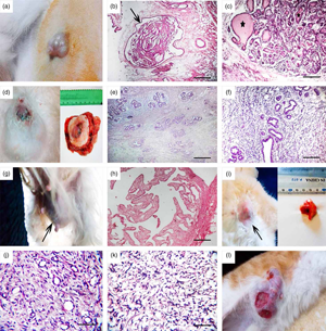
Mammary cancer is the second most common tumor worldwide. Small animal mammary neoplasms provide an outstanding model to study cancer in humans, as tumors in both share a similar environment, histopathologic features, and biological behavior. This study aims to investigate the percentage and microscopy of breast tumors in affected dogs and cats; its relationship to breed, age, and sex; and the immunohistochemical expression of estrogen receptor (ER), progesterone receptor (PR), Ki-67, and cytokeratin 8. Twenty-four females (12 dogs and 12 cats) and one male were examined from February 2018 to February 2020. The highest percentage of mammary neoplasia from the highest to the lowest manifested as tubular carcinoma, leiomyosarcoma, fibroadenoma, and cystic papillary carcinoma. The current study reported the second micropapillary invasive carcinoma in a male cat and the third lipid-rich carcinoma in a female cat. Although tubular carcinoma was the most common mammary neoplasm in cats, leiomyosarcoma was the most common in dogs. The immunohistochemical staining revealed diffuse and intense cytoplasmic immunoreactivity for cytokeratin 8 in lipid-rich carcinomas. However, moderate expression of ER in benign tumors and slight to moderate ER expression in malignant mammary lesions were reported. On the contrary, there was a negative PR expression in benign lesion. It could be concluded that a close relationship between ER expression and nuclear antigen Ki-67 was found.

Type
Biological Applications
Copyright
Copyright © The Author(s), 2021. Published by Cambridge University Press on behalf of the Microscopy Society of America

Access options

Get access to the full version of this content by using one of the access options below. (Log in options will check for institutional or personal access. Content may require purchase if you do not have access.)

References

Abdelmegeed, SM & Mohammed, S (2018). Canine mammary tumors as a model for human disease. Oncology Letters 15(6), 81958205.Google ScholarPubMed
Al-Mansour, MA, Kubba, MA, Al-Azreg, SA & Dribika, SA (2018). Comparative histopathology and immunohistochemistry of human and canine mammary tumors. Open Vet J 8(3), 243249.CrossRefGoogle ScholarPubMed
Andrade, FH, Figueiroa, FC, Bersano, PR, Bissacot, DZ & Rocha, NS (2010). Malignant mammary tumor in female dogs: Environmental contaminants. Diagn Pathol 5(1), 15.CrossRefGoogle ScholarPubMed
Badowska-Kozakiewicz, AM (2012). Prospective study of tumor markers as prognostic factors in the histopathological differential diagnosis of mammary gland neoplasms in female canines. In A Bird's-Eye View of Veterinary Medicine, Perez-Marin CC (Ed.), p. 199. London, UK: Intechopen.Google Scholar
Bancroft, JD & Gamble, M (Eds.) (2008). Theory and Practice of Histological Techniques. Amsterdam, Netherlands: Elsevier Health Sciences.Google Scholar
Bergman, PJ (2003). Clinical techniques in small animal molecular oncology. Clin Tech Small Anim Pract 18(2), 8891.CrossRefGoogle ScholarPubMed
Campos, CBD, Gamba, CDO, Damasceno, KA, Lavalle, GE & Cassali, GD (2016). Glycogen-rich clear cell carcinoma of the feline mammary gland: Case report. Arq Bras Med Vet Zootec 68(5), 11171120.CrossRefGoogle Scholar
Cassali, GD, de Campos, CB, Bertagnolli, AC, Estrela-Lima, A, Lavalle, GE, Damasceno, KA, Di Nardi, AB, Cogliati, B, da Costa, FV, Sobral, R & Di Santis, GW (2018). Consensus for the diagnosis, prognosis and treatment of feline mammary tumors. Braz J Vet Res Anim Sci 55(2), e135084.CrossRefGoogle Scholar
Chetty, R, Cooper, K & Gown, AM (2016). Antibodies. In Leong's Manual of Diagnostic Antibodies for Immunohistology, Chetty, R, Cooper, K & Gown, AM (Eds.), 3rd ed. pp. 397398. Cambridge, UK: Cambridge University Press.CrossRefGoogle Scholar
Chi, D, Berchuck, A, Dizon, DS & Yashar, CM (2017). Principles and Practice of Gynecologic Oncology. Baltimore, USA: Lippincott Williams & Wilkins.Google Scholar
De Las Mulas, JM, Millán, Y & Dios, R (2005). A prospective analysis of immunohistochemically determined estrogen receptor α and progesterone receptor expression and host and tumor factors as predictors of disease-free period in mammary tumors of the dog. Vet Pathol 42(2), 200212.CrossRefGoogle ScholarPubMed
Erin, E (2019). Cat Diagnosed with Mammary Gland Fibroadenomatous Hyperplasia. Texas A&M Veterinary Medical Diagnostic Laboratory.Google Scholar
Filgueira, KD, de Macêdo, LB, de Medeiros Oliveira, IVP, Pimentel, MML, da Costa Reis, PFC & Júnior, AR (2015). Histopathological features of mammary gland tumors in native domestic female cats from the State of Rio Grande do Norte, Brazil. Acta Sci Vet 43, 1304.Google Scholar
Gal, AF, Cătoi, C, Taulescu, M, Miclăuș, V, Nagy, A, Marica, R, Popa, R & Tăbăran, F (2017). A metastatic lipid-rich carcinoma of the mammary gland in a female cat: Clinicopathological. histopa-tho logical and immunohistochemical features. Bull UASVM Vet Med 74, 2.Google Scholar
Gama, A (2011). A novel myoepithelial cell marker in canine mammary tissue. Vet J 190(3), 303304.CrossRefGoogle ScholarPubMed
Geraldes, M, Gärtner, F & Schmitt, F (2000). Immunohistochemical study of hormonal receptors and cell proliferation in normal canine mammary glands and spontaneous mammary tumours. Vet Rec 146(14), 403406.CrossRefGoogle ScholarPubMed
Gizinski, S, Boryczko, Z, Katkiewicz, M & Bostedt, H (2003). Ki-67 protein as a prognostic factor in mammary gland tumors in female dogs. Med Weter 59(10), 888891.Google Scholar
Goldschmidt, M, Peña, L, Rasotto, R & Zappulli, V (2011). Classification and grading of canine mammary tumors. Vet Pathol 48(1), 117131.CrossRefGoogle ScholarPubMed
Gupta, N & Tiwari, SK (2009). Study on incidence. Histopathological features and surgical management of neoplasms in canine. Vet World 2, 10.Google Scholar
Hassan, E, El-Neweshy, M, Hassan, M & Noreldin, A (2019). Thymoquinone attenuates testicular and spermotoxicity following subchronic lead exposure in male rats: Possible mechanisms are involved. Life Sci 230, 132140.CrossRefGoogle ScholarPubMed
Hsu, SM, Raine, L & Fanger, HX (1981). Use of avidin-biotin-peroxidase complex (ABC) in immunoperoxidase techniques: A comparison between ABC and unlabeled antibody (PAP) procedures. J Histochem Cytochem 29(4), 577580.CrossRefGoogle ScholarPubMed
Kabel, AM & Baali, FH (2015). Breast cancer: Insights into risk factors, pathogenesis, diagnosis and management. J Cancer Res Treat 3(2), 2833.Google Scholar
Kamstock, DA, Fredrickson, R & Ehrhart, EJ (2005). Lipid-rich carcinoma of the mammary gland in a cat. Vet Pathol 42(3), 360362.CrossRefGoogle ScholarPubMed
Khafaga, AF, Noreldin, AE & Taha, AE (2019). The adaptogenic anti-ageing potential of resveratrol against heat stress-mediated liver injury in aged rats: Role of HSP70 and NF-kB signalling. J Therm Biol 83, 821.CrossRefGoogle ScholarPubMed
Kutzler, M (2020). Overview of Mammary Tumors. Kenilworth, NJ, USA: Merck Sharp & Dohme Corp., a subsidiary of Merck & Co, Inc.Google Scholar
Lana, SE, Rutteman, GR & Withrow, SJ (2007). Tumors of the mammary gland. In Withrow & MacEwen's Small Animal Clinical Oncology, Withrow, SJ (Ed.), pp. 619636. Philadelphia, USA: WB Saunders.CrossRefGoogle Scholar
Lichtenbeld, HC, Barendsz-Janson, AF, van Essen, H, Struijker Boudier, H, Griffioen, AW & Hillen, HF (1998). Angiogenic potential of malignant and non-malignant human breast tissues in an in vivo angiogenesis model. Int J Cancer 77(3), 455459.3.0.CO;2-5>CrossRefGoogle Scholar
Little, S (2013). Management of reproduction and related disorders. In BSAVA Manual of Feline Practice, Scherk, M, Little, S, Maddison, JE, Murrell, J, Hibbert, A & Taylor, S (Eds.), pp. 399412. Gloucester, UK: BSAVA Library.CrossRefGoogle Scholar
Mayayo, SL, Bo, S & Pisu, MC (2018). Mammary fibroadenomatous hyperplasia in a male cat. J Feline Med Surg Open Rep 4(1), 2055116918760155.Google Scholar
Metawea, OR, Abdelmoneem, MA, Haiba, NS, Khalil, HH, Teleb, M, Elzoghby, AO, Khafaga, AF, Noreldin, AE, Albericio, F & Khattab, SN (2021). A novel “smart” PNIPAM-based copolymer for breast cancer targeted therapy: Synthesis, and characterization of dual pH/temperature-responsive lactoferrin-targeted PNIPAM-co-AA. Colloids Surf B 202, 111694.CrossRefGoogle ScholarPubMed
Millanta, F, Calandrella, M, Bari, G, Niccolini, M, Vannozzi, I & Poli, A (2005). Comparison of steroid receptor expression in normal, dysplastic, and neoplastic canine and feline mammary tissues. Res Vet Sci 79(3), 225232.CrossRefGoogle ScholarPubMed
Millanta, F, Silvestri, G, Vaselli, C, Citi, S, Pisani, G, Lorenzi, D & Poli, A (2006). The role of vascular endothelial growth factor and its receptor Flk-1/KDR in promoting tumour angiogenesis in feline and canine mammary carcinomas: A preliminary study of autocrine and paracrine loops. Res Vet Sci 81(3), 350357.CrossRefGoogle ScholarPubMed
Mills, SW, Musil, KM, Davies, JL, Hendrick, S, Duncan, C, Jackson, B, Kidney, H, Philibert, B, Wobeser, K, Simko, E & Simko, E (2015). Prognostic value of histologic grading for feline mammary carcinoma: A retrospective survival analysis. Vet Pathol 52(2), 238249.CrossRefGoogle ScholarPubMed
Mishra, P, Vagha, S, Shukla, S, Acharya, S & Goyal, A (2020). Assessment of cytokeratin expression in carcinoma breast. J Evol Med Dental Sci 35(9), 25452549.CrossRefGoogle Scholar
Morris, JS, Nixon, C, King, OJ, Morgan, IM & Philbey, AW (2009). Expression of TopBP1 in canine mammary neoplasia in relation to histological type, Ki67, ERα and p53. Vet J 179(3), 422429.CrossRefGoogle ScholarPubMed
Morrison, WB (2002). Cancer in Dogs and Cats: Medical and Surgical Management. Wyoming, USA: Teton NewMedia.Google Scholar
Muto, T, Wakui, S, Takahashi, H, Maekawa, S, Masaoka, T, Ushigome, S & Furusato, M (2000). P53 gene mutations occurring in spontaneous benign and malignant mammary tumors of the dog. Vet Pathol 37(3), 248253.CrossRefGoogle ScholarPubMed
Nieto, A, Pena, L, Pérez-Alenza, MD, Sanchez, MA, Flores, JM & Castano, M (2000). Immunohistologic detection of estrogen receptor alpha in canine mammary tumors: Clinical and pathologic associations and prognostic significance. Vet Pathol 37(3), 239247.CrossRefGoogle ScholarPubMed
Novosad, CA (2003). Principles of treatment for mammary gland tumors. Clin Tech Small Anim Pract 18(2), 107109.CrossRefGoogle ScholarPubMed
Peleteiro, MC (1994). Tumores mamários na cadela e na gata. Rev Port Ciênc Vet 89(509), 1029.Google Scholar
Pires, I, Silva, F & Queiroga, FL (2008). Invasive micropapillary mammary carcinoma in a male cat: First report. Vet Pathol 45(5), 723.CrossRefGoogle Scholar
Rutteman, GR, Misdorp, W, Blankenstein, MA & Van den Brom, WE (1988). Oestrogen (ER) and progestin receptors (PR) in mammary tissue of the female dog: Different receptor profile in non-malignant and malignant states. Br J Cancer 58(5), 594599.CrossRefGoogle ScholarPubMed
Sánchez, J, Buendía, AJ, Vilafranca, M, Velarde, R, Altimara, J, Martínez, CM & Navarro, JA (2005). Canine carcinosarcomas in the head. Vet Pathol 42(6), 828833.CrossRefGoogle ScholarPubMed
Scholzen, T & Gerdes, J (2000). The Ki-67 protein: From the known and the unknown. J Cell Physiol 182(3), 311322.3.0.CO;2-9>CrossRefGoogle ScholarPubMed
Seixas, F, Palmeira, C, Pires, MA & Lopes, C (2007). Mammary invasive micropapillary carcinoma in cats: Clinicopathologic features and nuclear DNA content. Vet Pathol 44(6), 842848.CrossRefGoogle ScholarPubMed
Shafiee, R, Javanbakh, J, Atyabi, N, Kheradmand, P, Kheradmand, D, Bahrami, A, Daraei, H & Khadivar, F (2016). Diagnosis, classification and grading of canine mammary tumours as a model to study human breast cancer: An clinico-cytohistopathological study with environmental factors influencing public health and medicine (Retraction of Vol 13, Art No 79, 2013).Google Scholar
Smith, MA, Ethridge, SB, Gibson, AN, Schmidt, KT & Sharp, JL (2020). The effects of artificially induced proestrus on heroin intake: A critical role for estradiol. Experimental and Clinical Psychopharmacology. doi: 10.1037/pha0000428.CrossRefGoogle ScholarPubMed
Sontas, BH, Ozyogurtcu, H, Gurel, A & Ekici, H (2009). Evaluation of clinical and pathological characteristics of 155 canines with mammary tumours: A retrospective study. Arch Med Vet 41(1), 5359.CrossRefGoogle Scholar
Sorenmo, KU, Kristiansen, VM, Cofone, MA, Shofer, FS, Breen, AM, Langeland, M, Mongil, CM, Grondahl, AM, Teige, J & Goldschmidt, MH (2009). Canine mammary gland tumours; a histological continuum from benign to malignant; clinical and histopathological evidence. Vet Comp Oncol 7(3), 162172.CrossRefGoogle ScholarPubMed
Sorenmo, KU, Shofer, FS & Goldschmidt, MH (2000). Effect of spaying and timing of spaying on survival of dogs with mammary carcinoma. J Vet Intern Med 14(3), 266270.CrossRefGoogle ScholarPubMed
Szczubial, M & Lopuszanski, W (2002). Prognosis in mammary gland tumors in bitches. Vet Med 58(40), 261264.Google Scholar
Taneja, P, Maglic, D, Kai, F, Zhu, S, Kendig, RD, Elizabeth, AF & Inoue, K (2010). Classical and novel prognostic markers for breast cancer and their clinical significance. Clin Med Insights: Oncol 4, CMO-S4773.CrossRefGoogle ScholarPubMed
Telang, NT, Katdare, M, Bradlow, HL & Osborne, MP (1997). Estradiol metabolism: An endocrine biomarker for modulation of human mammary carcinogenesis. Environ Health Perspect 105(Suppl 3), 559564.Google ScholarPubMed
Thuróczy, J, Reisvaag, GJK, Perge, E, Tibold, A, Szilágyi, J & Balogh, L (2007). Immunohistochemical detection of progesterone and cellular proliferation in canine mammary tumours. J Comp Pathol 137(2–3), 122129.CrossRefGoogle ScholarPubMed
Timmermans-Sprang, EP, Gracanin, A & Mol, JA (2017). Molecular signaling of progesterone, growth hormone, wnt, and HER in mammary glands of dogs, rodents, and humans: New treatment target identification. Front Vet Sci 4, 53.CrossRefGoogle ScholarPubMed
Toniti, W, Buranasinsup, S, Kongcharoen, A, Charoonrut, P, Puchadapirom, P & Kasorndorkbua, C (2009). Immunohistochemical determination of estrogen and progesterone receptors in canine mammary tumors. Asian Pac J Cancer Prev 10(5), 907911.Google ScholarPubMed
Van Dijk, JE, Gruys, E & Mouwen, JMV (2007). Color atlas of veterinary pathology: General morphological reactions of organs and tissues (No. V600 DIJc).Google Scholar
Veerle, F (2005). Immunohistochemical Study of Canine Mammary Gland Tumors. Uppsala, Sweden: Uppsala University.Google Scholar
Wey, N, Gutberlet, K & Khon, B (2000). Mammatumore bei der huding: Hormolle abhangigkeit unter besonderer berucksichtigung von 17 ß ostradiol und progesteron. Kleintierpraxis 45, 1931.Google Scholar
Yang, WY, Liu, CH, Chang, CJ, Lee, CC, Chang, KJ & Lin, CT (2006). Proliferative activity, apoptosis and expression of oestrogen receptor and Bcl-2 oncoprotein in canine mammary gland tumours. J Comp Pathol 134(1), 7079.CrossRefGoogle ScholarPubMed
Zuccari, DA, Santana, AE & Rocha, NS (2002). Expression of intermediate filaments in canine mammary tumor diagnosis. Arq Bras Med Vet Zootec 54, 586591.CrossRefGoogle Scholar