No CrossRef data available.
Published online by Cambridge University Press: 20 June 2022

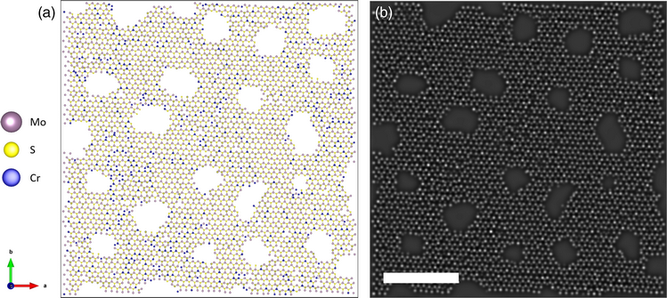
In recent years, atomic resolution imaging of two-dimensional (2D) materials using scanning transmission electron microscopy (STEM) has become routine. Individual dopant atoms in 2D materials can be located and identified using their contrast in annular dark-field (ADF) STEM. However, in order to understand the effect of these dopant atoms on the host material, there is now the need to locate and quantify them on a larger scale. In this work, we analyze STEM images of MoS2 monolayers that have been ion-implanted with chromium at ultra-low energies. We use functions from the open-source TEMUL Toolkit to create and refine an atomic model of an experimental image based on the positions and intensities of the atomic columns in the image. We then use the refined model to determine the likely composition of each atomic site. Surface contamination stemming from the sample preparation of 2D materials can prevent accurate quantitative identification of individual atoms. We disregard atomic sites from regions of the image with hydrocarbon surface contamination to demonstrate that images acquired using contaminated samples can give significant atom statistics from their clean regions, and can be used to calculate the retention rate of the implanted ions within the host lattice. We find that some of the implanted chromium ions have been successfully integrated into the MoS2 lattice, with 4.1% of molybdenum atoms in the transition metal sublattice replaced with chromium.
Present address: Max Planck Institute for the Science of Light and Max-Planck-Zentrum für Physik und Medizin, Erlangen, Germany.