Hostname: page-component-78c5997874-mlc7c Total loading time: 0 Render date: 2024-11-10T08:24:46.443Z Has data issue: false hasContentIssue false

Ultra-Microtome for the Preparation of TEM Specimens from Battery Cathodes

Published online by Cambridge University Press:  01 September 2020

Hanlei Zhang
Affiliation:
Engineering Research Center of Nano-Geomaterials of Ministry of Education, Faculty of Materials Science and Chemistry, China University of Geosciences, No. 68 Jincheng Street, East Lake High-Tech Development Zone, Wuhan430078, Hubei, P. R. China Materials Science and Engineering Program & Department of Mechanical Engineering, State University of New York, Binghamton, NY13902, USA NorthEast Center for Chemical Energy Storage, State University of New York, Binghamton, NY13902, USA
Chongmin Wang
Affiliation:
Environmental Molecular Sciences Laboratory, Pacific Northwest National Laboratory, Richland, WA99352, USA
Guangwen Zhou*
Affiliation:
Materials Science and Engineering Program & Department of Mechanical Engineering, State University of New York, Binghamton, NY13902, USA NorthEast Center for Chemical Energy Storage, State University of New York, Binghamton, NY13902, USA
*
*Author for correspondence: Guangwen Zhou, E-mail: gzhou@binghamton.edu
Get access

Abstract

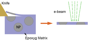
With the wide application of ultra-microtome sectioning in the preparation of transmission electron microscopy (TEM) specimens with bio- and organic materials, here, we report an ultra-microtome-based method for the preparation of TEM specimens from cathodes of Li-ion batteries. The ultra-microtome sectioning reduces the sample thickness to tens of nanometers and yields atomic resolution from the core region of particles of hundreds of nanometers. Analysis indicates that the mechanical cross-sectioning introduces no observable microstructural artifacts or structural damage, such as microcracking and nanoporosity. These results demonstrate the high efficiency of the ultra-microtome approach in preparing well-thinned specimens of particulate materials that allow for atomic-scale TEM imaging of a large number of sectioned particles in one single TEM specimen, thereby providing statistically significant results of the TEM analysis.

Type
Materials Science Applications
Copyright
Copyright © Microscopy Society of America 2020

Access options

Get access to the full version of this content by using one of the access options below. (Log in options will check for institutional or personal access. Content may require purchase if you do not have access.)

References

Ahn, JH & Peacor, DR (1986). Transmission electron microscope data for rectorite: Implications for the origin and structure of “fundamental particles”. Clays Clay Miner 34, 180186.Google Scholar
Antonovsky, A (1995). Ultramicrotomy of industrial alumina. Microsc Res Tech 31, 300307.CrossRefGoogle ScholarPubMed
Barna, A, Pécz, B & Menyhard, M (1999). TEM sample preparation by ion milling/amorphization. Micron 30, 267276.CrossRefGoogle Scholar
Bashir, A, Millar, RW, Gallacher, K, Paul, DJ, Darbal, AD, Stroud, R, Ballabio, A, Frigerio, J, Isella, G & MacLaren, I (2019). Strain analysis of a Ge micro disk using precession electron diffraction. J Appl Phys 126, 235701.CrossRefGoogle Scholar
Becker, O & Bange, K (1993). Ultramicrotomy: An alternative cross section preparation for oxidic thin films on glass. Ultramicroscopy 52, 7384. http://www.sciencedirect.com/science/article/pii/030439919390023Q.CrossRefGoogle Scholar
Boesenberg, U, Sokaras, D, Nordlund, D, Weng, T-C, Gorelov, E, Richardson, TJ, Kostecki, R & Cabana, J (2019). Electronic structure changes upon lithium intercalation into graphite–Insights from ex situ and operando x-ray Raman spectroscopy. Carbon 143, 371377.CrossRefGoogle Scholar
Brown, AP, Brydson, RMD & Hondow, NS (2014). Measuring in vitro cellular uptake of nanoparticles by transmission electron microscopy. J Phys Conf Ser 522, 12058.CrossRefGoogle Scholar
Burghardt, RC & Droleskey, R (2006). Transmission electron microscopy. Curr Protoc Microbiol 3, 2B.1.1.Google Scholar
Buseck, P, Cowley, J & Eyring, L (1989). High-Resolution Transmission Electron Microscopy and Associated Techniques. Oxford University Press.Google Scholar
Dai, JY, Tee, SF, Tay, CL, Song, ZG, Ansari, S, Er, E & Redkar, S (2001). Development of a rapid and automated TEM sample preparation method in semiconductor failure analysis and the study of the relevant TEM artifact. Microelectronics J 32, 221226.CrossRefGoogle Scholar
Flom, Y & Arsenault, RJ (1989). Effect of particle size on fracture toughness of SiC/Al composite material. Acta Metall 37, 24132423.CrossRefGoogle Scholar
Giannuzzi, LA & Stevie, FA (1999). A review of focused ion beam milling techniques for TEM specimen preparation. Micron 30, 197204.CrossRefGoogle Scholar
Glanvill, SR (1995). Ultramicrotomy of semiconductors and related materials. Microsc Res Tech 31, 275284.CrossRefGoogle ScholarPubMed
Graf, CM, Meinke, MC, Gao, Q, Hadam, S, Raabe, J, Sterry, W, Blume-Peytavi, U, Lademann, JM, Ruhl, E & Vogt, A (2009). Qualitative detection of single submicron and nanoparticles in human skin by scanning transmission X-ray microscopy. J Biomed Opt 14, 21015.CrossRefGoogle ScholarPubMed
Griffiths, AJV & Walther, T (2010). Quantification of carbon contamination under electron beam irradiation in a scanning transmission electron microscope and its suppression by plasma cleaning. J Phys Conf Ser 241, 12017.CrossRefGoogle Scholar
Harris, KM, Perry, E, Bourne, J, Feinberg, M, Ostroff, L & Hurlburt, J (2006). Uniform serial sectioning for transmission electron microscopy. J Neurosci 26, 1210112103.CrossRefGoogle ScholarPubMed
Haswell, R, McComb, DW & Smith, W (2003). Preparation of site-specific cross-sections of heterogeneous catalysts prepared by focused ion beam milling. J Microsc 211, 161166.CrossRefGoogle ScholarPubMed
He, J, Zhao, Y, Wang, Y, Wang, J, Zheng, J, Zhang, H, Zhou, G, Wang, C, Wang, S & Ma, X (2017). A Fe5C2 nanocatalyst for the preferential synthesis of ethanol via dimethyl oxalate hydrogenation. Chem Commun 53, 53765379.CrossRefGoogle ScholarPubMed
Hochella, MF Jr, Moore, JN, Golla, U & Putnis, A (1999). A TEM study of samples from acid mine drainage systems: Metal-mineral association with implications for transport. Geochim Cosmochim Acta 63, 33953406.CrossRefGoogle Scholar
Hughes, AE, Trinchi, A, Chen, FF, Yang, YS, Cole, IS, Sellaiyan, S, Carr, J, Lee, PD, Thompson, GE & Xiao, TQ (2014). The application of multiscale quasi 4D CT to the study of SrCrO4 distributions and the development of porous networks in epoxy-based primer coatings. Prog Org Coat 77, 19461956.CrossRefGoogle Scholar
Hwang, S, Chang, W, Kim, SM, Su, D, Kim, DH, Lee, JY, Chung, KY & Stach, EA (2014). Investigation of changes in the surface structure of LixNi0. 8Co0. 15Al0. 05O2 cathode materials induced by the initial charge. Chem Mater 26, 10841092.CrossRefGoogle Scholar
Ishitani, T & Yaguchi, T (1996). Cross-sectional sample preparation by focused ion beam: A review of ion-sample interaction. Microsc Res Tech 35, 320333.3.0.CO;2-Q>CrossRefGoogle ScholarPubMed
Karki, K, Huang, Y, Hwang, S, Gamalski, AD, Whittingham, MS, Zhou, G & Stach, EA (2016). Tuning the activity of oxygen in LiNi0. 8Co0. 15Al0. 05O2 battery electrodes. ACS Appl Mater Interfaces 8, 2776227771.CrossRefGoogle ScholarPubMed
Keller, LP, Messenger, S, Flynn, GJ, Clemett, S, Wirick, S & Jacobsen, C (2004). The nature of molecular cloud material in interplanetary dust. Geochim Cosmochim Acta 68, 25772589.CrossRefGoogle Scholar
Kisch, HJ (1991). Illite crystallinity: Recommendations on sample preparation, X-ray diffraction settings, and interlaboratory samples. J Metamorph Geol 9, 665670.CrossRefGoogle Scholar
Klepeis, SJ, Benedict, JP & Anderson, RM (1987). A grinding/polishing tool for TEM sample preparation. MRS Online Proceedings Library Archive 115, 179.CrossRefGoogle Scholar
Kumlutaş, D, Tavman, IH & Çoban, MT (2003). Thermal conductivity of particle filled polyethylene composite materials. Compos Sci Technol 63, 113117.CrossRefGoogle Scholar
Lezec, HJ, Musil, CR, Melngailis, J, Mahoney, LJ & Woodhouse, JD (1991). Dose-rate effects in focused-ion-beam implantation of Si into GaAs. J Vac Sci Technol B Microelectron Nanometer Struct Process Meas Phenom 9, 27092713.CrossRefGoogle Scholar
Li, J, Malis, T & Dionne, S (2006). Recent advances in FIB–TEM specimen preparation techniques. Mater Charact 57, 6470. http://www.sciencedirect.com/science/article/pii/S1044580305002780.CrossRefGoogle Scholar
Lin, F, Markus, IM, Nordlund, D, Weng, T-C, Asta, MD, Xin, HL & Doeff, MM (2014 a). Surface reconstruction and chemical evolution of stoichiometric layered cathode materials for lithium-ion batteries. Nat Commun 5, 3529.CrossRefGoogle ScholarPubMed
Lin, F, Nordlund, D, Markus, IM, Weng, T-C, Xin, HL & Doeff, MM (2014 b). Profiling the nanoscale gradient in stoichiometric layered cathode particles for lithium-ion batteries. Energy Environ Sci 7, 30773085.CrossRefGoogle Scholar
Liu, Y, Wang, R, Guo, X & Dai, J (2007). A cross-sectional TEM sample preparation method for films deposited on metallic substrates. Mater Charact 58, 666669.CrossRefGoogle Scholar
Lu, L, Han, X, Li, J, Hua, J & Ouyang, M (2013). A review on the key issues for lithium-ion battery management in electric vehicles. J Power Sources 226, 272288.CrossRefGoogle Scholar
Luft, JH (1961). Improvements in epoxy resin embedding methods. J Cell Biol 9, 409414.CrossRefGoogle ScholarPubMed
Malis, TF & Steele, D (1990). Ultramicrotomy for materials science. MRS Proceedings 199, 3. https://www.cambridge.org/core/article/ultramicrotomy-for-materials-science/1B9BFDF4FD1AFCBE26B6460374053D48.CrossRefGoogle Scholar
May, BM, Serrano-Sevillano, J, Dauphin, AL, Nazib, A, Lima, N, Casas-Cabanas, M & Cabana, J (2018). Effect of synthetic parameters on defects, structure, and electrochemical properties of layered oxide LiNi0. 80Co0. 15Al0. 05O2. J Electrochem Soc 165, A3537A3543.CrossRefGoogle Scholar
Mayer, J, Giannuzzi, LA, Kamino, T & Michael, J (2007). TEM sample preparation and FIB-induced damage. MRS Bull 32, 400407.CrossRefGoogle Scholar
McCaffrey, JP, Phaneuf, MW & Madsen, LD (2001). Surface damage formation during ion-beam thinning of samples for transmission electron microscopy. Ultramicroscopy 87, 97104.CrossRefGoogle ScholarPubMed
Mcmahon, G & Malis, T (1995). Ultramicrotomy of nanocrystalline materials. Microsc Res Tech 31, 267274.CrossRefGoogle ScholarPubMed
Messenger, S, Keller, LP, Stadermann, FJ, Walker, RM & Zinner, E (2003). Samples of stars beyond the solar system: Silicate grains in interplanetary dust. Science 300, 105108.CrossRefGoogle ScholarPubMed
Monticelli, O, Musina, Z, Russo, S & Bals, S (2007). On the use of TEM in the characterization of nanocomposites. Mater Lett 61, 34463450.CrossRefGoogle Scholar
Mori, D, Kobayashi, H, Shikano, M, Nitani, H, Kageyama, H, Koike, S, Sakaebe, H & Tatsumi, K (2009). Bulk and surface structure investigation for the positive electrodes of degraded lithium-ion cell after storage test using X-ray absorption near-edge structure measurement. J Power Sources 189, 676680.CrossRefGoogle Scholar
Mori, H, Miyoshi, H, Takeda, K, Yoneyama, H, Fujita, H, Iwata, Y, Otsuka, Y & Murata, Y (1992). TEM observation of iron oxide pillars in montmorillonite. J Mater Sci 27, 31973199.CrossRefGoogle Scholar
Mori, H, Sawada, M, Higuchi, T, Hasebe, K, Otsuka, N & Terano, M (1999). Direct observation of MgCl2-supported Ziegler catalysts by high resolution transmission electron microscopy. Macromol Rapid Commun 20, 245250.3.0.CO;2-R>CrossRefGoogle Scholar
Nanda, J, Remillard, J, O'Neill, A, Bernardi, D, Ro, T, Nietering, KE, Go, J & Miller, TJ (2011). Local state-of-charge mapping of lithium-ion battery electrodes. Adv Funct Mater 21, 32823290.CrossRefGoogle Scholar
Puttick, KE, Whitmore, LC, Chao, CL & Gee, AE (1994). Transmission electron microscopy of nanomachined silicon crystals. Philos Mag A 69, 91103.CrossRefGoogle Scholar
Quintana, C (1997). Ultramicrotomy for cross-sections of nanostructure. Micron 28, 217219. http://www.sciencedirect.com/science/article/pii/S0968432897000231.CrossRefGoogle Scholar
Reymond, OL & Pickett-Heaps, JD (1983). A routine flat embedding method for electron microscopy of microorganisms allowing selection and precisely orientated sectioning of single cells by light microscopy. J Microsc 130, 7984.CrossRefGoogle ScholarPubMed
Schaffer, M, Pfeffer, S, Mahamid, J, Kleindiek, S, Laugks, T, Albert, S, Engel, BD, Rummel, A, Smith, AJ, Baumeister, W & Plitzko, JM (2019). A cryo-FIB lift-out technique enables molecular-resolution cryo-ET within native Caenorhabditis elegans tissue. Nat Methods 16, 757762.CrossRefGoogle ScholarPubMed
Schaffer, M, Schaffer, B & Ramasse, Q (2012). Sample preparation for atomic-resolution STEM at low voltages by FIB. Ultramicroscopy 114, 6271.CrossRefGoogle ScholarPubMed
Schertel, A, Snaidero, N, Han, H-M, Ruhwedel, T, Laue, M, Grabenbauer, M & Möbius, W (2013). Cryo FIB-SEM: Volume imaging of cellular ultrastructure in native frozen specimens. J Struct Biol 184, 355360. http://www.sciencedirect.com/science/article/pii/S1047847713002657.CrossRefGoogle ScholarPubMed
Schrand, AM, Schlager, JJ, Dai, L & Hussain, SM (2010). Preparation of cells for assessing ultrastructural localization of nanoparticles with transmission electron microscopy. Nat Protoc 5, 744.CrossRefGoogle ScholarPubMed
Siu, C, Seymour, I, Britto, S, Zhang, H, Rana, J, Feng, J, Omenya, FO, Zhou, H, Chernova, N & Zhou, G (2018). Enabling multi-electron reaction of ɛ-VOPO4 to reach theoretical capacity for lithium-ion batteries. Chem Commun 54, 78027805.CrossRefGoogle ScholarPubMed
Stamenkovic, VR, Strmcnik, D, Lopes, PP & Markovic, NM (2017). Energy and fuels from electrochemical interfaces. Nat Mater 16, 5769.CrossRefGoogle Scholar
Sun, BB, Wang, YB, Wen, J, Yang, H, Sui, ML, Wang, JQ & Ma, E (2005). Artifacts induced in metallic glasses during TEM sample preparation. Scr Mater 53, 805809.CrossRefGoogle Scholar
Sun, F, Li, H, Leifer, K & Gamstedt, EK (2017). Rate effects on localized shear deformation during nanosectioning of an amorphous thermoplastic polymer. Int J Solids Struct 129, 4048.CrossRefGoogle Scholar
Tucker, DS, Jenkins, EJ & Hren, JJ (1985). Sectioning spherical aluminum oxide particles for transmission electron microscopy. J Electron Microsc Tech 2, 2933.CrossRefGoogle Scholar
Volkert, CA & Minor, AM (2007). Focused ion beam microscopy and micromachining. MRS Bull 32, 389399.CrossRefGoogle Scholar
Wang, K, Strunk, K, Zhao, G, Gray, JL & Zhang, P (2012). 3D structure determination of native mammalian cells using cryo-FIB and cryo-electron tomography. J Struct Biol 180, 318326. http://www.sciencedirect.com/science/article/pii/S1047847712001955.CrossRefGoogle ScholarPubMed
Williams, DB & Carter, CB (2009). Transmission Electron Microscopy: A Textbook for Materials Science. New York: Springer.CrossRefGoogle Scholar
Xiao, J, Chen, X, Sushko, PV, Sushko, ML, Kovarik, L, Feng, J, Deng, Z, Zheng, J, Graff, GL & Nie, Z (2012). High-performance LiNi0. 5Mn1. 5O4 spinel controlled by Mn3+ concentration and site disorder. Adv Mater 24, 21092116.CrossRefGoogle ScholarPubMed
Yan, P, Zheng, J, Gu, M, Xiao, J, Zhang, J-G & Wang, C-M (2017). Intragranular cracking as a critical barrier for high-voltage usage of layer-structured cathode for lithium-ion batteries. Nat Commun 8, 14101.CrossRefGoogle ScholarPubMed
Yang, H-S, Kim, D-J & Kim, H-J (2003). Rice straw–wood particle composite for sound absorbing wooden construction materials. Bioresour Technol 86, 117121.CrossRefGoogle ScholarPubMed
Yoshida, J, Nakanishi, S, Iba, H, Abe, H & Naito, M (2013). Thermal behavior of delithiated Li1−xMnPO4 (0≤ x<1) structure for lithium-ion batteries. Int J Appl Ceram Technol 10, 764772.CrossRefGoogle Scholar
Zachman, MJ, Tu, Z, Archer, LA & Kourkoutis, LF (2020). Nanoscale elemental mapping of intact solid–liquid interfaces and reactive materials in energy devices enabled by cryo-FIB/SEM. ACS Energy Lett 5, 12241232.CrossRefGoogle Scholar
Zachman, MJ, Tu, Z, Choudhury, S, Archer, LA & Kourkoutis, LF (2018). Cryo-STEM mapping of solid–liquid interfaces and dendrites in lithium-metal batteries. Nature 560, 345349.CrossRefGoogle ScholarPubMed
Zhang, B & Howes, TD (1994). Material-removal mechanisms in grinding ceramics. CIRP Ann 43, 305308.CrossRefGoogle Scholar
Zhang, H, Karki, K, Huang, Y, Whittingham, MS, Stach, EA & Zhou, G (2017 a). Atomic insight into the layered/spinel phase transformation in charged LiNi0. 80Co0. 15Al0. 05O2 cathode particles. J Phys Chem C 121, 14211430.CrossRefGoogle Scholar
Zhang, H, May, BM, Serrano-Sevillano, J, Casas-Cabanas, M, Cabana, J, Wang, C & Zhou, G (2018). Facet-dependent rock-salt reconstruction on the surface of layered oxide cathodes. Chem Mater 30, 692699.CrossRefGoogle Scholar
Zhang, H, Omenya, F, Whittingham, MS, Wang, C & Zhou, G (2017 b). Formation of an anti-core–shell structure in layered oxide cathodes for Li-ion batteries. ACS Energy Lett 2, 25982606.CrossRefGoogle Scholar
Zhang, H, Omenya, F, Yan, P, Luo, L, Whittingham, MS, Wang, C & Zhou, G (2017 c). Rock-salt growth-induced (003) cracking in a layered positive electrode for Li-ion batteries. ACS Energy Lett 2, 26072615.CrossRefGoogle Scholar
Zhang, J, Ji, G, Huang, X, Xu, W & Sun, F (2016). An improved cryo-FIB method for fabrication of frozen hydrated lamella. J Struct Biol 194, 218223. http://www.sciencedirect.com/science/article/pii/S1047847716300284.CrossRefGoogle ScholarPubMed
Zhang, Z, Wu, Y, Guo, D & Huang, H (2011). Phase transformation of single crystal silicon induced by grinding with ultrafine diamond grits. Scr Mater 64, 177180.CrossRefGoogle Scholar
Zhao, H, Qiu, B, Guo, H, Jia, K, Liu, Z & Xia, Y (2017). Characterization of Li-rich layered oxides by using transmission electron microscope. Green Energy Environ 2, 174185.CrossRefGoogle Scholar