Nanosurf Announces the Acquisition of Scuba Probe Technologies
In January 2021 Nanosurf acquired Scuba Probe Technologies, a start-up company that develops atomic force microscopy probes for electrical measurements in liquid. This acquisition underlines Nanosurf's dedication to the clean energy challenge that requires understanding of nanoscale electrochemical phenomena. Scuba Probe Technologies was founded by Dr. Paul Ashby and Dr. Dominik Ziegler in the Molecular Foundry at Lawrence Berkeley National Laboratory.
Nanosurf

ZEISS Donates Powerful Primo Star Digital Classroom Microscope to White Plains High School
The Primo Star features an integrated HD streaming camera and Labscope imaging software, an easy-to-use imaging app that enables teachers to connect several classrooms to a network. White Plains High School science teachers are using the equipment in biology, environment, and other classes to enhance lab activities for both students in the classroom and those working remotely. The donation was made as part of ZEISS's Science Classroom Outreach Program for Educators (SCOPEs) Grant.
ZEISS
https://www.zeiss.com/microscopy/us/local/scopes-grant.html

Interphase Analysis in Multiphase CoRe Alloy
Preparing polyphase materials can be problematic for electron backscatter diffraction (EBSD) analysis due to a variation in each phase's polishing response. This often causes unwanted topography that can cause shadowing artifacts when acquiring EBSD data. The latest GATAN experiment brief highlights the PECS™ II broad beam ion mill's ability to successfully minimize the topography and ensure an optimum surface where all phases and grain boundaries can be successfully analyzed with the EDAX Velocity™ Super EBSD analysis system.
Gatan

Greater Pharmaceutical Adoption of Raman Spectroscopy with New USP Chapters
The NanoRam family of handheld Raman instruments is used globally for raw material identification in the pharmaceutical industry. The pharmacopeial chapters for Raman spectroscopy have followed and added more information and guidance for system testing. The replacement of USP <1120> Raman spectroscopy with two new chapters, USP <858> Raman Spectroscopy and USP <1858> Raman Spectroscopy – Theory and Practice are now in effect and reflect the regulatory recognition of Raman spectroscopy.
B&W Tek

Changes in the Management Board of 3D-Micromac AG
3D-Micromac AG, a leading specialist in laser micromachining and roll-to-roll laser processing systems, has undergone successful strategic realignment with a consortium of US investors acquiring a majority stake in the company. The founder and previous majority shareholder, Tino Petsch, has sold part of his share package and will withdraw from the operative business. His former fellow board member, Uwe Wagner, will continue to manage the business of 3D-Micromac AG with overall responsibility.
3D-Micromac

JEOL Names Soquelec as Sales Representative in Canada
On April 1st Soquelec (Montreal, Quebec), with more than 40 years of experience in scientific sales, will become the Canadian sales representative for JEOL's electron microscope line. Service will remain part of JEOL Canada, also located in Montreal. JEOL offers a broad range of electron microscopy products ranging from tabletop scanning electron microscopes (SEMs) to ultrahigh resolution SEMs, transmission electron microscopes (TEMs), cryo-electron microscopes (CRYO-EMs), electron probe microanalysis (EPMA), and focused-ion beam (FIB) systems.
JEOL USA and Soquelec Ltd.
www.jeolusa.com, www.soquelec.com

2021 NIGHTSEA/EMS KEY Award
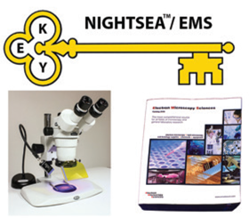
The application period for the 7th annual NIGHTSEA/EMS KEY Award for New Faculty is open. The award is an annual equipment grant to an individual entering their first faculty position at a college or university. Eligibility has been expanded to those entering any academic institution worldwide. The award consists of a stereo microscope fluorescence adapter system with two excitation/emission combinations and $750 in equipment or supplies selected from the full EMS catalog.
Electron Microscopy Sciences

NT-MDT: Atomic Force Microscopy (AFM) Specialists
NT-MDT Spectrum Instruments, an AFM and Raman manufacturer, has been in the market for 30 years. NT-MDT systems implement the latest technology and cover a wide range of applications in areas such as material science, biological studies, polymer studies, semiconductors, and others. Providing standard AFM techniques and integrated solutions, NT-MDT AFM-Raman systems support simultaneous topographical and chemical analysis, tip-enhanced Raman scattering, and nano-IR techniques used to conduct chemical analysis with resolution beyond diffraction limits.
NT-MDT

Metrology Partnership Brings Vision to Coordinated Measuring Machine (CMM) Market
Vision Engineering has partnered with metrology innovator Aberlink to add the first contact-only measurement system, DELTRON, to its range of metrology systems. Designed for robustness, reliability, affordability, and ease of use, Deltron is a hardened non-Cartesian CMM with an innovative delta robotic mechanism, known for repeatable motion and fast acceleration with measurement accuracy. The delta mechanism on Deltron operates with a maximum vector speed of 500 mm/sec and a maximum vector acceleration of 750 mm/sec2 with a measuring accuracy of 2.6 + 0.4L/100 µm at 0.1 µm resolution.
Vision Engineering Limited

Astronomy and Pathology: A Novel Collaboration with Johns Hopkins University
Akoya Biosciences has established a collaboration with Johns Hopkins to develop, validate, and clinically implement novel multiplexed immunofluorescence signatures to facilitate more effective drug development using Akoya's Phenoptics™ technology and Johns Hopkins's AstroPath™ platform. The AstroPath™ program leverages principles from immunology, pathology, computer science, and astronomy to analyze large mIF datasets with celestial object–mapping algorithms.
Akoya Biosciences

Austrian Research Institutions Form Imaging Node Within the Euro-BioImaging Consortium
Austrian BioImaging/CMI, an Austrian research consortium comprised of nine universities and research institutions in the field of biological and (pre)clinical imaging, has successfully applied to be Austria's official imaging node within the European Research Infrastructure Consortium (ERIC) Euro-BioImaging. ERIC gives researchers access to imaging instruments, expertise, training capability, and data processing services, which they would be unlikely to find in their home universities or among its collaborative partners.
Austrian BioImaging/CMI
https://www.bioimaging-austria.at

NenoVision Releases LiteScope 2.0
NenoVision has released LiteScope 2.0 for unprecedented correlative analyses, merging AFM and SEM capabilities. Compared to its predecessor, this unique AFM designed for plug-and-play integration into SEMs was upgraded in nearly every aspect. Combining advantages of both microscopic techniques, LiteScope 2.0 opens new possibilities of complex sample analysis that was difficult or impossible by conventional separate AFM and SEM instrumentation. The most powerful feature of LiteScope is Correlative Probe and Electron Microscopy (CPEM), which enables simultaneous acquisition of data from SEM and AFM systems.
Nenovision

Using SEM to Analyze Microplastics in Sea Water
The pollution of our oceans with plastic has become a worldwide problem. Microplastics are now found almost everywhere and are the predominant form of plastic debris found in seawater. Traditional methods of analysis typically separate and study individual particles, which is a slow and time-consuming process. As interest increases in identifying and analyzing these pollutants, a rapid and accurate method is required. SEM with EDS can yield particle size and chemistry in an effort to mitigate the problem. The study “Using SEM to Analyze Microplastics in Sea Water” is available at https://www.azom.com/article.aspx?ArticleID=19602.
COXEM

RJ Lee Group Introduces the Second-Generation IntelliSEM
In April 2021, RJ Lee Group introduced the second-generation IntelliSEM software for automated identification and analysis of particles. IntelliSEM software applies high-performance instrument control and data analysis to scanning electron microscopy (SEM) and energy dispersive X-ray spectroscopy (EDX) to provide a rich set of tools for data visualization and collaboration. The flexibility, speed, and data management provided by IntelliSEM secures its key role in areas as diverse as steel inclusion analysis, environmental air particulates, and additive manufacturing.
RJ Lee Group
https://go.rjlg.com/intellisem

Sideband Kelvin Probe Force Microscopy for Advanced Materials Characterization
Sideband Kelvin probe force microscopy (KPFM) uses the intermodulation of an electrostatic drive force and a mechanical drive force to upconvert the electrostatic frequency to the first flexural resonance, where the high-quality factor of the resonance yields a more sensitive measurement. The sideband KPFM signal is calculated using a local interaction between the tip apex and the sample rather than a total interaction between the cantilever and the sample, improving the spatial resolution over other technique variations.
Park Systems

Janelia's Optical Interest Group Will Host a New Lecture Series
Janelia's Optical Interest Group (OIG) will host a new lecture series partnering with the Australian Biomedical Group (ABG). The ABG consists of six universities in Australia. The OIG-ABG Educational Lectures will specifically focus on Education in Optics & Technology, Optics & Biology, and Optics & Disease. The lectures will be open to all Janelian and Australian Biomedical Groups (ANU, University of Adelaide, UQ, UWA, UNSW, Monash, and Garvan).
Optical Interest Group (OIG)
https://www.janelia.org/content/oig-abg

Thermo Scientific Automated 3D Acquisition with Tomography 5 Software
Tomography 5 Software facilitates automated 3D image acquisition of vitrified samples and is compatible with Thermo Scientific™ Krios™, Glacios™, and Talos™ Cryo-TEMs. The in situ capabilities of cryo-electron tomography allow it to bridge the gap between single-particle analysis and structural cell biology. With a new user-friendly interface, Tomography 5 Software enables automated 3D data acquisition, cassette mapping, and dose symmetric acquisition schemes, and is fully Selectrin Imaging Filter integrated.
Thermo Scientific

Antigen Retriever Program from EMS
The 2100 Retriever is a fully automatic unit that allows reproducible antigen recovery for 108 slides in one go, in up to 6 individual buffers if need be. It fully preserves cells and tissue morphology during recovery, is easy to operate, and does not require training. In combination with a unique R-Universal buffer that reduces chemical modifications of the formalin cross-linking, the 2100 Retriever is usable for most epitopes.
Electron Microscopy Sciences

Stunning Images and Smart Features from Olympus DP Microscope Cameras
Two new DP series cameras share advanced, time-saving features enabling users to choose the most suitable model based on the level of resolution required. With 4K ultra-high-definition image quality, the DP28 camera enables users to share and review sample images in fine detail. The DP23 camera's 6.4-megapixel resolution and 45 frames per second (fps) rate make it easy to quickly capture images with the detail needed for most life science imaging applications.
Olympus
https://www.olympusamerica.com

Cutting-Edge of Raman-Based Microparticle Characterization Gets Even Sharper
WITec's ParticleScout has been enhanced with new features including integration time optimization that uses the signal-to-noise ratio to determine how long each particle is measured for identification. This greatly reduces overall measurement time and minimizes the effects of fluorescence. The updates to ParticleScout are driven by direct feedback from researchers and their specific requirements in laboratories focused on environmental research, food science, pharma, and many other applications.
WITec

EMAN2.9/SPHIRE1.4/SPARX Has Been Released
EMAN2 is a greyscale scientific image processing suite with a primary focus on processing data from transmission electron microscopes. EMAN2 is capable of processing large data sets (>100,000 particle) very efficiently. The new tomography pipeline in EMAN2 has been significantly improved since its initial release. Data compression is now a standard part of the package for both CryoEM and CryoET data processing pipelines, providing dramatic reductions in disk usage and transfer times with no reduction in quality.
NIH Free Software
https://blake.bcm.edu/emanwiki/EMAN2

Cryogenic Plunger from Neptune Fluid Flow Systems
Neptune Fluid Flow Systems offer a manual cryogenic plunge freezer for the preparation of vitrified biological and material science samples for cryogenic electron microscopy studies. The plunger itself has a base footprint of 12" by 12" with a height of 18". The entire weight is a little over 10 lbs. The base and most of the parts are made of aluminum, and the trigger bar is brass. The foot plunger is small and can be placed in a convenient location.
Neptune Fluid Flow Systems

Abbelight SAFe RedSTORM: Solution for Multi-Color 3D Nanoscopy
Abbelight has designed a new product, SAFe RedSTORM, to simplify simultaneous multi-color imaging. The SAFe RedSTORM uses spectral demixing to separate far-red dyes, limiting the excitation to one laser and decreasing chromatic aberrations. Used to facilitate single-molecule imaging, the SAFe RedSTORM module can be adapted to any inverted microscope to perform SMLM with homogenous TIRF/HiLo/Epi illumination over a large field of view of 150 × 150 um2 vs. the usual 50 × 50 um2.
Abbelight

New Flexible and Powerful Atomic Force Microscopy
The Nano-Observer AFM combines performance and ease of use. The USB controller offers integrated lock-in for better measurement capability (phase detection, Piezo-Response Mode). A low-noise laser and a pre-alignment system provide high resolution on a compact AFM head. Its intuitive software simplifies all settings to allow quick and safe AFM acquisitions. Laser alignment is unnecessary with the pre-positioned tip system. A top and side view of the tip/sample, combined with vertical motorized control, simplifies the pre-approach.
Concept Scientific Instruments

Next Generation of Dragonfly Software for Advanced Segmentation
From quick, efficient deep learning models to robust analysis workflows and bringing real-world materials to life with stunning visualizations, Dragonfly 2021.1 enhances understanding of 3D scientific, industrial, and biomedical data. This major software release features: 1) enhancements for the Segmentation Wizard that allow quick deep learning and machine learning segmentation of multi-dimensional images, 2) deep learning architectures for training semantic segmentation models, and 3) new pre-processing options for reconstructing projection data and additional support for importing micro-CT data.
Dragonfly
https://www.theobjects.com/dragonfly/index.html

Light Sheet Microscopy from Miltenyi Biotec
When studying complex biological systems, scientists need to see the big picture while still being able to analyze all the details. Miltenyi Biotec's imaging systems, such as the UltraMicroscope II light sheet system, can be used to visualize whole biological systems and systems with ultrahigh-content to unravel nature's mysteries and take research further and faster.
Miltenyi Biotec
https://www.miltenyibiotec.com

NanoRack Sample Stretching Stage for the Jupiter XR AFM
The NanoRack stage enables direct measurement of nanoscale properties of polymer materials, thin films, and biological samples such as tissues or bones while under stressed conditions. Stress and/or strain information is extracted from stretched or compressed samples in two dimensions, providing a more comprehensive understanding of materials under real-world experimental conditions. Jupiter XR is the first and only large-sample AFM to offer high-speed and high-resolution imaging in one scanner configuration.
Oxford Instruments-Asylum Research
https://afm.oxinst.com/products/jupiter-family-of-afms/jupiter-xr-afm

UniFlow CE AireStream Energy-Saving Laboratory Fume Hood
The UniFlow CE AireStream is a full-duty fume hood in a compact size, offering 50% energy savings. It's equipped with the exclusive vector slotted rear VaraFlow baffle system. The hoods are offered in 30″, 36″, 48″, and 72″ widths and can be equipped with a wide selection of accessories to meet specific processing needs. They are constructed of composite resin for superior chemical resistance and can be supplied with or without an exhaust.
HEMCO

Flexible Light Sources for Biophotonics
The latest member of the iChrome family, the iChrome FLE, is designed as a “flexible laser engine” to serve the numerous and very diverse requirements in biophotonics. Up to 7 wavelengths (between 405 and 785 nm) and two fiber outputs can be integrated into the iChrome FLE. The fiber outputs can be configured separately, so that individual wavelengths (for example, UV or IR) can be corrected separately in the application. Alternatively, a fiber switch/splitter can be installed, allowing each wavelength to be switched separately to one of the two fibers, or to distribute the power between both fibers.
TOPTICA Photonics
https://www.toptica.com/flexible

Culture Cells on a 3D Gel with Defined Flow
ibidi's new μ-Slide I Luer 3D is an innovative slide with one channel and three wells for 3D cell culture under flow. The Luer provides easy sample preparation by filling of the three wells with a gel (for example, collagen, Matrigel, or fibrinogen). The cells are then seeded into the channel where they adhere to the gel. For applying defined shear stress, the slide is connected to a pump. The cells can be imaged using high-resolution microscopy.
ibidi

Inspect Liquid Samples in a Scanning Electron Microscope
FlowVIEW has developed a novel chip system that incorporates microfluid control and system integration. It allows observation of both wet and dry samples in a SEM, including living cells, with a resolution up to 7 nm. The platform features include dynamic fluid, temperature, and gas control.
FlowView
https://www.flowviewtek.com/index.php?lang=en

The World's First Event-Based Camera for High-Throughput Cryo-EM
Direct Electron announces Apollo, a new event-based direct detection camera for electron cryo-microscopy (cryo-EM). This next-generation sensor and camera architecture in electron counting is the highest throughput direct detector for cryo-EM. Using a software-based approach implemented in existing cameras, electron counting is done by identifying electron detection events on the sensor. It generates super-resolution (67 megapixel) frames at high-speed and is optimized for downstream cryo-EM image processing before electrons leave the camera.
Direct Electron
https://www.directelectron.com


