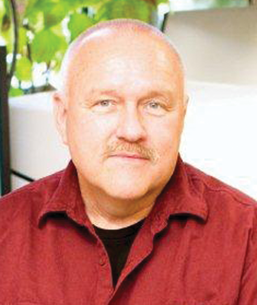
As many of my colleagues know, I recently experienced a “vaccination breakthrough” case and paid my COVID-19 dues with a stay in an intensive care unit in August. While too numerous to name here, I would like to thank all of those from the microscopy and microanalysis communities who provided support through their thoughts and prayers during that difficult time. While in the ICU and in recovery, I realized how important the MSA and MAS friendships I have established over the years are in my life. The support from all of you certainly helped in my recovery.
My stay in the ICU provided me with some unexpected free time, and, not able to do much more than stare at the ceiling, I began to think about the important role that microscopy and microanalysis have played in combating the COVID-19 pandemic. From the early cryo-EM 3D reconstructions of the coronavirus presented on the news media to raise public awareness, to the development of vaccines as detailed in the M&M 2021 Plenary presentation by Jason McLellan, imaging has been at the forefront of understanding COVID-19 structure and function. Many sessions at the meeting including Imaging, Microscopy, and Micro/Nano-Analysis of Pharmaceutical, Biopharmaceutical, and Medical Health Products - Research, Development, Analysis, Regulation, and Commercialization; Cryo-EM in Drug Discovery; and Challenges and Advances in Electron Microscopy Research and Diagnosis of Diseases in Humans, Plants and Animals also addressed topics relevant to studying COVID-19 and other diseases.
Many MSA Focused Interest Groups (FIG), including 3D EM in the Biological Sciences; Cryopreparation; Diagnostic and Biomedical Microscopy; and the Pharmaceuticals group, also use a range of imaging techniques to study viruses as well as other microbes and diseases. If your research and interests align with any of these or other MSA FIGs, please consider joining and contributing to the important research being performed in these groups. A full list of FIGs can be found on the MSA website at Communities | Focused Interest Groups (https://www.microscopy.org/communities/fig.cfm). I can personally vouch for the personal and professional benefits that being a member of these communities provides, and I encourage all to join the group(s) that fit your interests and research.


