Hostname: page-component-745bb68f8f-5r2nc Total loading time: 0 Render date: 2025-01-13T12:48:46.606Z Has data issue: false hasContentIssue false

The biology of ergothioneine, an antioxidant nutraceutical

Published online by Cambridge University Press:  13 February 2020

Irina Borodina
Affiliation:
The Novo Nordisk Foundation Center for Biosustainability, Building 220, Chemitorvet 200, Technical University of Denmark, 2800Kongens Lyngby, Denmark
Louise C. Kenny
Affiliation:
Department of Women’s and Children’s Health, Institute of Translational Medicine, University of Liverpool, Crown Street, LiverpoolL8 7SS, UK
Cathal M. McCarthy
Affiliation:
Irish Centre for Fetal and Neonatal Translational Research (INFANT), Cork University Maternity Hospital, Cork, Republic of Ireland Department of Pharmacology and Therapeutics, Western Gateway Building, University College Cork, Cork, Republic of Ireland
Kalaivani Paramasivan
Affiliation:
The Novo Nordisk Foundation Center for Biosustainability, Building 220, Chemitorvet 200, Technical University of Denmark, 2800Kongens Lyngby, Denmark
Etheresia Pretorius
Affiliation:
Department of Physiological Sciences, Faculty of Science, Stellenbosch University, Stellenbosch, Private Bag X1 Matieland, 7602, South Africa
Timothy J. Roberts
Affiliation:
Department of Physiological Sciences, Faculty of Science, Stellenbosch University, Stellenbosch, Private Bag X1 Matieland, 7602, South Africa Department of Biochemistry, Institute of Integrative Biology, Faculty of Health and Life Sciences, University of Liverpool, Crown Street, LiverpoolL69 7ZB, UK
Steven A. van der Hoek
Affiliation:
The Novo Nordisk Foundation Center for Biosustainability, Building 220, Chemitorvet 200, Technical University of Denmark, 2800Kongens Lyngby, Denmark
Douglas B. Kell*
Affiliation:
The Novo Nordisk Foundation Center for Biosustainability, Building 220, Chemitorvet 200, Technical University of Denmark, 2800Kongens Lyngby, Denmark Department of Physiological Sciences, Faculty of Science, Stellenbosch University, Stellenbosch, Private Bag X1 Matieland, 7602, South Africa Department of Biochemistry, Institute of Integrative Biology, Faculty of Health and Life Sciences, University of Liverpool, Crown Street, LiverpoolL69 7ZB, UK
*
*Corresponding author: Douglas B. Kell, email dbk@liv.ac.uk
Rights & Permissions [Opens in a new window]

Abstract

Ergothioneine (ERG) is an unusual thio-histidine betaine amino acid that has potent antioxidant activities. It is synthesised by a variety of microbes, especially fungi (including in mushroom fruiting bodies) and actinobacteria, but is not synthesised by plants and animals who acquire it via the soil and their diet, respectively. Animals have evolved a highly selective transporter for it, known as solute carrier family 22, member 4 (SLC22A4) in humans, signifying its importance, and ERG may even have the status of a vitamin. ERG accumulates differentially in various tissues, according to their expression of SLC22A4, favouring those such as erythrocytes that may be subject to oxidative stress. Mushroom or ERG consumption seems to provide significant prevention against oxidative stress in a large variety of systems. ERG seems to have strong cytoprotective status, and its concentration is lowered in a number of chronic inflammatory diseases. It has been passed as safe by regulatory agencies, and may have value as a nutraceutical and antioxidant more generally.

Type
Review Article
Creative Commons
Creative Common License - CCCreative Common License - BY
This is an Open Access article, distributed under the terms of the Creative Commons Attribution licence (http://creativecommons.org/licenses/by/4.0/), which permits unrestricted re-use, distribution, and reproduction in any medium, provided the original work is properly cited.
Copyright
© The Authors 2020

Introduction

Most of the classical vitamins such as vitamins A, B1, B2, B3, C, D, etc., were discovered by means of the fact that an inadequacy in their supply led to overt forms of deficiency disease such as blindness, beri-beri, pellagra, scurvy, rickets and so on. Consequently, it was easy to establish those food sources that contained such vitamins, since they relieved or prevented the relevant syndromes(Reference Kraemer, Semba and Eggersdorfer1,Reference Semba2) . It is correspondingly hard, by these means, to detect the presence of a vitamin if it is present in virtually every foodstuff that an individual consumes. Recently, however, l-(+)-ergothioneine, hereafter ergothioneine (ERG), has emerged(Reference Paul and Snyder3Reference Beelman, Kalaras and John10) as an important nutrient, and indeed possible vitamin(Reference Paul and Snyder3), that has precisely these properties of a very widespread occurrence coupled, commonly, to a functional undersupply.

A related class of nutrient, which has not been demonstrated as necessary or essential for life yet provides health benefits when added at levels greater than a normal diet generally provides, has come to be known as nutraceuticals, a coinage based on an amalgamation of ‘nutrition’ and ‘pharmaceutical’(Reference Cencic and Chingwaru11). Interest in such nutraceuticals, also known as ‘functional foods’, has increased enormously over the last few decades(Reference Cencic and Chingwaru11Reference Spindler, Mote and Flegal22) as our understanding of the important roles of diet in health has improved. However, the enthusiasm for such products has not always been matched by the extent or quality of the evidence for their efficacy(Reference Sharif, Khalid, Holban and Grumezescu20,Reference Poddar, Kolge and Bezman23Reference Orr28) .

Since ERG classes as a nutraceutical, it seems timely to bring together the extensive but widespread knowledge of its biology so that it may be made more widely available, and that is the purpose of this review.

Discovery and structure

ERG is a somewhat unusual betaine amino acid. It was discovered by Charles Tanret in 1909 while investigating the ergot fungus Claviceps purpurea (Reference Tanret29,Reference Tanret30) . It is also known as 2-mercaptohistidine trimethylbetaine, and its formal International Union of Pure and Applied Chemistry (IUPAC) name is (2S)-3-(2-thioxo-2,3-dihydro-1H-imidazol-4-yl)-2-(trimethylammonio)propanoate. It is an l-histidine derivative that is Nα,Nα,Nα-trimethyl-l-histidine in which the hydrogen at position 2 on the imidazole ring is replaced by a mercapto group. Its structure(Reference Barger and Ewins31), and those of some related molecules, is given in Fig. 1, indicating that is a tautomer that has both a thiol and a thione form. Although it is a thiol, and hence an antioxidant(Reference Servillo, Castaldo and Casale32,Reference Franzoni, Colognato and Galetta33) , the thione tautomer is predominant at physiological pH(Reference Hand, Taylor and Honek34,Reference Hand and Honek35) , and this makes it unusually resistant to autoxidation, i.e. simple oxidation by molecular O2 (Reference Servillo, Castaldo and Casale32,Reference Fahey36Reference Fahey38) . Its midpoint potential for a thiol is consequently unusually high, being +0·06 V v. −0·2 to −0·4 V for typical thiols including glutathione(Reference Cheah and Halliwell4,Reference Jacob39Reference Walz41) and mycothiol(Reference Reyes, Pedre and De Armas42,Reference Sharma, Van Laer and Messens43) , and −0·193 V for the also somewhat oxidising thiol cofactor coenzyme M, which is 2-mercaptoethanesulfonate(Reference Kell and Morris44). Its reaction with hydroxyl radicals (OH) is virtually instantaneous, while it reacts only more slowly with H2O2 and/or O2 •− (Reference Fahey38). Its Se equivalent is known as selenoneine and also has strong antioxidant properties(Reference Achouba, Dumas and Ouellet45Reference Yamashita, Yabu and Yamashita52), but is not otherwise discussed here.
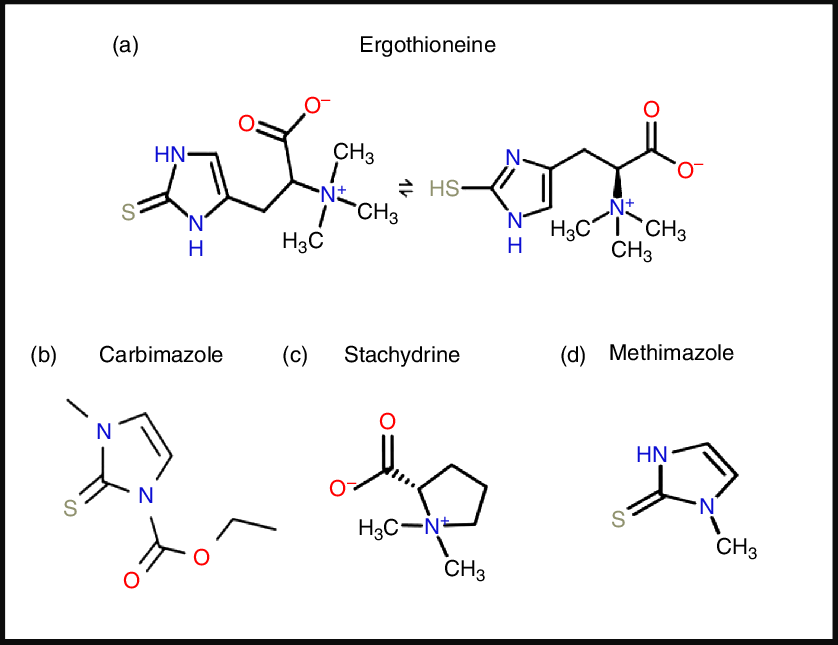
Fig. 1. Structures of ergothioneine and related molecules. For a colour figure, see the online version of the paper.

From a pharmaco-chemical point of view ERG is also unusual, since – using our standard substructure analysis(Reference O’Hagan and Kell53,Reference O’Hagan and Kell54) in KNIME(Reference O’Hagan and Kell55) – we note that just two drugs marketed for human consumption (the anti-thyroxine-production drug methimazole and its pro-drug carbimazole, Fig. 1), and no endogenous genome-encoded metabolites from Recon2(Reference Thiele, Swainston and Fleming56) contain the imidazole-2-thione substructure(Reference O’Hagan, Swainston and Handl57). This said, a good many fungicides do contain the benzimidazole substructure(Reference Lewis, Tzilivakis and Warner58), and a variety of benzothiazoles are used as dyes.

Biosynthesis and phylogenetic distribution

A particular feature of ERG is that although it is more or less universally distributed among higher organisms, none of them – as is consistent with the idea that it may in fact be a vitamin requiring exogeneous sources – can in fact biosynthesise it. The chief organisms capable of its synthesis are fungi and certain yeasts(Reference Melville, Genghof and Inamine59,Reference Jones, Doyle and Fitzpatrick60) , though actinobacteria and certain other micro-organisms(Reference Jones, Doyle and Fitzpatrick60Reference Trivedi, Singh and Bhat66), including the slime mould Physarum polycephalum (Reference Genghof65), cyanobacteria(Reference Narainsamy, Farci and Braun67Reference Liao and Seebeck71) and methylotrophs(Reference Alamgir, Masuda and Fujitani72) are also naturally capable of its production. The related mycothiol is typically ten times more concentrated in actinobacteria than is ERG(Reference Rawat and Av-Gay73), and its biosynthetic pathway might provide an antitubercular drug target. Other organisms acquire ERG through transporter-mediated uptake. Thus higher plants contain it but do not biosynthesise it(Reference Melville and Eich74); instead they and other organisms(Reference Baran, Bowen and Price68,Reference Baran, Brodie and Mayberry-Lewis75) take it up from fungal production in the soil(Reference Melville76Reference Warren79), and possibly via actinobacterial(Reference Park, Lee and Kim80) or fungal(Reference Park, Lee and Kim80,Reference Guo, Lin and Wang81) symbionts. Animals are also considered not to biosynthesise it(Reference Melville, Otken and Kovalenko82,Reference Melville, Horner and Otken83) , and accumulate it using a particular transporter, detailed below, via the plants and animals that they eat. Although not easy, it is possible to raise animals such as pigs on a diet such as casein, sucrose, lard, butter and salts that is considered to lack ERG; such animals are said to have undetectable levels of the compound(Reference Eagles and Vars84), and rats treated similarly have reproduced(Reference Melville, Horner and Lubschez85,Reference Kawano, Otani and Takeyama86) . However, we do not know the minimum amount and its location that animals need, and these are old experiments that need to be repeated with modern techniques with lower detection limits. Only then might we have a definitive statement as to whether ERG is absolutely required as a true vitamin or not, and if so in what amounts for health. In a similar vein, ERG can be present in cell culture media and cells with organic cation transporter N1 (OCTN1)/solute carrier family 22, member 4 (SLC22A4) can accumulate it(Reference Tucker, Cheah and Halliwell87), a fact little considered to date in cell culture studies.

To the extent that ERG is a ‘secondary’ metabolite, defined(Reference Bu’Lock88) as a molecule whose synthesis has a relatively restricted distribution in different organisms, the biosynthetic pathways diverge from primary metabolism via the amino acids histidine, cysteine and methionine(Reference Melville, Eich and Ludwig89Reference Naowarojna, Cheng and Chen94). Thus (Fig. 2), histidine is trimethylated using S-adenosyl methionine to form trimethyl histidine, also known as hercynine(Reference Reinhold, Ishikawa and Melville95,Reference Melville, Ludwig and Inamine96) . This reacts oxidatively with cysteine to form hercynylcysteine sulfoxide(Reference Ishikawa, Israel and Melville97), which is converted to ERG. In some organisms, hercynine takes a more convoluted route via γ-glutamylhercynylcysteine sulfoxide (Fig. 2)(Reference Naowarojna, Cheng and Chen94). Table 1 provides references for different organisms. An excellent phylogenetic analysis is given by Jones et al. (Reference Jones, Doyle and Fitzpatrick60). In more recent work, it has been suggested that ERG was probably first biosynthesised by anaerobes using a slightly different route that converts hercynine directly to ERG(Reference Leisinger, Burn and Meury98Reference Ruszczycky and Liu100), and that was later repurposed.
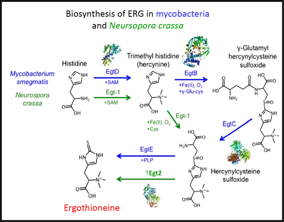
Fig. 2. The two main pathways of aerobic ergothioneine (ERG) biosynthesis, noting the relevant enzymes and thumbnails of three-dimensional structures where known. SAM, S-adenosyl methionine. For a colour figure, see the online version of the paper.

Table 1. Biosynthesis of ergothioneine in various non-recombinant micro-organisms

Three-dimensional structures are known for a number of the relevant enzymes, including mycobacterial EgtB(Reference Goncharenko, Vit and Blankenfeldt101) for example, PDB 4XBE, EgtC(Reference Vit, Mashabela and Blankenfeldt102) for example, PDB 4ZFJ, EgtD(Reference Vit, Misson and Blankenfeldt103Reference Misson, Burn and Vit105) for example, PDB 4PIM, and Neurospora crassa early G1 transcript 2 (egt2) which is like egtE(Reference Irani, Naowarojna and Tang106) for example, PDB 5UTS. Very recently, EgtB from Candidatus Chloracidobacterium thermophilum was crystallised(Reference Naowarojna, Irani and Hu107), and engineered towards Egt1 activity. Thumbnails are given in Fig. 2. Egt1 from N. crassa is 876 amino acids long(108), while egtD (from Mycobacterium tuberculosis (109)) is just 321 amino acids long; since the N-terminal sequences are well conserved (Fig. 3), this implies an extra C-terminal domain catalysing the production of hercynylcysteine sulfoxide from hercynine.

Fig. 3. Alignment of Neurospora crassa Egt1 and N-terminal part of Mycobacterium tuberculosis EgtD. For a colour figure, see the online version of the paper.

In addition, enantiopure l-ERG has been synthesised chemically(Reference Melville76,Reference Daunay, Lebel and Farescour110Reference Khonde and Jardine112) , and by fermentation of genetically engineered micro-organisms (Table 2). Initial efforts in ERG synthesis were carried out in Schizosaccahromyces pombe using egt1 overexpression under an inducible promoter. The N starvation and glucose starvation conditions causing long quiescence led to the maximum ERG production of 1606·3 µm while the wild-type strain produced 0·3 µm (Reference Pluskal, Ueno and Yanagida50). Methylobacterium aquaticum strain 22A was engineered by expressing an additional copy of egtBD genes and by deleting the gene encoding histidine ammonia lyase, which degrades an ERG precursor l-histidine. The resulting strain produced up to 7·0 mg EGT/g dry cell weight and 100 μg EGT/5 ml per 7 d in test-tubes(Reference Fujitani, Alamgir and Tani113). The filamentous fungus Aspergillus oryzae has also been engineered to produce ERG by expression of egt1 and egt2 genes from N. crassa, resulting in 231 mg ERG per kg of solid media(Reference Takusagawa, Satoh and Ohtsu114).

Table 2. Fermentative production of ergothioneine in recombinant micro-organisms
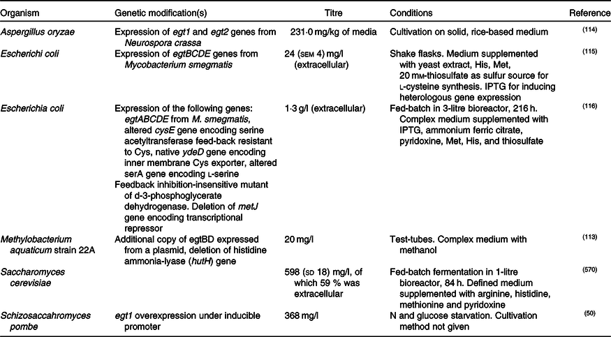
IPTG, isopropyl β- d-1-thiogalactopyranoside.

Expression of egtBCDE genes from Mycobacterium smegmatis in Escherichia coli and optimisation of medium composition has led to 24 mg/l or 104 μm of secreted ERG(Reference Osawa, Kamide and Satoh115). The egtA gene from M. smegmatis was not expressed because E. coli contains a homologous glutamate–cysteine ligase encoded by gshA and involved in glutathione biosynthesis. In a follow-up study, the authors expressed egtA from M. smegmatis and it had a positive effect on ERG production. Furthermore, they enhanced cysteine and S-adenosine methionine biosynthesis and obtained 1·3 g/l or ERG in a fed-batch fermentation(Reference Tanaka, Kawano and Satoh116), achieving currently the highest titre reported for heterologous ERG production.

Recently, we reported the engineering of baker’s yeast Saccharomyces cerevisiae for the production of ERG(Reference van der Hoek, Darbani and Zugaj117). S. cerevisiae has a generally recognised as safe (GRAS) status and has been exploited for the commercial production of several nutraceutical compounds(Reference Li and Borodina118); it is thus a highly attractive host for the production of ERG. We have tested sixteen different pathway variants, nine containing only fungal genes, one with bacterial genes from M. smegmatis, and six hybrid pathway variants containing both fungal and bacterial transgenes. The best-performing strain contained egt1 from N. crassa and egt2 from C. purpurea. The composition of the medium was improved using a fractional factorial design. Fed-batch cultivation resulted in 598 (sd 18) mg/l ERG after an 84-h fermentation. Some 60 % of the measured ERG was extracellular and the rest accumulated in the cells. Table 2 summarises the various recombinant expression hosts that have been used.

The distribution of solute transporters between tissues in differentiated organisms is particularly heterogeneous(Reference O’Hagan, Wright Muelas and Day119), and it is to be expected that both SLC22A4 and ERG might also be distributed heterogeneously as well. This is indeed the case, their distribution being especially high in tissues that are considered to have the potential for oxidative stress(Reference Cheah and Halliwell4), such as erythrocytes(Reference Chaleckis, Ebe and Pluskal120Reference Mitsuyama and May129), bone marrow(Reference Gründemann, Harlfinger and Golz130), liver and kidney(Reference Melville, Horner and Lubschez85,Reference Tang, Cheah and Yew131) , seminal fluid(Reference Nikodemus, Lazic and Bach132,Reference Kaneko, Takeuchi and Yamaoka133) and the lens and cornea of the eyes(Reference Shires, Brummel and Pulido134). It may also be accumulated in the CNS(Reference Crossland, Mitchell and Woodruff135,Reference Nakamichi, Taguchi and Hosotani136) .

Finally, here, we note – as with the activity of the ‘master Fe regulator’ hepcidin(Reference Vermeulen and Vermeersch137Reference Reichert, da Cunha and Levy141), that acts chiefly via the ferrous Fe transporter ferroportin – that the action of a transporter in concentrating a substance in one tissue will typically lead to its depletion from another. Consequently, it is necessary to measure all relevant compartments to assess whether a molecule such as ERG, whose distribution is strictly transporter-mediated, is protective against a particular disease/effect or otherwise in a particular place or case.

SLC22A4: the ergothioneine transporter

Although this view remains controversial, even hydrophobic molecules do not normally ‘float across’ whatever phospholipid bilayer portion of cells may be untrammelled by proteins. Xenobiotics in particular need to ‘hitchhike’ on protein transporters that have presumably evolved for ‘natural’ substrates but that are capable of their uptake(Reference Kell, Swainston and Pir142Reference Dickens, Rädisch and Chiduza152). While transporters seem to have remained somewhat understudied(Reference César-Razquin, Snijder and Frappier-Brinton153), those transporters involved in uptake and encoded by the human genome are now catalogued formally as SLC for solute carriers(Reference Hediger, Clemencon and Burrier154,155) , with efflux transporters mainly being classed as ABC families(Reference Chen, Shi and Zhang156).

One solute carrier, previously known as organic cation transporter N1 (OCTN1)(Reference Koepsell157,Reference Pochini, Galluccio and Scalise158) , and now known as SLC22A4 (the human version is Uniprot Q9H015), a 551-amino-acid transporter with three glycosylation sites, is of special interest. It had been designated as a transporter of carnitine and of the (non-physiological) tetraethylammonium cation. However, in a really groundbreaking paper, Gründemann et al. (Reference Gründemann, Harlfinger and Golz130) recognised that the rates observed (using radioisotopes) were too small to be physiologically meaningful, and using a method that we would now refer to as ‘untargeted metabolomics’(Reference Garg, Kapono and Lim159Reference Dunn, Erban and Weber164), they incubated two kinds of HEK293 cells in serum. The first were normal cells, that, as with many transporters(Reference O’Hagan, Wright Muelas and Day119), do not in fact express SLC22A4 at significant levels, while the second had been engineered to overexpress the transporter. They then simply looked for those molecules that were most differentially taken up, a molecule called stachydrine, also known as proline betaine, being the main one observed, Stachydrine is a constituent of citrus juices(Reference Heinzmann, Brown and Chan165Reference Lloyd, Beckmann and Favé167). Some elementary cheminformatics based on structure similarity searches(Reference O’Hagan, Swainston and Handl57,Reference Gasteiger168) indicated that ERG, as a betaine, was indeed similar to stachydrine. Incubating the cells just with ERG showed that it was taken up about 100 times more quickly than was tetraethylammonium, leading to the designation of SLC22A4 as ‘the’ ERG transporter(Reference Gründemann, Harlfinger and Golz130). Subsequent work(Reference Tucker, Cheah and Halliwell87,Reference Bacher, Giersiefer and Bach169Reference Tschirka, Kreisor and Betz172) has confirmed and reinforced this view of SLC22A4 and its homologues(Reference Shimizu, Masuo and Takahashi173) as having significant specificity for ERG, and weak activity for various drugs(Reference Yabuuchi, Tamai and Nezu174Reference Tamai177). It is concentrative, coupled in humans to influx of 2 or 3 Na+ ions per ERG transported(Reference Gründemann, Harlfinger and Golz130). Interestingly, it is up-regulated chronobiologically just before meal times(Reference Akamine, Koyanagi and Kusunose175). The rat and human orthologues are interchangeable(Reference Nakamura, Yoshida and Yabuuchi178). Tissue levels of ERG depend on an exogenous supply(Reference Darghouth, Giarratana and Oliveira179), but are then well correlated with the expression levels of SLC22A4(Reference Paul and Snyder3,Reference Taubert, Lazar and Grimberg180) . SLC22A4 expresses well even in microbial systems(Reference Indiveri, Galluccio and Scalise181), and is widely tolerant of amino acid substitutions(Reference Frigeni, Iacobazzi and Yin182). As yet, no other transporter with significant activity for ERG in humans is known, making it a potentially interesting drug target(Reference Nigam183,Reference Pochini, Scalise and Galluccio184) .

Expression patterns

SLC22A4 is known to express in the intestinal lumen(Reference Wolf, Paine and McQueen185), acting to take up ERG, as well as some xenobiotics including pyrilamine, quinidine and verapamil, and having multiple known but weak inhibitors.

Fig. 4 shows the expression of the transcript for SLC22A4 in fifty-six cell lines using previous data(Reference O’Hagan, Wright Muelas and Day119) taken from the human protein atlas(Reference Thul, Åkesson and Wiking186), indicating a range in expression levels between different cell lines of more than 4000-fold, a number not atypical for human transporters(Reference O’Hagan, Wright Muelas and Day119). Tissue expression data are given in Fig. S4 of O’Hagan et al. (Reference O’Hagan, Wright Muelas and Day119).
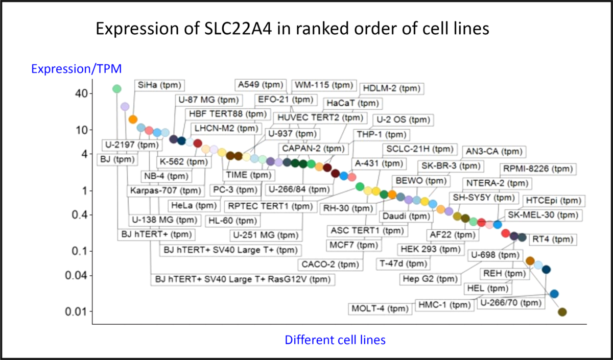
Fig. 4. Differences in expression of SLC22A4 transcript in a series of mammalian cell lines. Data are from Thul et al. (Reference Thul, Åkesson and Wiking186) and O’Hagan et al. (Reference O’Hagan, Wright Muelas and Day119). For a colour figure, see the online version of the paper.

The intracellular expression patterns are as yet uncertain, with early claims for a mitochondrial expression(Reference Kawano, Otani and Takeyama86,Reference Apostolova and Victor187Reference Xuan, Lamhonwah and Librach190) being based on very weak and contradictory evidence(Reference Kerley, McCarthy and Kell8). However, while the cellular uptake of ERG does require plasma membrane expression, the latest version of the protein atlas indicates mitochondrial expression as well(191). However, as is well known, antibody specificities are rarely either known or absolute(Reference Skogs, Stadler and Schutten192Reference Michaud, Salcius and Zhou198). Thus, relying on antibody evidence alone is rather hazardous, and, as mentioned before(Reference Kerley, McCarthy and Kell8), mitochondrial transporters have an SLC25 family designation(Reference Palmieri199,Reference Palmieri200) . Definitive measurements on the uptake or otherwise of ERG into isolated mitochondria, or indeed into other organisms that cannot make it, are eagerly awaited.

Evolution and phylogenetic distribution of SLC22A4

Phylogenetic analyses(201,202) indicate that homologues of SLC22A4, a relative of the major facilitator superfamily 2, exist only in vertebrate animals, especially mammals, birds and fish, with occasional examples in reptiles (for example, Xenopus spp.).

In practice, it appears that the transporters responsible for the uptake of some 85 % of pharmaceutical drugs actually evolved to take up exogenous natural products(Reference O’Hagan and Kell203). In the case of the cocaine transporter(Reference Chapy, Smirnova and Andre204), a simple narrative can serve to explain how a cocaine-mediated ability to outrun a predator such as a sabre-tooth tiger can rather obviously be selected provided the bioactive substance is actually taken up by the host. More generally, the ability to transport exogenous natural products is likely to be selected for when these confer fitness benefits on the host(Reference Danchin205), and this probably underpins the finding that successful, marketed drugs are indeed similar to (mainly ‘secondary’) natural products(Reference O’Hagan and Kell203).

Oxidative stress

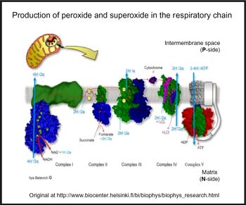
Oxidative stress is widespread to the point of ubiquity in chronic, inflammatory diseases(Reference Kell and Pretorius206,Reference Butterfield and Halliwell207) , with over fifty papers having the words ‘oxidative’, ‘stress’ and ‘review’ in their titles at PubMed in 2018 alone! It can occur when oxygen tension is low and respiratory chains are over-reduced such that they reduce O2 with one electron to superoxide or two electrons to H2O2, instead of the four that are used during the reduction of dioxygen to water by cytochrome oxidase(Reference Kell208) (Fig. 5). Peroxides are also produced in vivo by various oxidases and peroxidases, such as xanthine oxidase, by reduction of dioxygen (for example, Babior(Reference Babior209), Cave et al. (Reference Cave, Brewer and Narayanapanicker210) and Bedard & Krause(Reference Bedard and Krause211)).
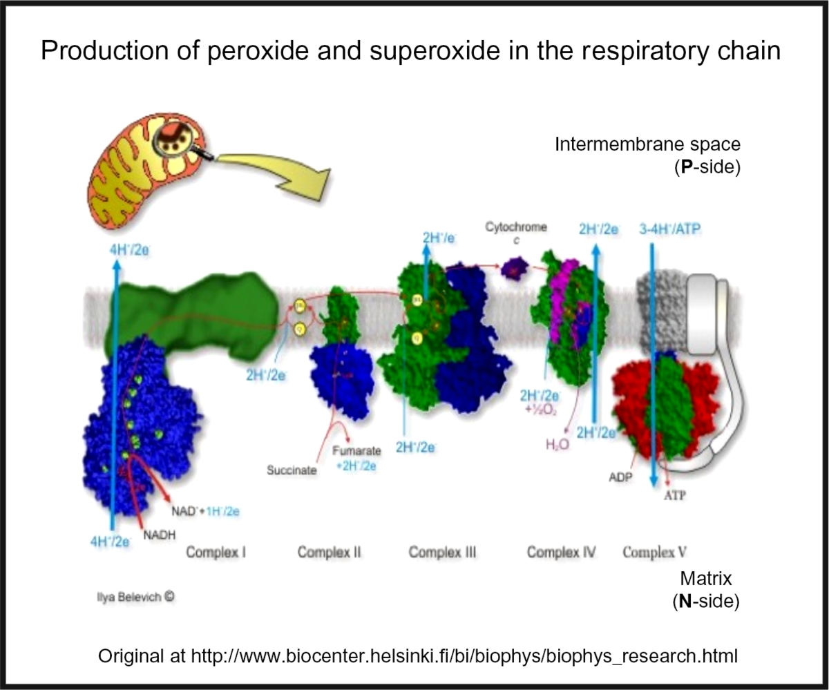
Fig. 5. Superoxide and peroxide are produced by 1- and 2-electron reduction of dioxygen by the mammalian respiratory chain. For a colour figure, see the online version of the paper.

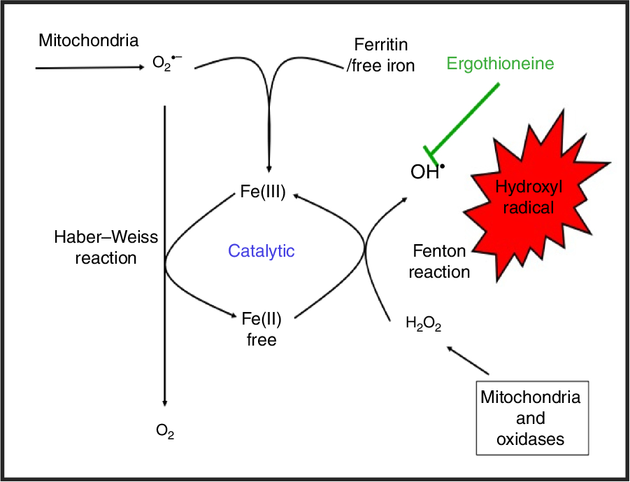
While H2O2 and superoxide are certainly capable of effecting unwanted oxidations, it is the hydroxyl radical that is the key. Thus an important reaction of H2O2 with (free or poorly liganded) Fe(II) is the Fenton reaction(Reference Kell208,Reference Wardman and Candeias212,Reference Kell213) , leading to the very reactive and damaging hydroxyl radical (OH):

(1) $${\rm{Fe}}\left( {{\rm{II}}} \right) + {{\rm{H}}_2}{{\rm{O}}_2} \to {\rm{Fe}}\left( {{\rm{III}}} \right) + {\rm{ O}}{{\rm{H}}^ - } + {\rm{ O}}{{\rm{H}}^ \bullet }$$

which can react within nanoseconds with anything adjacent. The role of Fe is absolutely vital here(Reference Kell208,Reference Kell213) . Superoxide can also react with ferric Fe in the Haber–Weiss reaction(Reference Winston, Feierman and Cederbaum214Reference Kehrer216) to produce Fe(II) again, thereby effecting redox cycling, and meaning the ‘iron’ is catalytic (Fig. 6):
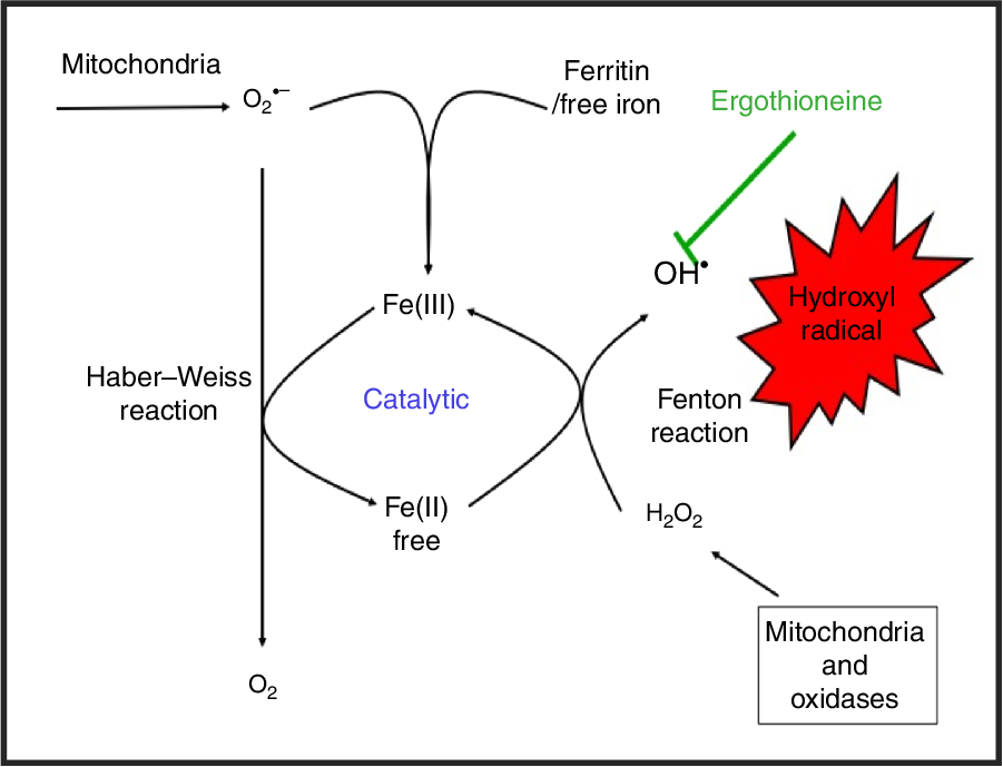
(2) $${{\rm{O}}_2}^{ \bullet - } + {\rm{ Fe}}\left( {{\rm{III}}} \right)\, \to {{\rm{O}}_2} + {\rm{ Fe}}\left( {{\rm{II}}} \right)$$

Fig. 6. Catalytic roles of unliganded iron in hydroxyl radical production via the Fenton and Haber–Weiss reactions. This can be stopped by ensuring that iron is fully liganded. For a colour figure, see the online version of the paper.

In addition O2 •− can release ‘catalytic’ Fe from Fe-S clusters in certain proteins and from ferritin(Reference Kell208,Reference Kell and Pretorius217) , another way in which it can promote the Fenton reaction. Note that other reactions can produce OH anaerobically(Reference Valachová, Mach and Dubovický218). Because OH is so reactive it is not really observable in its free form; its action is detected via products of molecules with which it has reacted. These include 8-oxo-guanine derivatives(Reference Migliore, Fontana and Colognato219), nitrotyrosine(Reference Ahsan220Reference Ryberg and Caidahl222) (itself formed from peroxynitrite(Reference Rahman223,Reference Aruoma, Whiteman and England224) , possibly formed more commonly via superoxide(Reference Halliwell225,Reference Ferrer-Sueta, Campolo and Trujillo226) ), 4-hydroxy-nonenal(Reference Petersen and Doorn227), and many others reviewed previously(Reference Kell208). In evaluating the antioxidant potency of ERG or anything else, it is molecules such as these that are normally assessed. Although the literature is somewhat scattered and heterogeneous, it seems clear that as well as hydroxyl radicals(Reference Akanmu, Cecchini and Aruoma228Reference Chaudière and Ferrari-Iliou232), ERG can also react with and detoxify, or prevent the formation of, singlet oxygen(Reference Oumari, Goldfuss and Stoffels233Reference Stoffels, Oumari and Perrou242), ozone(Reference He, Krone and Scherl243), superoxide(Reference Jang, Aruoma and Jen231,Reference Obayashi, Kurihara and Okano241,Reference Markova, Karaman-Jurukovska and Dong244Reference Servillo, D’Onofrio and Casale246) , peroxide(Reference Servillo, Castaldo and Casale32,Reference Li, Yang and Sit124,Reference Bello, Barrera-Perez and Morin247,Reference Hartman248) , hypochlorite(Reference Servillo, Castaldo and Casale32,Reference Chaudière and Ferrari-Iliou232,Reference Asahi, Wu and Shimoda249) and peroxynitrite(Reference Aruoma, Whiteman and England224,Reference Jang, Aruoma and Jen231,Reference Whiteman and Halliwell250,Reference Aruoma, Spencer and Mahmood251) . Consequently, it is a potent antioxidant.

Roles in the producer

Although it is not a priori certain that they would be the same in both producer and consumer organisms, it is of interest, before looking at higher organisms, to consider the roles of ERG in the producer organisms themselves. In the case of C. purpurea, the ERG serves as an antioxidant to neutralise a plant host defence response based on H2O2 that would otherwise inhibit the production of its conidia(Reference Garay252,Reference Garay253) . In M. tuberculosis and other mycobacteria(Reference Sao Emani, Williams and Wiid254), and also in other actinobacteria(Reference Nakajima, Satoh and Yanashima255) and in fungi(Reference Bello, Barrera-Perez and Morin247,Reference Liu, Zhao and Guo256,Reference Sheridan, Lechner and Keeffe257) , it is clear that ERG can have a role as an antioxidant(Reference Trivedi, Singh and Bhat66,Reference Ta, Buchmeier and Newton258Reference Cumming, Chinta and Reddy260) and also act as a buffer against reductive stress(Reference Farhana, Guidry and Srivastava261). In nature many organisms can be subjected to oxidative stress, and produce a variety of molecules to combat it(Reference Kurutas262Reference Davey and Kell270). This also seems true of mushrooms(Reference Savoie, Minvielle and Largeteau271,Reference Yokota, Frison and Marcante272) , where ERG is typically the main antioxidant(Reference Kozarski, Klaus and Jakovljevic273Reference Dubost, Ou and Beelman275), and where it may also inhibit the oxidative enzyme tyrosinase(Reference Liao, Wu and Tsai276). Given suggestions that the ‘purpose’ of secondary metabolite formation is to serve as a signalling molecule in different cells of the producer organism, i.e. as a pheromone(Reference Kell, Kaprelyants and Grafen277), it is interesting to note that this may also involve crosstalk of ERG(Reference Sheridan, Dolan and Doyle37), due in part to the complex networks in which it may be embedded(Reference Gallagher, Owens and Dolan278). The same is true of the imidazole thiol-containing ovothiol(Reference Song, Her and Raso279,Reference Castellano and Seebeck280) . In a similar vein, and although outwith our scope here, we note the potential of other antioxidant natural products such as curcumin(Reference Vázquez-Fresno, Rosana and Sajed281Reference Tomeh, Hadianamrei and Zhao286).

Nutritional sources

Betaines are generally seen as nutritionally beneficial(Reference Pekkinen, Rosa-Sibakov and Micard287), and many are ‘compatible solutes’(Reference Kempf and Bremer288Reference Fahnert293), defined as solutes whose accumulation assists the survival of the organism when undergoing various kinds of stress such as osmotic or thermal stress. However, of these, only ERG is seen as a major antioxidant. Although a variety of foodstuffs such as oats(Reference Ey, Schömig and Taubert294,Reference Lee, Alexander and Jonnalgadda295) contain ERG because they take it up from exogenous sources, it is really mushrooms that are the prime sources for humans(Reference Rathore, Prasad and Sharma18,Reference Ey, Schömig and Taubert294) . Indeed, ERG has been proposed as a nutritional biomarker for mushroom consumption(Reference Wang, Gapstur and Carter296,Reference Pallister, Jennings and Mohney297) , albeit that different mushrooms typically contain different amounts(Reference Dubost, Ou and Beelman275,Reference Dubost, Beelman and Peterson298Reference Kalaras, Richie and Calcagnotto300) , and these can vary with physiological or environmental conditions(Reference Tepwong and Ohshima301Reference Kalaras, Beelman and Holick305). Those with the highest amounts include oyster mushrooms (Pleurotus spp., up to 4 mg/g DM)(Reference Liang, Ho and Huang306), the golden oyster Pleurotus citrinopileatus with 10·65 mg/g DM(Reference Lin, Chien and Wang307,Reference Lin, Chien and Wang308) , and shiitake (Lentinula edodes, about 1 mg/g DM), while of those more common outside Asia, porcini or ceps (Boletus edulis, > 7 mg/g DM), stand out(Reference Halliwell, Cheah and Tang6,Reference Ey, Schömig and Taubert294,Reference Kalaras, Richie and Calcagnotto300) . However, even common field or ‘button’ mushrooms, Agaricus bisporus, contain some 0·4 mg/g DM(Reference Dubost, Ou and Beelman275,Reference Dubost, Beelman and Royse299,Reference Kalaras, Richie and Calcagnotto300,Reference Chen, Ho and Hsieh309) . Note too that tempe(h), the result of a solid substrate Rhizopus oligosporus fermentation(Reference Peñaloza, Davey and Hedger310Reference Nout and Kiers314), also contains high levels of ERG(Reference Halliwell, Cheah and Tang6). Mushrooms may also be a significant benefit to those seeking a meat-free diet as they can be made to share certain organoleptic features with meat(Reference Guinard, Myrdal Miller and Mills315,Reference Myrdal Miller, Mills and Wong316) . Notably, ‘the production of cultivated, edible mushrooms worldwide has increased more than 30-fold since 1978, whereas the population has only increased by about 1·7-fold during the same period’(Reference Beelman, Kalaras and John10,Reference Royse, Baars, Tan, Zied and Pardo-Giménez317) .

Some studies that have demonstrated nutritional/health benefits of mushrooms and their antioxidant activity(Reference Weigand-Heller, Kris-Etherton and Beelman125,Reference Savoie, Minvielle and Largeteau271,Reference Gallego, Rojas and Falcón318Reference Zembron-Lacny, Gajewski and Naczk351) ) did not always seek to deconstruct these into their constituents such as ERG, but ERG is clearly the chief mushroom antioxidant. We note too that some effects may be dependent on the composition of the gut microflora(Reference Jayachandran, Xiao and Xu352), that are of course themselves likely to be changed by ERG, just as they are by many other non-antibiotic drugs(Reference Maier, Pruteanu and Kuhn353).

Safety

Producer organisms such as mushrooms are well known to make many secondary metabolites, some of which can be highly toxic(Reference Schmutz, Carron and Yersin354Reference Verma, Bhalla and Kumar356) and by various mechanisms(Reference White, Weinstein and De Haro357). Notwithstanding the highly variable intake between individuals(Reference Ramirez-Martinez, Wesolek and Yadan358), however, a number of high-dose studies have indicated that ERG is safe for mammalian consumption at levels far in excess of those likely to be encountered in foodstuffs(Reference Weigand-Heller, Kris-Etherton and Beelman125,Reference Tang, Cheah and Yew131,Reference Marone, Trampota and Weisman359,Reference Cheah, Tang and Yew360) , and it has been declared safe by relevant committees such as those of the European Food Standards Agency(Reference Turck, Bresson and Burlingame361,Reference Turck, Bresson and Burlingame362) . It also lacks any detectable mutagenicity or genotoxicity in such assays, even at very high doses(Reference Schauss, Vértesi and Endres363,Reference Schauss, Béres and Vértesi364) .

Analytics

Leaving aside early efforts such as the colorimetric methods of Hunter(Reference Hunter365), of Melville and colleagues(Reference Melville76,Reference Melville, Horner and Lubschez85,Reference Melville and Lubschez366) and of Carlsson et al. (Reference Carlsson, Kierstan and Brocklehurst367), a variety of analytical methods have been proposed(Reference Cheah and Halliwell4), mostly involving capillary electrophoresis(Reference Sotgia, Arru and Sotgiu368,Reference Sotgia, Pisanu and Pintus369) or chromatography(Reference Sotgia, Arru and Sotgiu368,Reference Zhou, Liu and Jiang370Reference Sotgia, Zinellu and Arru372) coupled to absorbance(Reference Liu, Zhang and Wang373,Reference Muda, Pelizzoni and Sello374) , fluorescence detection(Reference Newton, Dorian and Fahey375Reference Sotgia, Pisanu and Cambedda378), electrochemical detection(Reference Kuninori and Nishiyama379) or MS(Reference Alamgir, Masuda and Fujitani72,Reference Wang, Thuya and Toh127,Reference Liu, Zhao and Guo256,Reference Sotgia, Arru and Sotgiu368,Reference Sotgia, Pisanu and Cambedda378,Reference Kroepfl, Francesconi and Schwerdtle380Reference Sotgia, Zinellu and Pintus382) . A useful feature is that ERG is unusually stable, in that anhydrous ERG decomposes only at 275–276°C(Reference Melville76), allowing its isolation at temperatures close to that of boiling water(Reference Alamgir, Masuda and Fujitani72). As judged by the reversibility of its acid–base titration(Reference Sakurai and Takeshima383), it is also stable to extremes of pH.

Industrial purification of glycine betaine is done by extraction with water(Reference Bessieres, Gibon and Lefeuvre384) and subsequent ion exchange chromatography(Reference Bessieres, Gibon and Lefeuvre384,Reference Kar and Singhal385) , which can be done in simulated moving bed fashion(Reference Bubnik, Pour and Gruberova386). Glycine betaine can then be crystallised(Reference Bessieres, Gibon and Lefeuvre384). As glycine betaine is structurally similar to ERG, this straightforward industrial process could potentially be adapted for ERG.

Serum and other concentrations

While most ERG is inside erythrocytes in whole blood(Reference Halliwell, Cheah and Tang6,Reference McMenamy, Lund and Wallach121,Reference McMenamy, Lund and Neville122,Reference Mitsuyama and May129,Reference Kumosani387) , there have been a number of measurements of ERG concentrations in serum. Unsurprisingly it varies with diet(Reference Baldridge and Lewis388,Reference Baldridge389) , starvation(Reference Teruya, Chaleckis and Takada390), age(Reference Cheah, Feng and Tang391,Reference Yan, Wu and Lin392) and other factors, including diseases of oxidative stress(Reference Dang, Shi and Werstuck393), with typical values of 20–100 µg/ml. A detailed list is provided by Cheah & Halliwell(Reference Cheah and Halliwell4); a smaller listing is given in Table 3. Interestingly, ERG is also present in seminal fluid(Reference Leone and Mann394Reference Strzeżek, Koziorowska-Gilun and Kielczewski396) and human breast milk(Reference Halliwell, Cheah and Tang6). Any possible correlation with male fertility(Reference Kenny and Kell397) seems not to have been established, though there were no negative effects(Reference Forster, Spézia and Papineau398), and ERG improved oocyte quality and maturation in cows and sheep(Reference Mishra, Reddy and Dhali399). ERG is also present in eye lens, where its concentration is lower in individuals with cataracts(Reference Shukla, Kulshrestha and Khuteta400).

Table 3. Concentrations of ergothioneine in human serum

* Molecular weight = 229·3, so 1 mm = 229 mg/l.

Metabolism and excretion

ERG is metabolised and excreted only slowly(Reference Cheah, Tang and Yew360,Reference Mayumi, Kawano and Sakamoto371,Reference Kato, Kubo and Iwata401,Reference Chaleckis, Murakami and Takada402) . In a recent and detailed study, Cheah et al. (Reference Cheah, Tang and Yew360) administered 5–25 mg daily doses of ERG to human volunteers for 7 d. There was little urinary excretion (<4 %), and the main metabolites were hercynine, plus lesser amounts of S-methyl-ERG, whose concentrations were well correlated with the level of ERG and the dose of ERG given. Similar observations were made in mice(Reference Tang, Cheah and Yew131). Various other biomarkers of oxidative stress (for example, 8-iso-PGF2α from lipid peroxidation) were lowered concomitantly in the human study, attesting to the antioxidant functions of ERG in vivo, although in this case the healthy young subjects were probably not suffering from oxidative stress. There was also quite some variation in uptake between individuals, presumably reflecting variation in their expression of SLC22A4. Agrobacterium radiobacter (Reference Jose403) and other bacteria(Reference Kelly and Appleman404Reference Maurer, Leisinger and Lim409) contain an ergothionase that degrades ERG to thiolurocanic acid (3-(1H-imidazol-5-yl)prop-2-enethioic S-acid) and trimethylamine, also implying that such cells possess one or more transporters for ERG. The thiolurocanic acid can be further degraded to glutamate(Reference Booth and Appleman410).

Apparent fitness benefits and bioactivities of ERG and the role of SLC22A4

Given the great technical difficulties associated, because of its ubiquity, with withholding ergothoneine from a human or animal diet, one means of ‘removing’ ERG from a host is to remove the ERG transporter by genetic means. This has in fact been done in mice(Reference Kato, Kubo and Iwata401); such SCL22A4–/– mice had immeasurably low levels of ERG relative to controls, and were much more sensitive to oxidative stress than were the wild type. Similar effects were observed in Caenorhabditis elegans (Reference Cheah, Ong and Gruber411). Polymorphisms in SLC22A4, of which there can be many(Reference Tamai177,Reference Ben Said, Grati and Ishimoto412Reference Toh, Cheung and Murray415) , under selection(Reference Mathieson, Lazaridis and Rohland416), have also been associated with ischaemic stroke(Reference Yamase, Horibe and Ueyama417), erythroid differentiation(Reference Nakamura, Sugiura and Kobayashi418), hearing loss(Reference Ben Said, Grati and Ishimoto412), rheumatoid arthritis(Reference Reglinski, Smith and Wilson126,Reference Taubert, Lazar and Grimberg180,Reference Maeda, Hirayama and Kobayashi419Reference Lee, Bae and Kim427) , lupus(Reference Orozco, Sánchez and Gómez428), Crohn’s disease(Reference Kato, Kubo and Iwata401,Reference Taubert, Grimberg and Jung429Reference Leung, Hong and Fraser436) , hearing loss(Reference Ben Said, Grati and Ishimoto412), type 1 diabetes(Reference Santiago, Martinez and de la Calle437) and diabetic embryopathy(Reference Zhao and Reece438). The expression of SLC22A4 can itself be modulated by other factors, including by PPAR-α activity(Reference Wada, Koyanagi and Kusunose439). The very diversity of these diseases speaks naturally to a broad and common underlying cause, the easiest of which involves mechanisms of oxidative stress, inflammation and cell death.

Mechanisms of action

It has become common to discover a binding of a small molecule to another molecule such as a protein, and assume that this interaction, leading to activation or inhibition of the target, constitutes ‘the’ mechanism of action of the small molecule at a physiological level. Unfortunately this is rarely the case, and known drugs, despite often being selected for inhibiting potently a specific molecular target(Reference Kell147), have, on average, six known binding targets(Reference Mestres, Gregori-Puigjané and Valverde440). When these interactions ramify through a complex and non-linear biochemical network it can be hard to apportion the effects of a small exogenous molecule between the various interactions(Reference Kell441Reference Sastry, Gao and Szubin443).

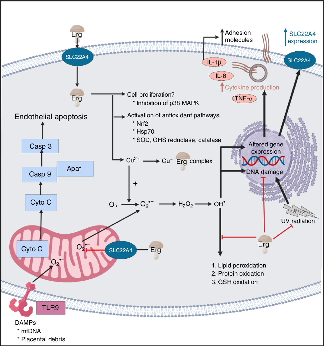
A standard view of systems or network biology (for example, Kell(Reference Kell444) and Kell & Knowles(Reference Kell, Knowles, Szallasi, Stelling and Periwal445)) develops these ideas in four stages. In stages 1 and 2 we simply recognise the actors and the interactions between them at a qualitative level. Stages 3 and 4 then seek to describe the equations reflecting individual steps and the values of the parameters of those equations. Armed with these we can make an ordinary or, if spatial resolution within a compartment is required, a partial differential equation model of the system. This can then be run and the sensitivities of each step determined(Reference Ihekwaba, Broomhead and Grimley446Reference Rand448). We are very far from this last part, and so studies of the effects of ERG have in general(Reference Kell and Oliver449) been rather descriptive in nature. Many have been at the level of processes rather than mechanisms, and they have been reviewed in detail(Reference Paul and Snyder3,Reference Cheah, Tang and Yew360) . Table 4 and Fig. 7 provide a selection of determinands that have been shown to change their concentrations or activities when ERG is added to the system of interest. In many cases it is not at all clear what the proximate mechanisms are. Note as just one example that the highly promiscuous transcription factor NF-κB(Reference Ledoux and Perkins450Reference Shih, Wang and Yang452), whose frequency-dependent activity directly affects the expression of hundreds of enzymes(Reference Nelson, Ihekwaba and Elliott453,Reference Ashall, Horton and Nelson454) , is itself redox-sensitive(Reference Buelna-Chontal and Zazueta455Reference Gao and Dudley458), and is affected by ERG(Reference Xiao, Zhao and Li459,Reference Rahman, Gilmour and Jimenez460) , while NF-κB increases the rates of SC22A4 transcription(Reference Maeda, Hirayama and Kobayashi419). Thus, deconstructing the many possible direct and consequential interactions of ERG with proteins, v. whether these are simply a consequence of its provision of a more reducing environment, is likely to be a formidable task. In a similar vein, the effects of ERG on the microbiomes of the hosts are likely to be significant, but do not yet seem to have been studied.

Table 4. Biological properties whose expression or activity varies on exposure of a biological system to ergothioneine (ERG) or a modulation of SLC22A4 activity
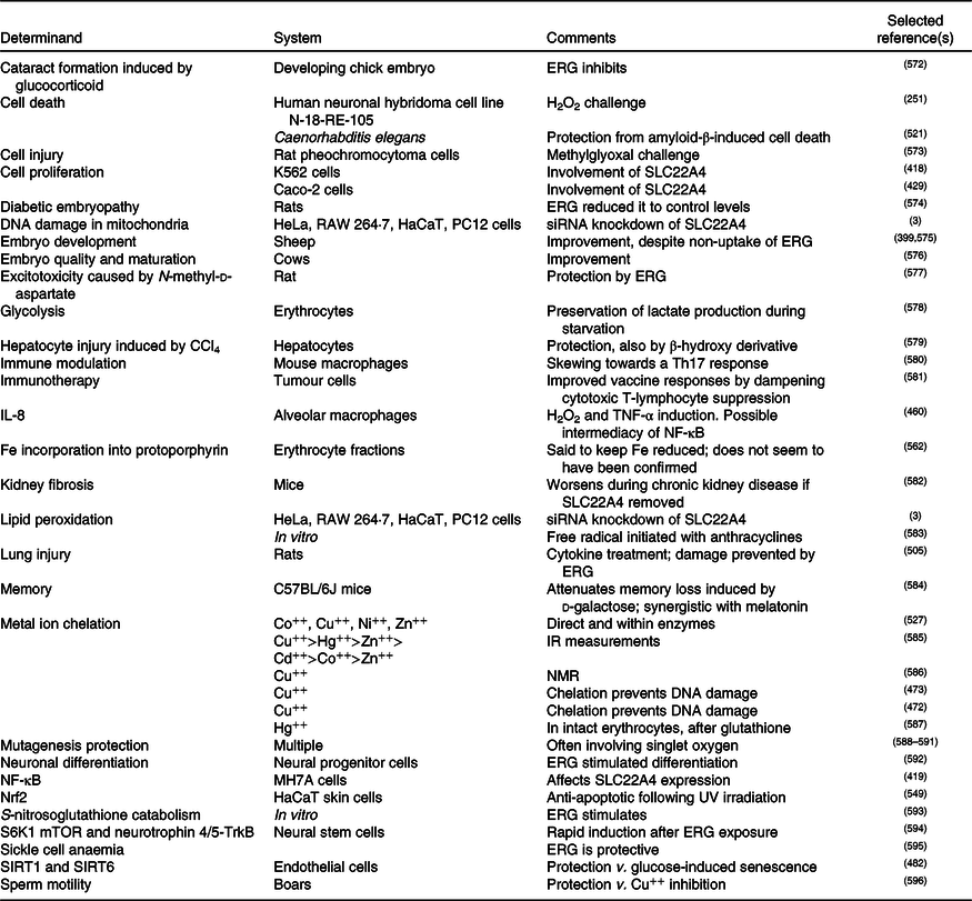
siRNA, small interfering RNA; mTOR, mammalian target of rapamycin; SIRT, sirtuin.
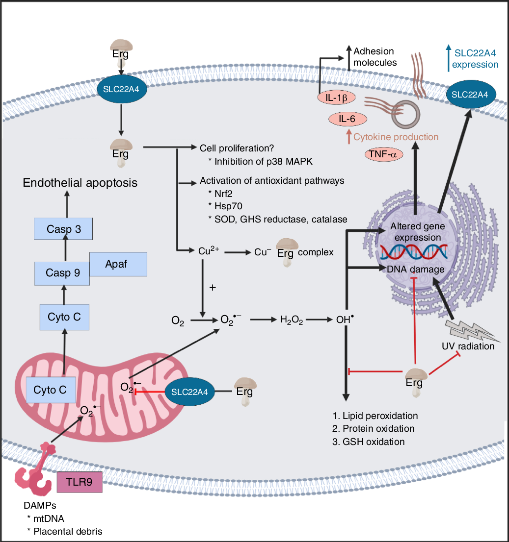
Fig. 7. Overview of some of the effects of ergothioneine in mammalian systems. For a colour figure, see the online version of the paper.

It seems clear that the chief role of ERG, via a variety of mechanisms, including directly, is to serve as an antioxidant and cellular protectant against various kinds of reactive oxygen and N species.

Cytoprotection

At a high level, ERG is seen as an excellent cytoprotectant against all kinds of cellular insults(Reference Paul and Snyder3,Reference Cheah and Halliwell4,Reference Halliwell, Cheah and Tang6,Reference Li, Yang and Sit124) . We split some of the more detailed analyses into subdivisions in the following few sections.

Oxidative stress

Oxidative stress can be defined and measured in many ways(Reference Agarwal, Aponte-Mellado and Premkumar461Reference Halliwell and Gutteridge468), but is broadly taken to involve a dysregulation in the various redox systems of the organism of interest, coupled to the production of various ‘reactive oxygen species’, principally peroxide, superoxide, hydroxyl radical, and singlet oxygen. ERG has been shown to decrease oxidative stress in the liver and kidney of rats(Reference Deiana, Rosa and Casu469), rescued cells from β-amyloid-induced apoptotic death(Reference Jang, Aruoma and Jen231), protected against palmitic acid-induced cell death(Reference Laurenza, Colognato and Migliore470), mercuric chloride-induced cellular dysfunction(Reference Gökçe, Arun and Ertuna471), and prevented Cu-induced oxidative damage to DNA(Reference Rai, Chalana and Karri472,Reference Zhu, Mao and Fan473) . It is protective against the oxidation of various kinds of molecule(Reference Aruoma, Spencer and Mahmood251,Reference Gokce and Arun474) , including astaxanthin(Reference Pahila, Ishikawa and Ohshima475), and accumulates in a guinea-pig model of non-alcoholic fatty liver disease(Reference Cheah, Tang and Ye476), massively so in mouse models of myocardial infarction and heart failure(Reference Sansbury, DeMartino and Xie477), and in a rat model of optic nerve crush(Reference Agudo-Barriuso, Lahoz and Nadal-Nicolás478). It serves to resist H2O2-induced cell death(Reference Colognato, Laurenza and Fontana479), pyrogallol-induced toxicity(Reference Li, Yang and Sit124), cisplatin-(Reference Song, Chen and Liao480) or oxaliplatin-induced(Reference Nishida, Takeuchi and Hosoda481) toxicity, glucose-induced senescence(Reference Servillo, D’Onofrio and Casale246,Reference D’Onofrio, Servillo and Giovane482) , as well as lipopolysaccharide-induced inflammation(Reference Gunawardena, Bennett and Shanmugam483). In particular, it is protective against ischaemia–reperfusion injury(Reference Sakrak, Kerem and Bedirli484Reference Arduini, Eddy and Hochstein486), and may have uses in prolonging the lifetime of stored blood(Reference Zimring, Smith and Stowell487). Probably such antioxidant activities are at the core of its biological benefits.

Ergothioneine as a therapeutic for chronic inflammatory diseases?

Inflammation and oxidative stress go hand in hand(Reference Paul and Snyder3), since reactive oxygen species (and materials such as bacterial cell wall components that can lead to their generation(Reference Kell and Pretorius206,Reference Kell and Pretorius488) ), lead to the production of inflammatory cytokines. Although a great many chronic, inflammatory diseases are recognised as having an oxidative stress component(Reference Rahman223), the history of treating them with antioxidants such as ascorbate has largely been a litany of failure, with the treatment arm often even giving worse prognoses than the placebo(Reference Halliwell, Cheah and Tang6,Reference Rahman223,Reference Goodman, Bostick and Kucuk489Reference Bjelakovic, Nikolova and Gluud501) . Arguably this is because nominally antioxidant molecules such as ascorbate have complex, multi-electron redox chemistry, and can in fact act as pro-oxidants(Reference Mendes-da-Silva, Lopes-de-Morais and Bandim-da-Silva502,Reference Zhang and Omaye503) , especially in the presence of free Fe(Reference Kell208,Reference Kell213) or Cu(Reference Ullah, Khan and Zubair504). This is not an issue with ERG, however, partly because it can chelate them, and ERG levels are decreased, or ERG has been proposed as a useful antioxidant, in diseases such as acute respiratory distress syndrome(Reference Repine and Elkins505), CVD(Reference Martin506,Reference Servillo, D’Onofrio and Balestrieri507) , chronic obstructive pulmonary disease(Reference Rahman223), pre-eclampsia(Reference Kerley, McCarthy and Kell8) (see also Turner et al. (Reference Turner, Brewster and Simpson128)), overhydrated hereditary stomatocytosis(Reference Darghouth, Koehl and Heilier508), and is significantly lowered in others such as certain leukaemias(Reference McMenamy, Lund and Wallach121,Reference McMenamy, Lund and Neville122) . The evidence for this comes from a variety of sources, including metabolite measurements in human subjects(Reference McMenamy, Lund and Wallach121,Reference McMenamy, Lund and Neville122,Reference Smith, Ottosson and Hellstrand509) , and intervention studies in both animals(Reference Repine and Elkins505) and cell lines(Reference Paul and Snyder3,Reference Li, Yang and Sit124,Reference Martin506) . In particular, there is a notable relationship between ERG consumption and longevity (Fig. 6 in Beelman et al. (Reference Beelman, Kalaras and John10)), while in a 3236-participant Swedish study, ERG was the metabolite most strongly connected to a ‘health conscious food pattern’ and was associated with a lower risk of coronary disease (hazard ratio (HR) per 1 sd increment of ERG, HR = 0·85; P = 0·01), cardiovascular mortality (HR = 0·79; P = 0·002) and overall mortality (HR = 0·86; P = 4 × 10⁻⁵)(Reference Smith, Ottosson and Hellstrand509).

Neurological diseases and cognitive function

Mushrooms have been shown to have very substantial effects on cognitive function(Reference Mori, Inatomi and Ouchi341,Reference Feng, Cheah and Ng348,Reference Nurk, Refsum and Drevon510Reference Thangthaeng, Miller and Gomes513) , and this is mainly ascribed to their ERG content, that also deceases with the age of the consumer(Reference Cheah, Feng and Tang391). The kinds of evidence include both double-blind, placebo-controlled clinical trials(Reference Mori, Inatomi and Ouchi341) and observational (cross-sectional) studies in both humans(Reference Feng, Cheah and Ng348,Reference Nurk, Refsum and Drevon510Reference Phan, David and Sabaratnam512) and rodents(Reference Thangthaeng, Miller and Gomes513). Thus, consuming 1·5 mushroom servings per week was associated with a halving of the incidence of mild cognitive impairment (a precursor of Alzheimer’s dementia), while intake of nine servings per week was associated with a five-fold decrease(Reference Feng, Cheah and Ng348). Note, however, that at least one mushroom trial indicated no measurable benefits in healthy young physical education students(Reference Tsuk, Lev and Rotstein514). Brain and serum ERG levels are also markedly different in Parkinson’s disease(Reference Hatano, Saiki and Okuzumi515), reviewed in Hang et al. (Reference Hang, Basil and Lim516), Shao & Le(Reference Shao and Le517) and Shah & Duda(Reference Shah and Duda518), and even in sudden infant death syndrome(Reference Graham, Chevallier and Kumar519), and ERG has been shown to be protective against β-amyloid-induced neuronal injury(Reference Yang, Lin and Wu520) and cytotoxicity(Reference Cheah, Ng and Ng521). It can also act as an antidepressant(Reference Nakamichi, Nakayama and Ishimoto522). The evidence for this comes from direct studies(Reference Yang, Lin and Wu520) and feeding experiments(Reference Nakamichi, Nakayama and Ishimoto522) in mice, as well as via the reduction of β-amyloid peptide in a transgenic C. elegans model(Reference Cheah, Ng and Ng521). As mentioned above, SLC22A4 polymorphisms are associated with ischaemic stroke(Reference Yamase, Horibe and Ueyama417).

Use of ergothioneine as an antioxidant in processed foodstuffs

Just as living beings exploit antioxidants, most foodstuffs can also be oxidised to produce taints, rancidity or other undesirable products(Reference Logan, Nienaber and Pan523Reference Gülçin525), often via the Fenton reaction(Reference Kell208,Reference Perron and Brumaghim526) . ERG inhibits polyphenoloxidases(Reference Hanlon527), and thus ERG has been used in the feed of the shrimp Marsupenaeus japonicas to prevent melanosis during storage(Reference Encarnacion, Fagutao and Hirono528), while ERG-rich mushroom extract has also been used to prevent melanosis in post-harvest storage of the crab Chionoecetes japonicus (Reference Encarnacion, Fagutao and Hirayama529). Thus, one can also envisage a role for ERG, whether natural or added, in extending shelf lives and commercial value(Reference Nguyen, Nagasaka, Ohshima, Logan, Nienaber and Pan245,Reference Tang, Hoo and Tan328,Reference Pahila, Ishikawa and Ohshima475,Reference Encarnacion, Fagutao and Hirono528Reference Duy Bao, Ohshima, Nienaber and Pan539) . The thermostability of ERG is of particular importance here.

Use of ergothioneine in cosmetics

Just as processed foodstuffs ‘age’, so do tissues such as the skin, and although the same principles apply(Reference Pérez-Sánchez, Barrajón-Catalán and Herranz-López540), it is common to refer to nutraceuticals that are also aimed at having cosmetic benefits as ‘cosmeceuticals’(Reference Lee541Reference Taofiq, González-Paramás and Martins543). Here too, ERG has been widely used(Reference Taofiq, González-Paramás and Martins543Reference Souyoul, Saussy and Lupo547), since much skin damage is caused by UV-mediated reactive oxygen species production(Reference Norins548); indeed, ERG is known as a skin protectant(Reference Dong, Damaghi and Kibitel240Reference Markova, Karaman-Jurukovska and Dong244,Reference Hseu, Lo and Korivi549Reference Bazela, Solyga-Zurek and Debowska551) .

Role of ergothioneine as a cofactor?

Although it is possible that the role of ERG lies simply in being an antioxidant capable of mopping up hydroxyl radicals and other reactive oxygen species, especially in prokaryotes(Reference Fahey36,Reference Trivedi, Singh and Bhat66,Reference Richard-Greenblatt, Bach and Adamson93,Reference Sao Emani, Williams and Wiid254,Reference Nakajima, Satoh and Yanashima255,Reference Ta, Buchmeier and Newton258Reference Cumming, Chinta and Reddy260,Reference Sao Emani, Williams and Van Helden552,Reference Sao Emani, Williams and Van Helden553) , the roles of most other vitamins involve interaction with proteins, often as cofactors. This is also true for mycothiol(Reference Rawat and Av-Gay73,Reference Jothivasan and Hamilton554Reference Newton, Buchmeier and Fahey556) , though that molecule can also serve as a signal and nutrient resource(Reference Bzymek, Newton and Ta557). However, despite many hypotheses(Reference Brummel558,Reference Brummel559) , the only example of ERG acting as a cofactor known to date is an involvement in the biosynthesis of lincomycin(Reference Wang, Zhao and Liu560,Reference Zhao, Wang and Xu561) . An early paper(Reference Goldberg562) implying an involvement of ERG in the maintenance of a reduced state of Fe in Hb, although apparently accurate, does not seem to have been followed up to date.

Conclusions

There is increasing awareness that health may be enhanced via the consumption of substances that either have no recommended daily intake or are taken at levels greater than normal, and such substances are commonly referred to as nutraceuticals. ERG, a potent and effective antioxidant, seems to be an important nutraceutical, and we rehearse a very broad literature, involving a great many cells, tissues and organisms, to that effect. The chief source of ERG in the human diet is mushrooms (usually the fruiting bodies of Basidiomycetes). The fact that a specific transporter known as SLC22A4 has evolved and been selected to effect ERG uptake in all known animals suggests strongly that ERG is of benefit to its consumers. While the evidence that ERG may be a useful nutraceutical as a preventive or palliative for various inflammatory diseases is extensive, it is mostly circumstantial rather than definitive, though many examples exist of the benefits of mushrooms in combating the results of oxidative stress.

Without mechanisms, finding that the concentration of a dietary metabolite X is low in disease Y does not mean that giving it might be of benefit in the prevention, delay or cure of that disease, although cases can clearly be made when X is a vitamin, or oxidative stress is known to be a damaging component of Y(Reference Kerley, McCarthy and Kell8,Reference Feng, Cheah and Ng348) . Thus far, we lack examples in which ERG is found both to be low in individuals with a particular syndrome and where exogenous administration effects functional improvements, although – as reviewed above – we often have one or the other.

To assess definitively any health benefits of ERG, the ‘gold standard’ of randomised controlled trials may take time and money, but – as with mushrooms(Reference Feeney, Miller and Roupas335,563) – are beginning. One trial with pure ERG has been registered(Reference Cheah564).

Note added in proof

A recent paper indicates that ERG relieves the effects seen in a rat model of the pregnancy disorder pre-eclampsia(Reference Williamson, McCarthy and Manna597).

Acknowledgements

We thank the Biotechnology and Biological Sciences Research Council (BBSRC) (grants no. BB/R000093/1 and no. BB/P009042/1) and the Novo Nordisk Foundation (grant no. NNF10CC1016517) for financial support.

D. B. K. decided to bring together the multifaceted contributions of the various authors listed. Their previous contributions to ergothioneine and antioxidant biology may be seen in the references cited. All authors contributed to and approved the final manuscript.

I. B., S. A. v. d. H. and D. B. K. are named inventors on a patent application involving the biotechnological production of l-(+)-ergothioneine in yeast. For the other authors, there are no conflicts of interest.

References

Kraemer, K, Semba, RD, Eggersdorfer, M, et al. (2012) Introduction: the diverse and essential biological functions of vitamins. Ann Nutr Metab 61, 185191.CrossRefGoogle ScholarPubMed
Semba, RD (2012) The discovery of the vitamins. Int J Vitam Nutr Res 82, 310315.CrossRefGoogle ScholarPubMed
Paul, BD & Snyder, SH (2010) The unusual amino acid l-ergothioneine is a physiologic cytoprotectant. Cell Death Differ 17, 11341140.CrossRefGoogle ScholarPubMed
Cheah, IK & Halliwell, B (2012) Ergothioneine; antioxidant potential, physiological function and role in disease. Biochim Biophys Acta 1822, 784793.CrossRefGoogle Scholar
Halliwell, B, Cheah, IK & Drum, CL (2016) Ergothioneine, an adaptive antioxidant for the protection of injured tissues? A hypothesis. Biochem Biophys Res Commun 470, 245250.CrossRefGoogle ScholarPubMed
Halliwell, B, Cheah, IK & Tang, RMY (2018) Ergothioneine – a diet-derived antioxidant with therapeutic potential. FEBS Lett 592, 33573366.CrossRefGoogle ScholarPubMed
Ames, BN (2018) Prolonging healthy aging: longevity vitamins and proteins. Proc Natl Acad Sci U S A 115, 1083610844.CrossRefGoogle ScholarPubMed
Kerley, RN, McCarthy, C, Kell, DB, et al. (2018) The potential therapeutic effects of ergothioneine in pre-eclampsia. Free Radic Biol Med 117, 145157.CrossRefGoogle ScholarPubMed
Anonymous (2015) l-Ergothioneine. http://www.tetrahedron.fr/products/research/l-ergothioneine (accessed August 2019).Google Scholar
Beelman, RB, Kalaras, MD, John, P, et al. (2019) Micronutrients and bioactive compounds in mushrooms: a recipe for healthy aging? Nutr Today 54, 1622.CrossRefGoogle Scholar
Cencic, A & Chingwaru, W (2010) The role of functional foods, nutraceuticals, and food supplements in intestinal health. Nutrients 2, 611625.CrossRefGoogle ScholarPubMed
Bahadoran, Z, Mirmiran, P & Azizi, F (2013) Dietary polyphenols as potential nutraceuticals in management of diabetes: a review. J Diabetes Metab Disord 12, 43.CrossRefGoogle ScholarPubMed
Ogle, WO, Speisman, RB & Ormerod, BK (2013) Potential of treating age-related depression and cognitive decline with nutraceutical approaches: a mini-review. Gerontology 59, 2331.CrossRefGoogle ScholarPubMed
Ragle, RL & Sawitzke, AD (2012) Nutraceuticals in the management of osteoarthritis: a critical review. Drugs Aging 29, 717731.CrossRefGoogle ScholarPubMed
Chauhan, B, Kumar, G, Kalam, N, et al. (2013) Current concepts and prospects of herbal nutraceutical: A review. J Adv Pharm Technol Res 4, 48.Google ScholarPubMed
Borghi, C & Cicero, AF (2017) Nutraceuticals with a clinically detectable blood pressure-lowering effect: a review of available randomized clinical trials and their meta-analyses. Br J Clin Pharmacol 83, 163171.CrossRefGoogle ScholarPubMed
Aruoma, OI, Coles, LS, Landes, B, et al. (2012) Functional benefits of ergothioneine and fruit- and vegetable-derived nutraceuticals: overview of the supplemental issue contents. Prev Med 54, Suppl., S4–S8.CrossRefGoogle ScholarPubMed
Rathore, H, Prasad, S & Sharma, S (2017) Mushroom nutraceuticals for improved nutrition and better human health: a review. PharmaNutrition 5, 3546.CrossRefGoogle Scholar
Espín, JC, García-Conesa, MT & Tomás-Barberán, FA (2007) Nutraceuticals: facts and fiction. Phytochemistry 68, 29863008.CrossRefGoogle ScholarPubMed
Sharif, MK & Khalid, R (2018) Nutraceuticals: myths versus realities. In Therapeutic Foods, vol. 8, pp. 321 [Holban, AM and Grumezescu, AM, editors]. Cambridge, MA: Academy Press.CrossRefGoogle Scholar
Singh, S, Razak, MA, Sangam, SR, et al. (2018) The impact of functional food and nutraceuticals in health. In Therapeutic Foods, vol. 8, pp. 2347 [Holban, AM and Grumezescu, AM, editors]. Cambridge, MA: Academy Press.CrossRefGoogle Scholar
Spindler, SR, Mote, PL & Flegal, JM (2014) Lifespan effects of simple and complex nutraceutical combinations fed isocalorically to mice. Age (Dordr) 36, 705718.CrossRefGoogle ScholarPubMed
Poddar, K, Kolge, S, Bezman, L, et al. (2011) Nutraceutical supplements for weight loss: a systematic review. Nutr Clin Pract 26, 539552.CrossRefGoogle ScholarPubMed
Camfield, DA, Sarris, J & Berk, M (2011) Nutraceuticals in the treatment of obsessive compulsive disorder (OCD): a review of mechanistic and clinical evidence. Prog Neuropsychopharmacol Biol Psychiatry 35, 887895.CrossRefGoogle ScholarPubMed
Goggs, R, Vaughan-Thomas, A, Clegg, PD, et al. (2005) Nutraceutical therapies for degenerative joint diseases: a critical review. Crit Rev Food Sci Nutr 45, 145164.CrossRefGoogle ScholarPubMed
Naveen, J & Baskaran, V (2018) Antidiabetic plant-derived nutraceuticals: a critical review. Eur J Nutr 57, 12751299.CrossRefGoogle ScholarPubMed
Orr, SL & Venkateswaran, S (2014) Nutraceuticals in the prophylaxis of pediatric migraine: evidence-based review and recommendations. Cephalalgia 34, 568583.CrossRefGoogle ScholarPubMed
Orr, SL (2016) Diet and nutraceutical interventions for headache management: a review of the evidence. Cephalalgia 36, 11121133.CrossRefGoogle Scholar
Tanret, C (1909) A new base taken from rye ergot, ergothioneine. Ann Chim Phys 18, 114124.Google Scholar
Tanret, C (1909) The new base drawn from rye ergot, ergothioneine. Cr Hebd Acad Sci 149, 222224.Google Scholar
Barger, G & Ewins, AJ (1911) The constitution of ergothioneine: a betaine related to histidine. J Chem Soc 99, 23362341.CrossRefGoogle Scholar
Servillo, L, Castaldo, D, Casale, R, et al. (2015) An uncommon redox behavior sheds light on the cellular antioxidant properties of ergothioneine. Free Radic Biol Med 79, 228236.CrossRefGoogle ScholarPubMed
Franzoni, F, Colognato, R, Galetta, F, et al. (2006) An in vitro study on the free radical scavenging capacity of ergothioneine: comparison with reduced glutathione, uric acid and trolox. Biomed Pharmacother 60, 453457.CrossRefGoogle Scholar
Hand, CE, Taylor, NJ & Honek, JF (2005) Ab initio studies of the properties of intracellular thiols ergothioneine and ovothiol. Bioorg Med Chem Lett 15, 13571360.CrossRefGoogle ScholarPubMed
Hand, CE & Honek, JF (2005) Biological chemistry of naturally occurring thiols of microbial and marine origin. J Nat Prod 68, 293308.CrossRefGoogle ScholarPubMed
Fahey, RC (2013) Glutathione analogs in prokaryotes. Biochim Biophys Acta 1830, 31823198.CrossRefGoogle ScholarPubMed
Sheridan, KJ, Dolan, SK & Doyle, S (2015) Endogenous cross-talk of fungal metabolites. Front Microbiol 5, 732.CrossRefGoogle ScholarPubMed
Fahey, RC (2001) Novel thiols of prokaryotes. Annu Rev Microbiol 55, 333356.CrossRefGoogle ScholarPubMed
Jacob, C (2006) A scent of therapy: pharmacological implications of natural products containing redox-active sulfur atoms. Nat Prod Rep 23, 851863.CrossRefGoogle ScholarPubMed
Clark, WM (1960) Oxidation-reduction Potentials of Organic Systems. Baltimore, MD: The Williams and Wilkins Co.Google Scholar
Walz, D (1979) Thermodynamics of oxidation–reduction reactions and its application to bioenergetics. Biochim Biophys Acta 505, 279353.CrossRefGoogle ScholarPubMed
Reyes, AM, Pedre, B, De Armas, MI, et al. (2018) Chemistry and redox biology of mycothiol. Antioxid Redox Signal 28, 487504.CrossRefGoogle ScholarPubMed
Sharma, SV, Van Laer, K, Messens, J, et al. (2016) Thiol redox and pKa properties of mycothiol, the predominant low-molecular-weight thiol cofactor in the actinomycetes. Chembiochemistry 17, 16891692.CrossRefGoogle ScholarPubMed
Kell, DB & Morris, JG (1979) Oxidation–reduction properties of coenzyme M (2-mercaptoethane sulphonate) at the mercury electrode. FEBS Lett 108, 481484.CrossRefGoogle ScholarPubMed
Achouba, A, Dumas, P, Ouellet, N, et al. (2019) Selenoneine is a major selenium species in beluga skin and red blood cells of Inuit from Nunavik. Chemosphere 229, 549558.CrossRefGoogle ScholarPubMed
Little, M, Achouba, A, Dumas, P, et al. (2019) Determinants of selenoneine concentration in red blood cells of Inuit from Nunavik (Northern Quebec, Canada). Environ Int 127, 243252.CrossRefGoogle Scholar
Turrini, NG, Kroepfl, N, Jensen, KB, et al. (2018) Biosynthesis and isolation of selenoneine from genetically modified fission yeast. Metallomics 10, 15321538.CrossRefGoogle ScholarPubMed
Rohn, I, Kroepfl, N, Bornhorst, J, et al. (2019) Side-directed transfer and presystemic metabolism of selenoneine in a human intestinal barrier model. Mol Nutr Food Res 63, e1900080.CrossRefGoogle Scholar
Klein, M, Ouerdane, L, Bueno, M, et al. (2011) Identification in human urine and blood of a novel selenium metabolite, Se-methylselenoneine, a potential biomarker of metabolization in mammals of the naturally occurring selenoneine, by HPLC coupled to electrospray hybrid linear ion trap-orbital ion trap MS. Metallomics 3, 513520.CrossRefGoogle ScholarPubMed
Pluskal, T, Ueno, M & Yanagida, M (2014) Genetic and metabolomic dissection of the ergothioneine and selenoneine biosynthetic pathway in the fission yeast, S. pombe, and construction of an overproduction system. PLOS ONE 9, e97774.CrossRefGoogle ScholarPubMed
Yamashita, Y & Yamashita, M (2010) Identification of a novel selenium-containing compound, selenoneine, as the predominant chemical form of organic selenium in the blood of bluefin tuna. J Biol Chem 285, 1813418138.CrossRefGoogle ScholarPubMed
Yamashita, Y, Yabu, T & Yamashita, M (2010) Discovery of the strong antioxidant selenoneine in tuna and selenium redox metabolism. World J Biol Chem 1, 144150.CrossRefGoogle ScholarPubMed
O’Hagan, S & Kell, DB (2017) Analysis of drug-endogenous human metabolite similarities in terms of their maximum common substructures. J Cheminform 9, 18.CrossRefGoogle ScholarPubMed
O’Hagan, S & Kell, DB (2016) MetMaxStruct: a Tversky-similarity-based strategy for analysing the (sub)structural similarities of drugs and endogenous metabolites. Front Pharmacol 7, 266.Google ScholarPubMed
O’Hagan, S & Kell, DB (2015) The KNIME workflow environment and its applications in genetic programming and machine learning. Genetic Progr Evol Mach 16, 387391.CrossRefGoogle Scholar
Thiele, I, Swainston, N, Fleming, RMT, et al. (2013) A community-driven global reconstruction of human metabolism. Nat Biotechnol 31, 419425.CrossRefGoogle ScholarPubMed
O’Hagan, S, Swainston, N, Handl, J, et al. (2015) A ‘rule of 0.5’ for the metabolite-likeness of approved pharmaceutical drugs. Metabolomics 11, 323339.CrossRefGoogle Scholar
Lewis, KA, Tzilivakis, J, Warner, DJ, et al. (2016) An international database for pesticide risk assessments and management. Hum Ecol Risk Assess 22, 10501064.CrossRefGoogle Scholar
Melville, DB, Genghof, DS, Inamine, E, et al. (1956) Ergothioneine in microorganisms. J Biol Chem 223, 917.Google Scholar
Jones, GW, Doyle, S & Fitzpatrick, DA (2014) The evolutionary history of the genes involved in the biosynthesis of the antioxidant ergothioneine. Gene 549, 161170.CrossRefGoogle ScholarPubMed
Newton, GL, Arnold, K, Price, MS, et al. (1996) Distribution of thiols in microorganisms: mycothiol is a major thiol in most actinomycetes. J Bacteriol 178, 19901995.CrossRefGoogle ScholarPubMed
Dosanjh, M, Newton, GL & Davies, J (2008) Characterization of a mycothiol ligase mutant of Rhodococcus jostii RHA1. Res Microbiol 159, 643650.CrossRefGoogle ScholarPubMed
Genghof, DS & Van Damme, O (1964) Biosynthesis of ergothioneine and hercynine by mycobacteria. J Bacteriol 87, 852862.CrossRefGoogle ScholarPubMed
Genghof, DS & Van Damme, O (1968) Biosynthesis of ergothioneine from endogenous hercynine in Mycobacterium smegmatis . J Bacteriol 95, 340344.CrossRefGoogle ScholarPubMed
Genghof, DS (1970) Biosynthesis of ergothioneine and hercynine by fungi and Actinomycetales. J Bacteriol 103, 475478.CrossRefGoogle ScholarPubMed
Trivedi, A, Singh, N, Bhat, SA, et al. (2012) Redox biology of tuberculosis pathogenesis. Adv Microb Physiol 60, 263324.CrossRefGoogle ScholarPubMed
Narainsamy, K, Farci, S, Braun, E, et al. (2016) Oxidative-stress detoxification and signalling in cyanobacteria: the crucial glutathione synthesis pathway supports the production of ergothioneine and ophthalmate. Mol Microbiol 100, 1524.CrossRefGoogle ScholarPubMed
Baran, R, Bowen, BP, Price, MN, et al. (2013) Metabolic footprinting of mutant libraries to map metabolite utilization to genotype. ACS Chem Biol 8, 189199.CrossRefGoogle Scholar
Baran, R, Ivanova, NN, Jose, N, et al. (2013) Functional genomics of novel secondary metabolites from diverse cyanobacteria using untargeted metabolomics. Mar Drugs 11, 36173631.CrossRefGoogle ScholarPubMed
Pfeiffer, C, Bauer, T, Surek, B, et al. (2011) Cyanobacteria produce high levels of ergothioneine. Food Chem 129, 17661769.CrossRefGoogle Scholar
Liao, C & Seebeck, FP (2017) Convergent evolution of ergothioneine biosynthesis in cyanobacteria. Chembiochemistry 18, 21152118.CrossRefGoogle ScholarPubMed
Alamgir, KM, Masuda, S, Fujitani, Y, et al. (2015) Production of ergothioneine by Methylobacterium species. Front Microbiol 6, 1185.CrossRefGoogle ScholarPubMed
Rawat, M & Av-Gay, Y (2007) Mycothiol-dependent proteins in actinomycetes. FEMS Microbiol Rev 31, 278292.CrossRefGoogle ScholarPubMed
Melville, DB & Eich, S (1956) The occurrence of ergothioneine in plant material. J Biol Chem 218, 647651.Google ScholarPubMed
Baran, R, Brodie, EL, Mayberry-Lewis, J, et al. (2015) Exometabolite niche partitioning among sympatric soil bacteria. Nat Commun 6, 8289.CrossRefGoogle ScholarPubMed
Melville, DB (1959) Ergothioneine. Vitam Horm 17, 155204.CrossRefGoogle Scholar
Tan, CH & Audley, BG (1968) Ergothioneine and hercynine in Hevea brasiliensis latex. Phytochemistry 7, 109118.CrossRefGoogle Scholar
Audley, BG & Tan, CH (1968) Uptake of ergothioneine from soil into latex of Hevea brasiliensis . Phytochemistry 7, 19992000.CrossRefGoogle Scholar
Warren, CR (2013) Quaternary ammonium compounds can be abundant in some soils and are taken up as intact molecules by plants. New Phytol 198, 476485.CrossRefGoogle ScholarPubMed
Park, EJ, Lee, WY, Kim, ST, et al. (2010) Ergothioneine accumulation in a medicinal plant Gastrodia elata . J Med Lant Res 4, 11411147.Google Scholar
Guo, QL, Lin, S, Wang, YN, et al. (2016) Gastrolatathioneine, an unusual ergothioneine derivative from an aqueous extract of “tian ma”: a natural product co-produced by plant and symbiotic fungus. Chin Chem Lett 27, 15771581.CrossRefGoogle Scholar
Melville, DB, Otken, CC & Kovalenko, V (1955) On the origin of animal ergothioneine. J Biol Chem 216, 325331.Google ScholarPubMed
Melville, DB, Horner, WH, Otken, CC, et al. (1955) Studies on the origin of ergothioneine in animals. J Biol Chem 213, 6168.Google ScholarPubMed
Eagles, BA & Vars, HM (1928) The physiology of ergothioneine. J Biol Chem 80, 615622.Google Scholar
Melville, DB, Horner, WH & Lubschez, R (1954) Tissue ergothioneine. J Biol Chem 206, 221228.Google ScholarPubMed
Kawano, H, Otani, M, Takeyama, K, et al. (1982) Studies on ergothioneine. VI. Distribution and fluctuations of ergothioneine in rats. Chem Pharm Bull (Tokyo) 30, 17601765.CrossRefGoogle ScholarPubMed
Tucker, RAJ, Cheah, IK & Halliwell, B (2019) Specificity of the ergothioneine transporter natively expressed in HeLa cells. Biochem Biophys Res Commun 513, 2227.CrossRefGoogle ScholarPubMed
Bu’Lock, JD (1961) Intermediary metabolism and antibiotic synthesis. Adv Microbial Physiol 3, 293333.Google ScholarPubMed
Melville, DB, Eich, S & Ludwig, ML (1957) The biosynthesis of ergothioneine. J Biol Chem 224, 871877.Google ScholarPubMed
Askari, A & Melville, DB (1962) The reaction sequence in ergothioneine biosynthesis: hercynine as an intermediate. J Biol Chem 237, 16151618.Google ScholarPubMed
Seebeck, FP (2013) Thiohistidine biosynthesis. Chimia (Aarau) 67, 333336.CrossRefGoogle ScholarPubMed
Seebeck, FP (2010) In vitro reconstitution of mycobacterial ergothioneine biosynthesis. J Am Chem Soc 132, 66326633.CrossRefGoogle ScholarPubMed
Richard-Greenblatt, M, Bach, H, Adamson, J, et al. (2015) Regulation of ergothioneine biosynthesis and its effect on Mycobacterium tuberculosis growth and infectivity. J Biol Chem 290, 2306423076.CrossRefGoogle ScholarPubMed
Naowarojna, N, Cheng, R, Chen, L, et al. (2018) Mini-review: ergothioneine and ovothiol biosyntheses, an unprecedented trans-sulfur strategy in natural product biosynthesis. Biochemistry 57, 33093325.CrossRefGoogle ScholarPubMed
Reinhold, VN, Ishikawa, Y & Melville, DB (1970) Conversion of histidine to hercynine by Neurospora crassa . J Bacteriol 101, 881884.CrossRefGoogle ScholarPubMed
Melville, DB, Ludwig, ML, Inamine, E, et al. (1959) Transmethylation in the biosynthesis of ergothioneine. J Biol Chem 234, 11951198.Google Scholar
Ishikawa, Y, Israel, SE & Melville, DB (1974) Participation of an intermediate sulfoxide in the enzymatic thiolation of the imidazole ring of hercynine to form ergothioneine. J Biol Chem 249, 44204427.Google ScholarPubMed
Leisinger, F, Burn, R, Meury, M, et al. (2019) Structural and mechanistic basis for anaerobic ergothioneine biosynthesis. J Am Chem Soc 141, 69066914.CrossRefGoogle ScholarPubMed
Burn, R, Misson, L, Meury, M, et al. (2017) Anaerobic origin of ergothioneine. Angew Chem Int Ed Engl 56, 1250812511.CrossRefGoogle ScholarPubMed
Ruszczycky, MW & Liu, HW (2017) The surprising history of an antioxidant. Nature 551, 3738.CrossRefGoogle ScholarPubMed
Goncharenko, KV, Vit, A, Blankenfeldt, W, et al. (2015) Structure of the sulfoxide synthase EgtB from the ergothioneine biosynthetic pathway. Angew Chem Int Ed Engl 54, 28212824.CrossRefGoogle ScholarPubMed
Vit, A, Mashabela, GT, Blankenfeldt, W, et al. (2015) Structure of the ergothioneine-biosynthesis amidohydrolase EgtC. ChemBioChem 16, 14901496.CrossRefGoogle ScholarPubMed
Vit, A, Misson, L, Blankenfeldt, W, et al. (2014) Crystallization and preliminary X-ray analysis of the ergothioneine-biosynthetic methyltransferase EgtD. Acta Crystallogr F Struct Biol Commun 70, 676680.CrossRefGoogle ScholarPubMed
Vit, A, Misson, L, Blankenfeldt, W, et al. (2015) Ergothioneine biosynthetic methyltransferase EgtD reveals the structural basis of aromatic amino acid betaine biosynthesis. ChemBioChem 16, 119125.CrossRefGoogle ScholarPubMed
Misson, L, Burn, R, Vit, A, et al. (2018) Inhibition and regulation of the ergothioneine biosynthetic methyltransferase EgtD. ACS Chem Biol 13, 13331342.CrossRefGoogle ScholarPubMed
Irani, S, Naowarojna, N, Tang, Y, et al. (2018) Snapshots of C-S cleavage in Egt2 reveals substrate specificity and reaction mechanism. Cell Chem Biol 25, 519–529.e514.CrossRefGoogle Scholar
Naowarojna, N, Irani, S, Hu, WY, et al. (2019) Crystal structure of the ergothioneine sulfoxide synthase from Candidatus Chloracidobacterium thermophilum and structure-guided engineering to modulate its substrate selectivity. ACS Catal 9, 69556961.CrossRefGoogle ScholarPubMed
Uniprot (2019) Neurospora crassa EGT1. https://www.uniprot.org/uniprot/Q7RX33 (accessed August 2019).Google Scholar
Uniprot (2019) Mycobacterium tuberculosis EGTD. https://www.uniprot.org/uniprot/P9WN46 (accessed August 2019).Google Scholar
Daunay, S, Lebel, R, Farescour, L, et al. (2016) Short protecting-group-free synthesis of 5-acetylsulfanyl-histidines in water: novel precursors of 5-sulfanyl-histidine and its analogues. Org Biomol Chem 14, 1047310480.CrossRefGoogle ScholarPubMed
Xu, JZ & Yadan, JC (1995) Synthesis of l-(+)-ergothioneine. J Org Chem 60, 62966301.CrossRefGoogle Scholar
Khonde, PL & Jardine, A (2015) Improved synthesis of the super antioxidant, ergothioneine, and its biosynthetic pathway intermediates. Org Biomol Chem 13, 14151419.CrossRefGoogle ScholarPubMed
Fujitani, Y, Alamgir, KM & Tani, A (2018) Ergothioneine production using Methylobacterium species, yeast, and fungi. J Biosci Bioeng 126, 715722.CrossRefGoogle ScholarPubMed
Takusagawa, S, Satoh, Y, Ohtsu, I, et al. (2019) Ergothioneine production with Aspergillus oryzae . Biosci Biotechnol Biochem 83, 181184.CrossRefGoogle ScholarPubMed
Osawa, R, Kamide, T, Satoh, Y, et al. (2018) Heterologous and high production of ergothioneine in Escherichia coli . J Agric Food Chem 66, 11911196.CrossRefGoogle ScholarPubMed
Tanaka, N, Kawano, Y, Satoh, Y, et al. (2019) Gram-scale fermentative production of ergothioneine driven by overproduction of cysteine in Escherichia coli . Sci Rep 9, 1895.CrossRefGoogle ScholarPubMed
van der Hoek, SA, Darbani, B, Zugaj, K, et al. (2019) Engineering the yeast Saccharomyces cerevisiae for the production of l-(+)-ergothioneine. Front Bioeng Biotechnol 7, 262.CrossRefGoogle ScholarPubMed
Li, M & Borodina, I (2015) Application of synthetic biology for production of chemicals in yeast Saccharomyces cerevisiae . FEMS Yeast Res 15, 112.Google ScholarPubMed
O’Hagan, S, Wright Muelas, M, Day, PJ, et al. (2018) GeneGini: assessment via the Gini coefficient of reference “housekeeping” genes and diverse human transporter expression profiles. Cell Syst 6, 230244.CrossRefGoogle ScholarPubMed
Chaleckis, R, Ebe, M, Pluskal, T, et al. (2014) Unexpected similarities between the Schizosaccharomyces and human blood metabolomes, and novel human metabolites. Mol Biosyst 10, 25382551.CrossRefGoogle ScholarPubMed
McMenamy, RH, Lund, CC & Wallach, DF (1960) Unbound amino acid concentrations in plasma, erythrocytes, leukocytes and urine of patients with leukemia. J Clin Invest 39, 16881705.CrossRefGoogle ScholarPubMed
McMenamy, RH, Lund, CC, Neville, GJ, et al. (1960) Studies of unbound amino acid distributions in plasma, erythrocytes, leukocytes and urine of normal human subjects. J Clin Invest 39, 16751687.CrossRefGoogle ScholarPubMed
Arduini, A, Mancinelli, G, Radatti, GL, et al. (1992) Possible mechanism of inhibition of nitrite-induced oxidation of oxyhemoglobin by ergothioneine and uric acid. Arch Biochem Biophys 294, 398402.CrossRefGoogle ScholarPubMed
Li, RWS, Yang, C, Sit, ASM, et al. (2014) Uptake and protective effects of ergothioneine in human endothelial cells. J Pharmacol Exp Ther 350, 691700.CrossRefGoogle ScholarPubMed
Weigand-Heller, AJ, Kris-Etherton, PM & Beelman, RB (2012) The bioavailability of ergothioneine from mushrooms (Agaricus bisporus) and the acute effects on antioxidant capacity and biomarkers of inflammation. Prev Med 54, Suppl., S75–S78.CrossRefGoogle ScholarPubMed
Reglinski, J, Smith, WE, Wilson, R, et al. (1991) Clinical analysis in intact erythrocytes using 1H spin echo NMR. Clin Chim Acta 201, 4557.CrossRefGoogle ScholarPubMed
Wang, LZ, Thuya, WL, Toh, DS, et al. (2013) Quantification of l-ergothioneine in human plasma and erythrocytes by liquid chromatography–tandem mass spectrometry. J Mass Spectrom 48, 406412.CrossRefGoogle ScholarPubMed
Turner, E, Brewster, JA, Simpson, NA, et al. (2009) Imidazole-based erythrocyte markers of oxidative stress in preeclampsia – an NMR investigation. Reprod Sci 16, 10401051.CrossRefGoogle ScholarPubMed
Mitsuyama, H & May, JM (1999) Uptake and antioxidant effects of ergothioneine in human erythrocytes. Clin Sci (Lond) 97, 407411.CrossRefGoogle ScholarPubMed
Gründemann, D, Harlfinger, S, Golz, S, et al. (2005) Discovery of the ergothioneine transporter. Proc Natl Acad Sci U S A 102, 52565261.CrossRefGoogle ScholarPubMed
Tang, RMY, Cheah, IK, Yew, TSK, et al. (2018) Distribution and accumulation of dietary ergothioneine and its metabolites in mouse tissues. Sci Rep 8, 1601.CrossRefGoogle ScholarPubMed
Nikodemus, D, Lazic, D, Bach, M, et al. (2011) Paramount levels of ergothioneine transporter SLC22A4 mRNA in boar seminal vesicles and cross-species analysis of ergothioneine and glutathione in seminal plasma. J Physiol Pharmacol 62, 411419.Google ScholarPubMed
Kaneko, I, Takeuchi, Y, Yamaoka, Y, et al. (1980) Quantitative determination of ergothioneine in plasma and tissues by TLC-densitometry. Chem Pharm Bull (Tokyo) 28, 30933097.CrossRefGoogle ScholarPubMed
Shires, TK, Brummel, MC, Pulido, JS, et al. (1997) Ergothioneine distribution in bovine and porcine ocular tissues. Comp Biochem Physiol C Pharmacol Toxicol Endocrinol 117, 117120.CrossRefGoogle ScholarPubMed
Crossland, J, Mitchell, J & Woodruff, GN (1966) The presence of ergothioneine in the central nervous system and its probable identity with the cerebellar factor. J Physiol 182, 427438.CrossRefGoogle ScholarPubMed
Nakamichi, N, Taguchi, T, Hosotani, H, et al. (2012) Functional expression of carnitine/organic cation transporter OCTN1 in mouse brain neurons: possible involvement in neuronal differentiation. Neurochem Int 61, 11211132.CrossRefGoogle ScholarPubMed
Vermeulen, E & Vermeersch, P (2012) Hepcidin as a biomarker for the diagnosis of iron metabolism disorders: a review. Acta Clin Belg 67, 190197.Google Scholar
Ganz, T & Nemeth, E (2011) The hepcidin–ferroportin system as a therapeutic target in anemias and iron overload disorders. Hematol Am Soc Hematol Educ Program 2011, 538542.CrossRefGoogle ScholarPubMed
Ganz, T & Nemeth, E (2012) Hepcidin and iron homeostasis. Biochim Biophys Acta 1823, 14341443.CrossRefGoogle ScholarPubMed
Nemeth, E & Ganz, T (2009) The role of hepcidin in iron metabolism. Acta Haematol 122, 7886.CrossRefGoogle ScholarPubMed
Reichert, CO, da Cunha, J, Levy, D, et al. (2017) Hepcidin: homeostasis and diseases related to iron metabolism. Acta Haematol 137, 220236.CrossRefGoogle ScholarPubMed
Kell, DB, Swainston, N, Pir, P, et al. (2015) Membrane transporter engineering in industrial biotechnology and whole-cell biocatalysis. Trends Biotechnol 33, 237246.CrossRefGoogle ScholarPubMed
Dobson, PD & Kell, DB (2008) Carrier-mediated cellular uptake of pharmaceutical drugs: an exception or the rule? Nat Rev Drug Disc 7, 205220.CrossRefGoogle ScholarPubMed
Kell, DB & Dobson, PD (2009) The cellular uptake of pharmaceutical drugs is mainly carrier-mediated and is thus an issue not so much of biophysics but of systems biology. In Proceedings of the International Beilstein Symposium on Systems Chemistry, pp. 149168 [Hicks, MG and Kettner, C, editors]. Berlin: Logos Verlag.Google Scholar
Kell, DB, Dobson, PD & Oliver, SG (2011) Pharmaceutical drug transport: the issues and the implications that it is essentially carrier-mediated only. Drug Disc Today 16, 704714.CrossRefGoogle ScholarPubMed
Kell, DB, Dobson, PD, Bilsland, E, et al. (2013) The promiscuous binding of pharmaceutical drugs and their transporter-mediated uptake into cells: what we (need to) know and how we can do so. Drug Disc Today 18, 218239.CrossRefGoogle Scholar
Kell, DB (2013) Finding novel pharmaceuticals in the systems biology era using multiple effective drug targets, phenotypic screening, and knowledge of transporters: where drug discovery went wrong and how to fix it. FEBS J 280, 59575980.CrossRefGoogle ScholarPubMed
Kell, DB & Oliver, SG (2014) How drugs get into cells: tested and testable predictions to help discriminate between transporter-mediated uptake and lipoidal bilayer diffusion. Front Pharmacol 5, 231.CrossRefGoogle ScholarPubMed
Kell, DB (2015) The transporter-mediated cellular uptake of pharmaceutical drugs is based on their metabolite-likeness and not on their bulk biophysical properties: towards a systems pharmacology. Perspect Sci 6, 6683.CrossRefGoogle Scholar
Kell, DB (2016) How drugs pass through biological cell membranes – a paradigm shift in our understanding? Beilstein Magazine 2, no. 5.Google Scholar
Giacomini, KM, Huang, SM, Tweedie, DJ, et al. (2010) Membrane transporters in drug development. Nat Rev Drug Discov 9, 215236.Google ScholarPubMed
Dickens, D, Rädisch, S, Chiduza, GN, et al. (2018) Cellular uptake of the atypical antipsychotic clozapine is a carrier-mediated process. Mol Pharm 15, 35573572.CrossRefGoogle ScholarPubMed
César-Razquin, A, Snijder, B, Frappier-Brinton, T, et al. (2015) A call for systematic research on solute carriers. Cell 162, 478487.CrossRefGoogle Scholar
Hediger, MA, Clemencon, B, Burrier, RE, et al. (2013) The ABCs of membrane transporters in health and disease (SLC series): Introduction. Mol Aspects Med 34, 95107.CrossRefGoogle ScholarPubMed
Anonymous (2019) SLC Tables. http://www.bioparadigms.org/slc/intro.htm (accessed August 2019Google Scholar
Chen, Z, Shi, T, Zhang, L, et al. (2016) Mammalian drug efflux transporters of the ATP binding cassette (ABC) family in multidrug resistance: a review of the past decade. Cancer Lett 370, 153164.CrossRefGoogle ScholarPubMed
Koepsell, H (2013) The SLC22 family with transporters of organic cations, anions and zwitterions. Mol Aspects Med 34, 413435.CrossRefGoogle ScholarPubMed
Pochini, L, Galluccio, M, Scalise, M, et al. (2019) OCTN: a small transporter subfamily with great relevance to human pathophysiology, drug discovery, and diagnostics. SLAS Discov 24, 89110.Google ScholarPubMed
Garg, N, Kapono, C, Lim, YW, et al. (2015) Mass spectral similarity for untargeted metabolomics data analysis of complex mixtures. Int J Mass Spectrom 377, 719–717.CrossRefGoogle ScholarPubMed
Li, XS, Wang, Z, Cajka, T, et al. (2018) Untargeted metabolomics identifies trimethyllysine, a TMAO-producing nutrient precursor, as a predictor of incident cardiovascular disease risk. JCI Insight 3, 99096.CrossRefGoogle ScholarPubMed
Coene, KLM, Kluijtmans, LAJ, van der Heeft, E, et al. (2018) Next-generation metabolic screening: targeted and untargeted metabolomics for the diagnosis of inborn errors of metabolism in individual patients. J Inherit Metab Dis 41, 337353.CrossRefGoogle Scholar
Tautenhahn, R, Cho, K, Uritboonthai, W, et al. (2012) An accelerated workflow for untargeted metabolomics using the METLIN database. Nature Biotechnol 30, 826828.CrossRefGoogle ScholarPubMed
Dunn, WB, Broadhurst, D, Begley, P, et al. (2011) Procedures for large-scale metabolic profiling of serum and plasma using gas chromatography and liquid chromatography coupled to mass spectrometry. Nat Protoc 6, 10601083.CrossRefGoogle ScholarPubMed
Dunn, WB, Erban, A, Weber, RJM, et al. (2013) Mass appeal: metabolite identification in mass spectrometry-focused untargeted metabolomics. Metabolomics 9, Suppl. 1, S44S66.CrossRefGoogle Scholar
Heinzmann, SS, Brown, IJ, Chan, Q, et al. (2010) Metabolic profiling strategy for discovery of nutritional biomarkers: proline betaine as a marker of citrus consumption. Am J Clin Nutr 92, 436443.CrossRefGoogle ScholarPubMed
Lang, R, Lang, T, Bader, M, et al. (2017) High-throughput quantitation of proline betaine in foods and suitability as a valid biomarker for citrus consumption. J Agric Food Chem 65, 16131619.CrossRefGoogle ScholarPubMed
Lloyd, AJ, Beckmann, M, Favé, G, et al. (2011) Proline betaine and its biotransformation products in fasting urine samples are potential biomarkers of habitual citrus fruit consumption. Br J Nutr 106, 812824.CrossRefGoogle ScholarPubMed
Gasteiger, J (2003) Basic Chemoinformatics: A Textbook. Weinheim: Wiley/VCH.CrossRefGoogle Scholar
Bacher, P, Giersiefer, S, Bach, M, et al. (2009) Substrate discrimination by ergothioneine transporter SLC22A4 and carnitine transporter SLC22A5: gain-of-function by interchange of selected amino acids. Biochim Biophys Acta 1788, 25942602.CrossRefGoogle ScholarPubMed
Grigat, S, Harlfinger, S, Pal, S, et al. (2007) Probing the substrate specificity of the ergothioneine transporter with methimazole, hercynine, and organic cations. Biochem Pharmacol 74, 309316.CrossRefGoogle ScholarPubMed
Gründemann, D (2012) The ergothioneine transporter controls and indicates ergothioneine activity – a review. Prev Med 54, Suppl., S71S74.CrossRefGoogle ScholarPubMed
Tschirka, J, Kreisor, M, Betz, J, et al. (2018) Substrate selectivity check of the ergothioneine transporter. Drug Metab Dispos 46, 779785.CrossRefGoogle ScholarPubMed
Shimizu, T, Masuo, Y, Takahashi, S, et al. (2015) Organic cation transporter OCTN1-mediated uptake of food-derived antioxidant ergothioneine into infiltrating macrophages during intestinal inflammation in mice. Drug Metab Pharmacokinet 30, 231239.CrossRefGoogle ScholarPubMed
Yabuuchi, H, Tamai, I, Nezu, J, et al. (1999) Novel membrane transporter OCTN1 mediates multispecific, bidirectional, and pH-dependent transport of organic cations. J Pharmacol Exp Ther 289, 768773.Google ScholarPubMed
Akamine, T, Koyanagi, S, Kusunose, N, et al. (2015) Dosing time-dependent changes in the analgesic effect of pregabalin on diabetic neuropathy in mice. J Pharmacol Exp Ther 354, 6572.CrossRefGoogle ScholarPubMed
Shimizu, T, Kijima, A, Masuo, Y, et al. (2015) Gene ablation of carnitine/organic cation transporter 1 reduces gastrointestinal absorption of 5-aminosalicylate in mice. Biol Pharm Bull 38, 774780.CrossRefGoogle ScholarPubMed
Tamai, I (2013) Pharmacological and pathophysiological roles of carnitine/organic cation transporters (OCTNs: SLC22A4, SLC22A5 and Slc22a21). Biopharm Drug Dispos 34, 2944.CrossRefGoogle Scholar
Nakamura, T, Yoshida, K, Yabuuchi, H, et al. (2008) Functional characterization of ergothioneine transport by rat organic cation/carnitine transporter OCTN1 (SLC22A4). Biol Pharm Bull 31, 15801584.CrossRefGoogle Scholar
Darghouth, D, Giarratana, MC, Oliveira, L, et al. (2016) Bio-engineered and native red blood cells from cord blood exhibit the same metabolomic profile. Haematologica 101, e220e222.CrossRefGoogle ScholarPubMed
Taubert, D, Lazar, A, Grimberg, G, et al. (2006) Association of rheumatoid arthritis with ergothioneine levels in red blood cells: a case control study. J Rheumatol 33, 21392145.Google ScholarPubMed
Indiveri, C, Galluccio, M, Scalise, M, et al. (2013) Strategies of bacterial over expression of membrane transporters relevant in human health: the successful case of the three members of OCTN subfamily. Mol Biotechnol 54, 724736.CrossRefGoogle ScholarPubMed
Frigeni, M, Iacobazzi, F, Yin, X, et al. (2016) Wide tolerance to amino acids substitutions in the OCTN1 ergothioneine transporter. Biochim Biophys Acta 1860, 13341342.CrossRefGoogle ScholarPubMed
Nigam, SK (2018) The SLC22 transporter family: a paradigm for the impact of drug transporters on metabolic pathways, signaling, and disease. Annu Rev Pharmacol Toxicol 58, 663687.CrossRefGoogle ScholarPubMed
Pochini, L, Scalise, M, Galluccio, M, et al. (2013) OCTN cation transporters in health and disease: role as drug targets and assay development. J Biomol Screen 18, 851867.CrossRefGoogle ScholarPubMed
Wolf, KK & Paine, MF (2018) Metabolic barrier of the gastrointestinal tract. In Comprehensive Toxicology, 3rd ed., pp. 7498 [McQueen, C, editor]. Amsterdam: Elsevier.CrossRefGoogle Scholar
Thul, PJ, Åkesson, L, Wiking, M, et al. (2017) A subcellular map of the human proteome. Science 356, eaal3321.CrossRefGoogle ScholarPubMed
Apostolova, N & Victor, VM (2015) Molecular strategies for targeting antioxidants to mitochondria: therapeutic implications. Antioxid Redox Signal 22, 686729.CrossRefGoogle ScholarPubMed
Lamhonwah, AM & Tein, I (2006) Novel localization of OCTN1, an organic cation/carnitine transporter, to mammalian mitochondria. Biochem Biophys Res Commun 345, 13151325.CrossRefGoogle ScholarPubMed
Lamhonwah, AM, Hawkins, CE, Tam, C, et al. (2008) Expression patterns of the organic cation/carnitine transporter family in adult murine brain. Brain Dev 30, 3142.CrossRefGoogle ScholarPubMed
Xuan, W, Lamhonwah, AM, Librach, C, et al. (2003) Characterization of organic cation/carnitine transporter family in human sperm. Biochem Biophys Res Commun 306, 121128.CrossRefGoogle ScholarPubMed
Anonymous (2019) SLC22A4. https://www.proteinatlas.org/ENSG00000197208-SLC22A4/cell (accessed August 2019).Google Scholar
Skogs, M, Stadler, C, Schutten, R, et al. (2017) Antibody validation in bioimaging applications based on endogenous expression of tagged proteins. J Proteome Res 16, 147155.CrossRefGoogle ScholarPubMed
Edfors, F, Hober, A, Linderbäck, K, et al. (2018) Enhanced validation of antibodies for research applications. Nat Commun 9, 4130.CrossRefGoogle ScholarPubMed
Rabia, LA, Desai, AA, Jhajj, HS, et al. (2018) Understanding and overcoming trade-offs between antibody affinity, specificity, stability and solubility. Biochem Eng J 137, 365374.CrossRefGoogle ScholarPubMed
Jain, D & Salunke, DM (2019) Antibody specificity and promiscuity. Biochem J 476, 433447.CrossRefGoogle ScholarPubMed
Edwards, BM, Barash, SC, Main, SH, et al. (2003) The remarkable flexibility of the human antibody repertoire; isolation of over one thousand different antibodies to a single protein, BLyS. J Mol Biol 334, 103118.CrossRefGoogle ScholarPubMed
Vaughan, TJ, Osbourn, JK & Tempest, PR (1998) Human antibodies by design. Nat Biotechnol 16, 535539.CrossRefGoogle ScholarPubMed
Michaud, GA, Salcius, M, Zhou, F, et al. (2003) Analyzing antibody specificity with whole proteome microarrays. Nat Biotechnol 21, 15091512.CrossRefGoogle ScholarPubMed
Palmieri, F (2013) The mitochondrial transporter family SLC25: identification, properties and physiopathology. Mol Aspects Med 34, 465484.CrossRefGoogle ScholarPubMed
Palmieri, F (2014) Mitochondrial transporters of the SLC25 family and associated diseases: a review. J Inherit Metab Dis 37, 565575.CrossRefGoogle ScholarPubMed
Anonymous (2019) Q9H015 tree in phylome 533. http://phylomedb.org/?q=search_tree&seqid=Q9H015# (accessed August 2019).Google Scholar
Anonymous (2019) GeneTree Image. http://www.ensembl.org/Multi/GeneTree/Image?gt=ENSGT00940000154155 (accessed August 2019).Google Scholar
O’Hagan, S & Kell, DB (2017) Consensus rank orderings of molecular fingerprints illustrate the ‘most genuine’ similarities between marketed drugs and small endogenous human metabolites, but highlight exogenous natural products as the most important ‘natural’ drug transporter substrates. ADMET DMPK 5, 85125.CrossRefGoogle Scholar
Chapy, H, Smirnova, M, Andre, P, et al. (2014) Carrier-mediated cocaine transport at the blood–brain barrier as a putative mechanism in addiction liability. Int J Neuropsychopharmacol 18, pyu001.Google ScholarPubMed
Danchin, A (2018) Bacteria in the ageing gut: did the taming of fire promote a long human lifespan? Environ Microbiol 20, 19661987.CrossRefGoogle ScholarPubMed
Kell, DB & Pretorius, E (2018) No effects without causes. The iron dysregulation and dormant microbes hypothesis for chronic, inflammatory diseases. Biol Rev 93, 15181557.CrossRefGoogle ScholarPubMed
Butterfield, DA & Halliwell, B (2019) Oxidative stress, dysfunctional glucose metabolism and Alzheimer disease. Nat Rev Neurosci 20, 148160.CrossRefGoogle ScholarPubMed
Kell, DB (2009) Iron behaving badly: inappropriate iron chelation as a major contributor to the aetiology of vascular and other progressive inflammatory and degenerative diseases. BMC Med Genomics 2, 2.CrossRefGoogle ScholarPubMed
Babior, BM (2000) Phagocytes and oxidative stress. Am J Med 109, 3344.CrossRefGoogle ScholarPubMed
Cave, AC, Brewer, AC, Narayanapanicker, A, et al. (2006) NADPH oxidases in cardiovascular health and disease. Antioxid Redox Signal 8, 691728.CrossRefGoogle ScholarPubMed
Bedard, K & Krause, KH (2007) The NOX family of ROS-generating NADPH oxidases: physiology and pathophysiology. Physiol Rev 87, 245313.CrossRefGoogle ScholarPubMed
Wardman, P & Candeias, LP (1996) Fenton chemistry: an introduction. Rad Res 145, 523531.CrossRefGoogle Scholar
Kell, DB (2010) Towards a unifying, systems biology understanding of large-scale cellular death and destruction caused by poorly liganded iron: Parkinson’s, Huntington’s, Alzheimer’s, prions, bactericides, chemical toxicology and others as examples. Arch Toxicol 577, 825889.CrossRefGoogle Scholar
Winston, GW, Feierman, DE & Cederbaum, AI (1984) The role of iron chelates in hydroxyl radical production by rat liver microsomes, NADPH-cytochrome P-450 reductase and xanthine oxidase. Arch Biochem Biophys 232, 378390.CrossRefGoogle ScholarPubMed
Fong, A & Hieftje, GM (1995) Near-IR multiplex bandpass spectrometer utilizing liquid molecular filters. Appl Spectrosc 49, 493498.CrossRefGoogle Scholar
Kehrer, JP (2000) The Haber–Weiss reaction and mechanisms of toxicity. Toxicology 149, 4350.CrossRefGoogle ScholarPubMed
Kell, DB & Pretorius, E (2014) Serum ferritin is an important disease marker, and is mainly a leakage product from damaged cells. Metallomics 6, 748773.CrossRefGoogle ScholarPubMed
Valachová, K, Mach, M, Dubovický, M, et al. (2019) The importance of ergothioneine synthesis in ancient time by organisms living in oxygen free atmosphere. Med Hypotheses 123, 7273.CrossRefGoogle ScholarPubMed
Migliore, L, Fontana, I, Colognato, R, et al. (2005) Searching for the role and the most suitable biomarkers of oxidative stress in Alzheimer’s disease and in other neurodegenerative diseases. Neurobiol Aging 26, 587595.CrossRefGoogle ScholarPubMed
Ahsan, H (2013) 3-Nitrotyrosine: a biomarker of nitrogen free radical species modified proteins in systemic autoimmunogenic conditions. Hum Immunol 74, 13921399.CrossRefGoogle ScholarPubMed
Bartesaghi, S & Radi, R (2018) Fundamentals on the biochemistry of peroxynitrite and protein tyrosine nitration. Redox Biol 14, 618625.CrossRefGoogle ScholarPubMed
Ryberg, H & Caidahl, K (2007) Chromatographic and mass spectrometric methods for quantitative determination of 3-nitrotyrosine in biological samples and their application to human samples. J Chromatogr B 851, 160171.CrossRefGoogle ScholarPubMed
Rahman, I (2012) Pharmacological antioxidant strategies as therapeutic interventions for COPD. Biochim Biophys Acta 1822, 714728.CrossRefGoogle ScholarPubMed
Aruoma, OI, Whiteman, M, England, TG, et al. (1997) Antioxidant action of ergothioneine: assessment of its ability to scavenge peroxynitrite. Biochem Biophys Res Commun 231, 389391.CrossRefGoogle ScholarPubMed
Halliwell, B (1997) What nitrates tyrosine? Is nitrotyrosine specific as a biomarker of peroxynitrite formation in vivo? FEBS Lett 411, 157160.CrossRefGoogle ScholarPubMed
Ferrer-Sueta, G, Campolo, N, Trujillo, M, et al. (2018) Biochemistry of peroxynitrite and protein tyrosine nitration. Chem Rev 118, 13381408.CrossRefGoogle ScholarPubMed
Petersen, DR & Doorn, JA (2004) Reactions of 4-hydroxynonenal with proteins and cellular targets. Free Radic Biol Med 37, 937945.CrossRefGoogle ScholarPubMed
Akanmu, D, Cecchini, R, Aruoma, OI, et al. (1991) The antioxidant action of ergothioneine. Arch Biochem Biophys 288, 1016.CrossRefGoogle ScholarPubMed
Asmus, KD, Bensasson, RV, Bernier, JL, et al. (1996) One-electron oxidation of ergothioneine and analogues investigated by pulse radiolysis: redox reaction involving ergothioneine and vitamin C. Biochem J 315, 625629.CrossRefGoogle ScholarPubMed
Motohashi, N & Mori, I (1986) Thiol-induced hydroxyl radical formation and scavenger effect of thiocarbamides on hydroxyl radicals. J Inorg Biochem 26, 205212.CrossRefGoogle ScholarPubMed
Jang, JH, Aruoma, OI, Jen, LS, et al. (2004) Ergothioneine rescues PC12 cells from β-amyloid-induced apoptotic death. Free Radic Biol Med 36, 288299.CrossRefGoogle ScholarPubMed
Chaudière, J & Ferrari-Iliou, R (1999) Intracellular antioxidants: from chemical to biochemical mechanisms. Food Chem Toxicol 37, 949962.CrossRefGoogle ScholarPubMed
Oumari, M, Goldfuss, B, Stoffels, C, et al. (2019) Regeneration of ergothioneine after reaction with singlet oxygen. Free Radic Biol Med 134, 498504.CrossRefGoogle ScholarPubMed
Devasagayam, TPA, Sundquist, AR, Di Mascio, P, et al. (1991) Activity of thiols as singlet molecular oxygen quenchers. J Photochem Photobiol B 9, 105116.CrossRefGoogle ScholarPubMed
Hartman, PE, Hartman, Z & Ault, KT (1990) Scavenging of singlet molecular oxygen by imidazole compounds: high and sustained activities of carboxy terminal histidine dipeptides and exceptional activity of imidazole-4-acetic acid. Photochem Photobiol 51, 5966.CrossRefGoogle ScholarPubMed
van den Broeke, LT & Beyersbergen van Henegouwen, GM (1993) Thiols as potential UV radiation protectors: an in vitro study. J Photochem Photobiol B 17, 279286.CrossRefGoogle Scholar
Egorov, S, Kurella, EG, Boldyrev, AA, et al. (1997) Quenching of singlet molecular oxygen by carnosine and related antioxidants. Monitoring 1270-nm phosphorescence in aqueous media. Biochem Mol Biol Int 41, 687694.Google ScholarPubMed
Boldyrev, A & Abe, H (1999) Metabolic transformation of neuropeptide carnosine modifies its biological activity. Cell Mol Neurobiol 19, 163175.CrossRefGoogle ScholarPubMed
Dahl, TA, Midden, WR & Hartman, PE (1988) Some prevalent biomolecules as defenses against singlet oxygen damage. Photochem Photobiol 47, 357362.CrossRefGoogle ScholarPubMed
Dong, KK, Damaghi, N, Kibitel, J, et al. (2007) A comparison of the relative antioxidant potency of l-ergothioneine and idebenone. J Cosmet Dermatol 6, 183188.CrossRefGoogle ScholarPubMed
Obayashi, K, Kurihara, K, Okano, Y, et al. (2005) l-Ergothioneine scavenges superoxide and singlet oxygen and suppresses TNF-α and MMP-1 expression in UV-irradiated human dermal fibroblasts. J Cosmet Sci 56, 1727.Google ScholarPubMed
Stoffels, C, Oumari, M, Perrou, A, et al. (2017) Ergothioneine stands out from hercynine in the reaction with singlet oxygen: resistance to glutathione and TRIS in the generation of specific products indicates high reactivity. Free Radic Biol Med 113, 385394.CrossRefGoogle ScholarPubMed
He, QC, Krone, K, Scherl, D, et al. (2004) The use of ozone as an oxidizing agent to evaluate antioxidant activities of natural substrates. Skin Pharmacol Physiol 17, 183189.CrossRefGoogle ScholarPubMed
Markova, NG, Karaman-Jurukovska, N, Dong, KK, et al. (2009) Skin cells and tissue are capable of using l-ergothioneine as an integral component of their antioxidant defense system. Free Radic Biol Med 46, 11681176.CrossRefGoogle ScholarPubMed
Nguyen, TH, Nagasaka, R & Ohshima, T (2013) The natural antioxidant ergothioneine: resources, chemical characterization, and applications. In Lipid Oxidation: Challenges in Food Systems, pp. 381415 [Logan, A, Nienaber, U and Pan, X, editors]. Urbana, IL: AOCS Press.CrossRefGoogle Scholar
Servillo, L, D’Onofrio, N, Casale, R, et al. (2017) Ergothioneine products derived by superoxide oxidation in endothelial cells exposed to high-glucose. Free Radic Biol Med 108, 818.CrossRefGoogle ScholarPubMed
Bello, MH, Barrera-Perez, V, Morin, D, et al. (2012) The Neurospora crassa mutant NcΔEgt-1 identifies an ergothioneine biosynthetic gene and demonstrates that ergothioneine enhances conidial survival and protects against peroxide toxicity during conidial germination. Fungal Genet Biol 49, 160172.CrossRefGoogle ScholarPubMed
Hartman, PE (1990) Ergothioneine as antioxidant. Meth Enzymol 186, 310318.CrossRefGoogle ScholarPubMed
Asahi, T, Wu, X, Shimoda, H, et al. (2016) A mushroom-derived amino acid, ergothioneine, is a potential inhibitor of inflammation-related DNA halogenation. Biosci Biotechnol Biochem 80, 313317.CrossRefGoogle ScholarPubMed
Whiteman, M & Halliwell, B (1997) Thiols and disulphides can aggravate peroxynitrite-dependent inactivation of α1-antiproteinase. FEBS Lett 414, 497500.CrossRefGoogle ScholarPubMed
Aruoma, OI, Spencer, JPE & Mahmood, N (1999) Protection against oxidative damage and cell death by the natural antioxidant ergothioneine. Food Chem Toxicol 37, 10431053.CrossRefGoogle ScholarPubMed
Garay, AS (1956) On the effect of some protective and stimulatory substances in honey-dew on the germination of ergot conidia. Physiol Plantarum 9, 344349.CrossRefGoogle Scholar
Garay, AS (1956) Role of ergothioneine and catalase in infection by ergot fungus (Claviceps purpurea Tul.). Nature 177, 9192.CrossRefGoogle Scholar
Sao Emani, C, Williams, MJ, Wiid, IJ, et al. (2013) Ergothioneine is a secreted antioxidant in Mycobacterium smegmatis . Antimicrob Agents Chemother 57, 32023207.CrossRefGoogle ScholarPubMed
Nakajima, S, Satoh, Y, Yanashima, K, et al. (2015) Ergothioneine protects Streptomyces coelicolor A3(2) from oxidative stresses. J Biosci Bioeng 120, 294298.CrossRefGoogle ScholarPubMed
Liu, H, Zhao, X, Guo, M, et al. (2015) Growth and metabolism of Beauveria bassiana spores and mycelia. BMC Microbiol 15, 267.CrossRefGoogle ScholarPubMed
Sheridan, KJ, Lechner, BE, Keeffe, GO, et al. (2016) Ergothioneine biosynthesis and functionality in the opportunistic fungal pathogen, Aspergillus fumigatus . Sci Rep 6, 35306.CrossRefGoogle ScholarPubMed
Ta, P, Buchmeier, N, Newton, GL, et al. (2011) Organic hydroperoxide resistance protein and ergothioneine compensate for loss of mycothiol in Mycobacterium smegmatis mutants. J Bacteriol 193, 19811990.CrossRefGoogle ScholarPubMed
Cumming, BM, Lamprecht, DA, Wells, RM, et al. (2014) The physiology and genetics of oxidative stress in mycobacteria. Microbiol Spectr 2 (epublication 2 May 2014).CrossRefGoogle ScholarPubMed
Cumming, BM, Chinta, KC, Reddy, VP, et al. (2018) Role of ergothioneine in microbial physiology and pathogenesis. Antioxid Redox Signal 28, 431444.CrossRefGoogle ScholarPubMed
Farhana, A, Guidry, L, Srivastava, A, et al. (2010) Reductive stress in microbes: implications for understanding Mycobacterium tuberculosis disease and persistence. Adv Microb Physiol 57, 43117.CrossRefGoogle ScholarPubMed
Kurutas, EB (2016) The importance of antioxidants which play the role in cellular response against oxidative/nitrosative stress: current state. Nutr J 15, 71.CrossRefGoogle ScholarPubMed
Cardoso, SM (2019) Special issue: the antioxidant capacities of natural products. Molecules 24, 492495.CrossRefGoogle ScholarPubMed
Ooi, BK, Chan, K-G, Goh, BH, et al. (2018) The role of natural products in targeting cardiovascular diseases via Nrf2 pathway: novel molecular mechanisms and therapeutic approaches. Front Pharmacol 9, 1308.CrossRefGoogle ScholarPubMed
Urquiaga, I & Leighton, F (2000) Plant polyphenol antioxidants and oxidative stress. Biol Res 33, 5564.CrossRefGoogle ScholarPubMed
Chen, W, Jia, Z, Pan, MH, et al. (2016) Natural products for the prevention of oxidative stress-related diseases: mechanisms and strategies. Oxid Med Cell Longev 2016, 4628502.CrossRefGoogle ScholarPubMed
Salehi, B, Stojanović-Radić, Z, Matejić, J, et al. (2019) The therapeutic potential of curcumin: a review of clinical trials. Eur J Med Chem 163, 527545.CrossRefGoogle ScholarPubMed
Gonçalves, PB, Romeiro, NC (2019) Multi-target natural products as alternatives against oxidative stress in chronic obstructive pulmonary disease (COPD). Eur J Med Chem 163, 911931.CrossRefGoogle Scholar
Jia, ZQ, Anandh Babu, PV, Chen, W, et al. (2018) Natural products targeting on oxidative stress and inflammation: mechanisms, therapies, and safety assessment. Oxid Med Cell Longev 2018, 6576093.CrossRefGoogle ScholarPubMed
Davey, HM & Kell, DB (1996) Flow cytometry and cell sorting of heterogeneous microbial populations: the importance of single-cell analysis. Microbiol Rev 60, 641696.CrossRefGoogle Scholar
Savoie, J-M, Minvielle, N & Largeteau, ML (2008) Radical-scavenging properties of extracts from the white button mushroom, Agaricus bisporus . J Sci Food Agric 88, 870875.CrossRefGoogle Scholar
Yokota, ME, Frison, PS, Marcante, RC, et al. (2016) Iron translocation in Pleurotus ostreatus basidiocarps: production, bioavailability, and antioxidant activity. Genet Mol Res 15.CrossRefGoogle ScholarPubMed
Kozarski, M, Klaus, A, Jakovljevic, D, et al. (2015) Antioxidants of edible mushrooms. Molecules 20, 1948919525.CrossRefGoogle ScholarPubMed
Zhao, H, Zhang, M, Liu, Q, et al. (2018) A comprehensive screening shows that ergothioneine is the most abundant antioxidant in the wild macrofungus Phylloporia ribis Ryvarden. J Environ Sci Health C 36, 98111.CrossRefGoogle ScholarPubMed
Dubost, NJ, Ou, B & Beelman, RB (2007) Quantification of polyphenols and ergothioneine in cultivated mushrooms and correlation to total antioxidant capacity. Food Chem 105, 727735.CrossRefGoogle Scholar
Liao, WC, Wu, WH, Tsai, PC, et al. (2012) Kinetics of ergothioneine inhibition of mushroom tyrosinase. Appl Biochem Biotechnol 166, 259267.CrossRefGoogle ScholarPubMed
Kell, DB, Kaprelyants, AS & Grafen, A (1995) On pheromones, social behaviour and the functions of secondary metabolism in bacteria. Trends Ecol Evolution 10, 126129.CrossRefGoogle Scholar
Gallagher, L, Owens, RA, Dolan, SK, et al. (2012) The Aspergillus fumigatus protein GliK protects against oxidative stress and is essential for gliotoxin biosynthesis. Eukaryot Cell 11, 12261238.CrossRefGoogle ScholarPubMed
Song, H, Her, AS, Raso, F, et al. (2014) Cysteine oxidation reactions catalyzed by a mononuclear non-heme iron enzyme (OvoA) in ovothiol biosynthesis. Org Lett 16, 21222125.CrossRefGoogle Scholar
Castellano, I & Seebeck, FP (2018) On ovothiol biosynthesis and biological roles: from life in the ocean to therapeutic potential. Nat Prod Rep 35, 12411250.CrossRefGoogle ScholarPubMed
Vázquez-Fresno, R, Rosana, ARR, Sajed, T, et al. (2019) Herbs and spices- biomarkers of intake based on human intervention studies – a systematic review. Genes Nutr 14, 18.CrossRefGoogle ScholarPubMed
Menon, VP & Sudheer, AR (2007) Antioxidant and anti-inflammatory properties of curcumin. Adv Exp Med Biol 595, 105125.CrossRefGoogle ScholarPubMed
Abrahams, S, Haylett, WL, Johnson, G, et al. (2019) Antioxidant effects of curcumin in models of neurodegeneration, aging, oxidative and nitrosative stress: a review. Neuroscience 406, 121.CrossRefGoogle ScholarPubMed
Li, H, Sureda, A, Devkota, HP, et al. (2019) Curcumin, the golden spice in treating cardiovascular diseases. Biotechnol Adv 2019, S0734-9750(19)30010-2 (epublication ahead of print version 1 February 2019).Google Scholar
Sanei, M & Saberi-Demneh, A (2019) Effect of curcumin on memory impairment: a systematic review. Phytomedicine 52, 98106.CrossRefGoogle ScholarPubMed
Tomeh, MA, Hadianamrei, R & Zhao, X (2019) A review of curcumin and its derivatives as anticancer agents. Int J Mol Sci 20, E1033.CrossRefGoogle ScholarPubMed
Pekkinen, J, Rosa-Sibakov, N, Micard, V, et al. (2015) Amino acid-derived betaines dominate as urinary markers for rye bran intake in mice fed high-fat diet – a nontargeted metabolomics study. Mol Nutr Food Res 59, 15501562.CrossRefGoogle ScholarPubMed
Kempf, B & Bremer, E (1998) Uptake and synthesis of compatible solutes as microbial stress responses to high-osmolality environments. Arch Microbiol 170, 319330.CrossRefGoogle ScholarPubMed
Roberts, MF (2005) Organic compatible solutes of halotolerant and halophilic microorganisms. Saline Systems 1, 5.CrossRefGoogle ScholarPubMed
Wani, SH, Singh, NB, Haribhushan, A, et al. (2013) Compatible solute engineering in plants for abiotic stress tolerance – role of glycine betaine. Curr Genomics 14, 157165.CrossRefGoogle ScholarPubMed
Weinand, M, Krämer, R, Morbach, S (2007) Characterization of compatible solute transporter multiplicity in Corynebacterium glutamicum . Appl Microbiol Biotechnol 76, 701708.CrossRefGoogle ScholarPubMed
Imhoff, JF & Rodriguez-Valera, F (1984) Betaine is the main compatible solute of halophilic eubacteria. Bacteriology 160, 478479.CrossRefGoogle ScholarPubMed
Fahnert, B (2012) Using folding promoting agents in recombinant protein production: a review. Methods Mol Biol 824, 336.CrossRefGoogle ScholarPubMed
Ey, J, Schömig, E & Taubert, D (2007) Dietary sources and antioxidant effects of ergothioneine. J Agric Food Chem 55, 64666474.CrossRefGoogle ScholarPubMed
Lee, DP, Alexander, D & Jonnalgadda, SS (2013) Diversity of nutrient content in grains – a pilot metabolomics analysis. J Nutr Food Sci 3, 10.4172/2155-9600.1000191.Google Scholar
Wang, Y, Gapstur, SM, Carter, BD, et al. (2018) Untargeted metabolomics identifies novel potential biomarkers of habitual food intake in a cross-sectional study of postmenopausal women. J Nutr 148, 932943.CrossRefGoogle Scholar
Pallister, T, Jennings, A, Mohney, RP, et al. (2016) Characterizing blood metabolomics profiles associated with self-reported food intakes in female twins. PLOS ONE 11, e0158568.CrossRefGoogle ScholarPubMed
Dubost, NJ, Beelman, RB, Peterson, D, et al. (2005) Identification and quantification of ergothioneine in cultivated mushrooms by liquid chromatography–mass spectroscopy. Int J Med Mush 8, 215222.CrossRefGoogle Scholar
Dubost, NJ, Beelman, RB & Royse, DJ (2007) Influence of selected cultural factors and postharvest storage on ergothioneine content of common button mushroom Agaricus bisporus (J. Lge) Imbach (Agaricomycetideae). Int J Med Mush 9, 163176.CrossRefGoogle Scholar
Kalaras, MD, Richie, JP, Calcagnotto, A, et al. (2017) Mushrooms: a rich source of the antioxidants ergothioneine and glutathione. Food Chem 233, 429433.CrossRefGoogle ScholarPubMed
Tepwong, P & Ohshima, T (2009) Biosynthesis of ergothioneine during different stages of submerged fermentation of “Shiitake” (Lentinus edodes) mushroom and their bioactive properties. J Biosci Bioeng 108, S4S5.CrossRefGoogle Scholar
Tepwong, P, Giri, A & Ohshima, T (2012) Effect of mycelial morphology on ergothioneine production during liquid fermentation of Lentinula edodes . Mycoscience 53, 102112.CrossRefGoogle Scholar
Tepwong, P, Giri, A, Sasaki, F, et al. (2012) Mycobial enhancement of ergothioneine by submerged cultivation of edible mushroom mycelia and its application as an antioxidative compound. Food Chem 131, 247258.CrossRefGoogle Scholar
Kalaras, MD, Beelman, RB & Elias, RJ (2012) Effects of postharvest pulsed UV light treatment of white button mushrooms (Agaricus bisporus) on vitamin D-2 content and quality attributes. J Agric Food Chem 60, 220225.CrossRefGoogle ScholarPubMed
Kalaras, MD, Beelman, RB, Holick, MF, et al. (2012) Generation of potentially bioactive ergosterol-derived products following pulsed ultraviolet light exposure of mushrooms (Agaricus bisporus). Food Chem 135, 396401.CrossRefGoogle Scholar
Liang, CH, Ho, KJ, Huang, LY, et al. (2013) Antioxidant properties of fruiting bodies, mycelia, and fermented products of the culinary-medicinal king oyster mushroom, Pleurotus eryngii (higher Basidiomycetes), with high ergothioneine content. Int J Med Mushrooms 15, 267275.CrossRefGoogle Scholar
Lin, SY, Chien, SC, Wang, SY, et al. (2015) Submerged cultivation of mycelium with high ergothioneine content from the culinary-medicinal golden oyster mushroom, Pleurotus citrinopileatus (higher Basidiomycetes). Int J Med Mushrooms 17, 749761.CrossRefGoogle Scholar
Lin, SY, Chien, SC, Wang, SY, et al. (2016) Nonvolatile taste components and antioxidant properties of fruiting body and mycelium with high ergothioneine content from the culinary-medicinal golden oyster mushroom Pleurotus citrinopileatus (Agaricomycetes). Int J Med Mushrooms 18, 689698.CrossRefGoogle Scholar
Chen, SY, Ho, KJ, Hsieh, YJ, et al. (2012) Contents of lovastatin, γ-aminobutyric acid and ergothioneine in mushroom fruiting bodies and mycelia. LWT Food Sci Technol 47, 274278.CrossRefGoogle Scholar
Peñaloza, W, Davey, CL, Hedger, JN, et al. (1991) Stimulation by potassium ions of the growth of Rhizopus oligosporus during liquid- and solid-substrate fermentations. World J Microbiol Biotechnol 7, 260268.CrossRefGoogle ScholarPubMed
Peñaloza, W, Davey, CL, Hedger, JN, et al. (1992) Physiological studies on the solid-state quinoa tempe fermentation, using on-line measurements of fungal biomass production. J Sci Food Agric 59, 227235.CrossRefGoogle Scholar
Karyadi, D & Lukito, W (1996) Beneficial effects of tempeh in disease prevention and treatment. Nutr Rev 54, S94S98.CrossRefGoogle ScholarPubMed
Lukito, W (2001) Candidate foods in the Asia-Pacific region for cardiovascular protection: nuts, soy, lentils and tempe. Asia Pac J Clin Nutr 10, 128133.CrossRefGoogle ScholarPubMed
Nout, MJR & Kiers, JL (2005) Tempe fermentation, innovation and functionality: update into the third millenium. J Appl Microbiol 98, 789805.CrossRefGoogle ScholarPubMed
Guinard, JX, Myrdal Miller, A, Mills, K, et al. (2016) Consumer acceptance of dishes in which beef has been partially substituted with mushrooms and sodium has been reduced. Appetite 105, 449459.CrossRefGoogle ScholarPubMed
Myrdal Miller, A, Mills, K, Wong, T, et al. (2014) Flavor-enhancing properties of mushrooms in meat-based dishes in which sodium has been reduced and meat has been partially substituted with mushrooms. J Food Sci 79, S1795S1804.CrossRefGoogle ScholarPubMed
Royse, DJ, Baars, J & Tan, Q (2017) Current overview of mushroom production in the world. In Edible and Medicinal Mushrooms: Technology and Applications, pp. 514 [Zied, DC and Pardo-Giménez, A, editors]. New York: Wiley-Blackwell.CrossRefGoogle Scholar
Gallego, P, Rojas, A, Falcón, G, et al. (2019) Water-soluble extracts from edible mushrooms (Agaricus bisporus) as inhibitors of hepatitis C viral replication. Food Funct 10, 37583767.CrossRefGoogle ScholarPubMed
Schmitz, LK (2015) Bioavailability and antioxidant effect of ergothioneine in human blood. MaRBLe Res Pap 6, 174183.Google Scholar
Dogan, A, Dalar, A, Sadullahoglu, C, et al. (2018) Investigation of the protective effects of horse mushroom (Agaricus arvensis Schaeff.) against carbon tetrachloride-induced oxidative stress in rats. Mol Biol Rep 45, 787797.CrossRefGoogle ScholarPubMed
Khatun, K, Mahtab, H, Khanam, PA, et al. (2007) Oyster mushroom reduced blood glucose and cholesterol in diabetic subjects. Mymensingh Med J 16, 9499.CrossRefGoogle ScholarPubMed
Hess, J, Wang, Q, Gould, T, et al. (2018) Impact of Agaricus bisporus mushroom consumption on gut health markers in healthy adults. Nutrients 10, E1402.CrossRefGoogle ScholarPubMed
Chaturvedi, VK, Agarwal, S, Gupta, KK, et al. (2018) Medicinal mushroom: boon for therapeutic applications. 3 Biotech 8, 334.CrossRefGoogle ScholarPubMed
Moro, C, Palacios, I, Lozano, M, et al. (2012) Anti-inflammatory activity of methanolic extracts from edible mushrooms in LPS activated RAW 264.7 macrophages. Food Chem 130, 350355.CrossRefGoogle Scholar
Guillamón, E, García-Lafuente, A, Lozano, M, et al. (2010) Edible mushrooms: role in the prevention of cardiovascular diseases. Fitoterapia 81, 715723.CrossRefGoogle ScholarPubMed
Friedman, M (2015) Chemistry, nutrition, and health-promoting properties of Hericium erinaceus (lion’s mane) mushroom fruiting bodies and mycelia and their bioactive compounds. J Agric Food Chem 63, 71087123.CrossRefGoogle ScholarPubMed
Moon, B & Lo, YM (2014) Conventional and novel applications of edible mushrooms in today’s food industry. J Food Process Pres 38, 21462153.CrossRefGoogle Scholar
Tang, C, Hoo, PC, Tan, LT, et al. (2016) Golden needle mushroom: a culinary medicine with evidenced-based biological activities and health promoting properties. Front Pharmacol 7, 474.CrossRefGoogle ScholarPubMed
Jayakumar, T, Sakthivel, M, Thomas, PA, et al. (2008) Pleurotus ostreatus, an oyster mushroom, decreases the oxidative stress induced by carbon tetrachloride in rat kidneys, heart and brain. Chem-Biol Interact 176, 108120.CrossRefGoogle ScholarPubMed
Jayakumar, T, Thomas, PA, Sheu, JR, et al. (2011) In vitro and in vivo antioxidant effects of the oyster mushroom Pleurotus ostreatus . Food Res Int 44, 851861.CrossRefGoogle Scholar
Jayakumar, T, Thomas, PA, Ramesh, E, et al. (2010) An extract of the Pleurotus ostreatus mushroom bolsters the glutathione redox system in various organs of aged rats. J Med Food 13, 771778.CrossRefGoogle ScholarPubMed
Rahman, MA, Abdullah, N & Aminudin, N (2017) Corroborative assessment of mushroom as the graceful ageing and lifespan promoting agent. Biointerface Res App 7, 20722083.Google Scholar
Rahman, MA, Abdullah, N & Aminudin, N (2016) Interpretation of mushroom as a common therapeutic agent for Alzheimer’s disease and cardiovascular diseases. Crit Rev Biotechnol 36, 11311142.CrossRefGoogle ScholarPubMed
Feeney, MJ, Dwyer, J, Hasler-Lewis, CM, et al. (2014) Mushrooms and health summit proceedings. J Nutr 144, 1128S1136S.CrossRefGoogle ScholarPubMed
Feeney, MJ, Miller, AM, Roupas, P (2014) Mushrooms – biologically distinct and nutritionally unique: exploring a “third food kingdom”. Nutr Today 49, 301307.CrossRefGoogle Scholar
Kawaguchi, Y, Nirengi, S, Kotani, K, et al. (2017) Mushroom intake and advanced glycation end products in the skin among community-dwelling elderly subjects: preliminary data. J Biomed 2, 811.CrossRefGoogle Scholar
Benson, KF, Ager, DM, Landes, B, et al. (2012) Improvement of joint range of motion (ROM) and reduction of chronic pain after consumption of an ergothioneine-containing nutritional supplement. Prev Med 54, Suppl., S83S89.CrossRefGoogle ScholarPubMed
Gargano, ML, van Griensven, LJLD, Isikhuemhen, OS, et al. (2017) Medicinal mushrooms: valuable biological resources of high exploitation potential. Plant Biosystems 151, 548565.CrossRefGoogle Scholar
Khan, MA & Tania, M (2012) Nutritional and medicinal importance of Pleurotus mushrooms: an overview. Food Rev Int 28, 313329.CrossRefGoogle Scholar
Calvo, MS, Mehrotra, A, Beelman, RB, et al. (2016) A retrospective study in adults with metabolic syndrome: diabetic risk factor response to daily consumption of Agaricus bisporus (white button mushrooms). Plant Foods Hum Nutr 71, 245251.CrossRefGoogle Scholar
Mori, K, Inatomi, S, Ouchi, K, et al. (2009) Improving effects of the mushroom Yamabushitake (Hericium erinaceus) on mild cognitive impairment: a double-blind placebo-controlled clinical trial. Phytother Res 23, 367372.CrossRefGoogle ScholarPubMed
Roncero-Ramos, I & Delgado-Andrade, C (2017) The beneficial role of edible mushrooms in human health. Curr Opin Food Sci 14, 122128.CrossRefGoogle Scholar
Reis, FS, Martins, A, Vasconcelos, MH, et al. (2017) Functional foods based on extracts or compounds derived from mushrooms. Trends Food Sci Tech 66, 4862.CrossRefGoogle Scholar
Soković, M, Glamočlija, J, Ćirić, A, et al. (2018) Mushrooms as sources of therapeutic foods. In Therapeutic Foods, vol. 8, pp. 141178 [Holban, AM and Grumezescu, AM, editors]. Cambridge, MA: Academy Press.CrossRefGoogle Scholar
Trovato Salinaro, A, Pennisi, M, Di Paola, R, et al. (2018) Neuroinflammation and neurohormesis in the pathogenesis of Alzheimer’s disease and Alzheimer-linked pathologies: modulation by nutritional mushrooms. Immun Ageing 15, 8.CrossRefGoogle ScholarPubMed
Valverde, ME, Hernandez-Perez, T & Paredes-Lopez, O (2015) Edible mushrooms: improving human health and promoting quality life. Int J Microbiol 2015, 376387.CrossRefGoogle ScholarPubMed
Muszyńska, B, Grzywacz-Kisielewska, A, Kała, K, et al. (2018) Anti-inflammatory properties of edible mushrooms: a review. Food Chem 243, 373381.CrossRefGoogle ScholarPubMed
Feng, L, Cheah, IK, Ng, MM, et al. (2019) The association between mushroom consumption and mild cognitive impairment: a community-based cross-sectional study in Singapore. J Alzheimers Dis 68, 197203.CrossRefGoogle ScholarPubMed
Poddar, KH, Ames, M, Hsin-Jen, C, et al. (2013) Positive effect of mushrooms substituted for meat on body weight, body composition, and health parameters. A 1-year randomized clinical trial. Appetite 71, 379387.CrossRefGoogle ScholarPubMed
Hodge, AM & Calvo, MS (2019) Do bioactive components in non-animal food sources contribute to the beneficial health effect of a Japanese dietary pattern? Public Health Nutr 22, 24692471.CrossRefGoogle ScholarPubMed
Zembron-Lacny, A, Gajewski, M, Naczk, M, et al. (2013) Effect of shiitake (Lentinus edodes) extract on antioxidant and inflammatory response to prolonged eccentric exercise. J Physiol Pharmacol 64, 249254.Google ScholarPubMed
Jayachandran, M, Xiao, J & Xu, B (2017) A critical review on health promoting benefits of edible mushrooms through gut microbiota. Int J Mol Sci 18, E1934.CrossRefGoogle ScholarPubMed
Maier, L, Pruteanu, M, Kuhn, M, et al. (2018) Extensive impact of non-antibiotic drugs on human gut bacteria. Nature 555, 623628.CrossRefGoogle ScholarPubMed
Schmutz, M, Carron, PN, Yersin, B, et al. (2018) Mushroom poisoning: a retrospective study concerning 11-years of admissions in a Swiss emergency department. Intern Emerg Med 13, 5967.CrossRefGoogle Scholar
Chan, CK, Lam, HC, Chiu, SW, et al. (2016) Mushroom poisoning in Hong Kong: a ten-year review. Hong Kong Med J 22, 124130.Google Scholar
Verma, N, Bhalla, A, Kumar, S, et al. (2014) Wild mushroom poisoning in north India: case series with review of literature. J Clin Exp Hepatol 4, 361365.CrossRefGoogle ScholarPubMed
White, J, Weinstein, SA, De Haro, L, et al. (2019) Mushroom poisoning: a proposed new clinical classification. Toxicon 157, 5365.CrossRefGoogle ScholarPubMed
Ramirez-Martinez, A, Wesolek, N, Yadan, JC, et al. (2016) Intake assessment of l-ergothioneine in some European countries and in the United States. Hum Ecol Risk Assess 22, 667677.CrossRefGoogle Scholar
Marone, PA, Trampota, J, Weisman, S (2016) A safety evaluation of a nature-identical l-ergothioneine in Sprague Dawley rats. Int J Toxicol 35, 568583.CrossRefGoogle ScholarPubMed
Cheah, IK, Tang, RMY, Yew, TSZ, et al. (2017) Administration of pure ergothioneine to healthy human subjects: uptake, metabolism, and effects on biomarkers of oxidative damage and inflammation. Antioxid Redox Signal 26, 193206.CrossRefGoogle ScholarPubMed
Turck, D, Bresson, JL, Burlingame, B, et al. (2016) Safety of synthetic l-ergothioneine (Ergoneine (R)) as a novel food pursuant to Regulation (EC) No 258/97. EFSA J 14, 4629.Google Scholar
Turck, D, Bresson, JL, Burlingame, B, et al. (2017) Statement on the safety of synthetic l-ergothioneine as a novel food – supplementary dietary exposure and safety assessment for infants and young children, pregnant and breastfeeding women. EFSA J 15, 5060.Google ScholarPubMed
Schauss, AG, Vértesi, A, Endres, JR, et al. (2010) Evaluation of the safety of the dietary antioxidant ergothioneine using the bacterial reverse mutation assay. Toxicology 278, 3945.CrossRefGoogle ScholarPubMed
Schauss, AG, Béres, E, Vértesi, A, et al. (2011) The effect of ergothioneine on clastogenic potential and mutagenic activity: genotoxicity evaluation. Int J Toxicol 30, 405409.CrossRefGoogle ScholarPubMed
Hunter, G (1928) A new test for ergothioneine upon which is based a method for its estimation in simple solution and in blood-filtrates. Biochem J 22, 410.CrossRefGoogle ScholarPubMed
Melville, DB & Lubschez, R (1953) A method for the determination of ergothioneine in blood. J Biol Chem 200, 275285.Google ScholarPubMed
Carlsson, J, Kierstan, MP & Brocklehurst, K (1974) A convenient spectrophotometric assay for the determination of l-ergothioneine in blood. Biochem J 139, 237242.CrossRefGoogle ScholarPubMed
Sotgia, S, Arru, D, Sotgiu, E, et al. (2016) Simultaneous determination of the main amino thiol and thione in human whole blood by CE and LC. Bioanalysis 8, 945951.CrossRefGoogle ScholarPubMed
Sotgia, S, Pisanu, E, Pintus, G, et al. (2013) Plasma l-ergothioneine measurement by high-performance liquid chromatography and capillary electrophoresis after a pre-column derivatization with 5-iodoacetamidofluorescein (5-IAF) and fluorescence detection. PLOS ONE 8, e70374.CrossRefGoogle ScholarPubMed
Zhou, T, Liu, Q, Jiang, W, et al. (2012) A new strategy for quantitative analysis of ergothioneine in fermentation broth by RP-HPLC. Lect Notes Elec Eng 249, 313321.CrossRefGoogle Scholar
Mayumi, T, Kawano, H, Sakamoto, Y, et al. (1978) Studies on ergothioneine. V. Determination by high performance liquid chromatography and application to metabolic research. Chem Pharm Bull (Tokyo) 26, 37723778.CrossRefGoogle ScholarPubMed
Sotgia, S, Zinellu, A, Arru, D, et al. (2015) Amniotic fluid l-ergothioneine concentrations in pregnant sheep after natural mating and transfer of vitrified/thawed in-vitro produced embryos. Res Vet Sci 102, 238241.CrossRefGoogle ScholarPubMed
Liu, Q, Zhang, W, Wang, H, et al. (2016) Validation of a HILIC method for the analysis of ergothioneine in fermentation broth. J Chromatogr Sci 54, 934938.CrossRefGoogle ScholarPubMed
Muda, M, Pelizzoni, F, Sello, G, et al. (1988) Determination of ergothioneine in red blood cells by high-performance liquid chromatography. J Chromatogr 434, 191195.CrossRefGoogle ScholarPubMed
Newton, GL, Dorian, R & Fahey, RC (1981) Analysis of biological thiols: derivatization with monobromobimane and separation by reverse-phase high-performance liquid chromatography. Anal Biochem 114, 383387.CrossRefGoogle ScholarPubMed
Bello, MH, Mogannam, JC, Morin, D, et al. (2014) Endogenous ergothioneine is required for wild type levels of conidiogenesis and conidial survival but does not protect against 254 nm UV-induced mutagenesis or kill. Fungal Genet Biol 73, 120127.CrossRefGoogle ScholarPubMed
Fahey, RC & Newton, GL (1987) Determination of low-molecular-weight thiols using monobromobimane fluorescent labeling and high-performance liquid chromatography. Methods Enzymol 143, 8596.CrossRefGoogle ScholarPubMed
Sotgia, S, Pisanu, E, Cambedda, D, et al. (2014) Ultra-performance liquid chromatographic determination of l-ergothioneine in commercially available classes of cow milk. J Food Sci 79, C1683C1687.CrossRefGoogle ScholarPubMed
Kuninori, T & Nishiyama, J (1991) Measurement of biological thiols and disulfides by high-performance liquid chromatography and electrochemical detection of silver mercaptide formation. Anal Biochem 197, 1924.CrossRefGoogle ScholarPubMed
Kroepfl, N, Francesconi, KA, Schwerdtle, T, et al. (2019) Selenoneine and ergothioneine in human blood cells determined simultaneously by HPLC/ICPQQQ-MS. J Anal At Spectrom 34, 127134.CrossRefGoogle Scholar
Bashir, R (2017) Bio-analytical screening and characterization of antioxidant compounds using online liquid chromatography techniques and mass spectrometry. Thesis for Doctor of Philosophy, Swinburne University of Technology.Google Scholar
Sotgia, S, Zinellu, A, Pintus, G, et al. (2013) Quantification of l-ergothioneine in whole blood by hydrophilic interaction ultra-performance liquid chromatography and UV-detection. J Sep Sci 36, 10021006.CrossRefGoogle ScholarPubMed
Sakurai, H & Takeshima, S (1977) Acid dissociation of 2-mercaptohistamine and its related compounds. Talanta 24, 531532.CrossRefGoogle ScholarPubMed
Bessieres, MA, Gibon, Y, Lefeuvre, JC, et al. (1999) A single-step purification for glycine betaine determination in plant extracts by isocratic HPLC. J Agric Food Chem 47, 37183722.CrossRefGoogle ScholarPubMed
Kar, JR & Singhal, RS (2017) Pilot scale production, kinetic modeling, and purification of glycine betaine and trehalose produced from Actinopolyspora halophila (MTCC 263) using acid whey: a dairy industry effluent. Chem Eng Sci 163, 8391.CrossRefGoogle Scholar
Bubnik, Z, Pour, V, Gruberova, A, et al. (2004) Application of continuous chromatographic separation in sugar processing. J Food Eng 61, 509513.CrossRefGoogle Scholar
Kumosani, TA (2001) l-Ergothioneine level in red blood cells of healthy human males in the Western province of Saudi Arabia. Exp Mol Med 33, 2022.CrossRefGoogle ScholarPubMed
Baldridge, RC & Lewis, HB (1953) Diet and the ergothioneine content of blood. J Biol Chem 202, 169176.Google ScholarPubMed
Baldridge, RC (1955) Blood ergothioneine and dietary oats. J Nutr 56, 107113.CrossRefGoogle ScholarPubMed
Teruya, T, Chaleckis, R, Takada, J, et al. (2019) Diverse metabolic reactions activated during 58-hr fasting are revealed by non-targeted metabolomic analysis of human blood. Sci Rep 9, 854.CrossRefGoogle ScholarPubMed
Cheah, IK, Feng, L, Tang, RMY, et al. (2016) Ergothioneine levels in an elderly population decrease with age and incidence of cognitive decline; a risk factor for neurodegeneration? Biochem Biophys Res Commun 478, 162167.CrossRefGoogle Scholar
Yan, S, Wu, B, Lin, Z, et al. (2009) Metabonomic characterization of aging and investigation on the anti-aging effects of total flavones of Epimedium . Mol Biosyst 5, 12041213.CrossRefGoogle ScholarPubMed
Dang, VT, Shi, Y & Werstuck, G (2015) Comprehensive metabolomic analysis of diabetic atherosclerosis. Can J Cardiol 31, S229.CrossRefGoogle Scholar
Leone, E, Mann, T (1951) Ergothioneine in the seminal vesicle secretion. Nature 168, 205206.CrossRefGoogle ScholarPubMed
Strzeżek, R, Koziorowska-Gilun, M, Kowalowka, M, et al. (2009) Characteristics of antioxidant system in dog semen. Pol J Vet Sci 12, 5560.Google ScholarPubMed
Strzeżek, R, Koziorowska-Gilun, M, Kielczewski, K, et al. (2015) Effect of dialysis of dog semen on sperm characteristics and some biochemical components of seminal plasma. Pol J Vet Sci 18, 447448.CrossRefGoogle ScholarPubMed
Kenny, LC & Kell, DB (2018) Immunological tolerance, pregnancy and preeclampsia: the roles of semen microbes and the father. Front Med Obs Gynecol 4, 239.Google ScholarPubMed
Forster, R, Spézia, F, Papineau, D, et al. (2015) Reproductive safety evaluation of l-ergothioneine. Food Chem Toxicol 80, 8591.CrossRefGoogle ScholarPubMed
Mishra, A, Reddy, IJ, Dhali, A, et al. (2018) l-Ergothioneine improves the developmental potential of in vitro sheep embryos without influencing OCTN1-mediated cross-membrane transcript expression. Zygote 26, 149161.CrossRefGoogle ScholarPubMed
Shukla, Y, Kulshrestha, OP & Khuteta, KP (1981) Ergothioneine content in normal and senile human cataractous lenses. Indian J Med Res 73, 472473.Google ScholarPubMed
Kato, Y, Kubo, Y, Iwata, D, et al. (2010) Gene knockout and metabolome analysis of carnitine/organic cation transporter OCTN1. Pharm Res 27, 832840.CrossRefGoogle ScholarPubMed
Chaleckis, R, Murakami, I, Takada, J, et al. (2016) Individual variability in human blood metabolites identifies age-related differences. Proc Natl Acad Sci U S A 113, 42524259.CrossRefGoogle ScholarPubMed
Jose, JM (2011) Ergothioneine degradation and properties of ergothionase from Agrobacterium radiobacter. BSc Thesis, Penn State University.Google Scholar
Kelly, B, Appleman, MD (1961) Degradation of ergothioneine by cell-free extracts of Alcaligenes faecalis . J Bacteriol 81, 715720.CrossRefGoogle ScholarPubMed
Muramatsu, H, Matsuo, H, Okada, N, et al. (2013) Characterization of ergothionase from Burkholderia sp. HME13 and its application to enzymatic quantification of ergothioneine. Appl Microbiol Biotechnol 97, 53895400.CrossRefGoogle ScholarPubMed
Yanasugondha, D & Appleman, MD (1957) Degradation of ergothioneine by Alcaligenes faecalis . J Bacteriol 74, 381385.CrossRefGoogle ScholarPubMed
Fennema, D, Phillips, IR & Shephard, EA (2016) Trimethylamine and trimethylamine N-oxide, a flavin-containing monooxygenase 3 (FMO3)-mediated host-microbiome metabolic axis implicated in health and disease. Drug Metab Dispos 44, 18391850.CrossRefGoogle ScholarPubMed
Wolff, JB (1962) Ergothionase from Escherichia coli . J Biol Chem 237, 874881.Google ScholarPubMed
Maurer, A, Leisinger, F, Lim, D, et al. (2019) Structure and mechanism of ergothionase from Treponema denticola . Chemistry 25, 1029810303.CrossRefGoogle ScholarPubMed
Booth, JS, Appleman, MD (1963) Degradation of ergothioneine by cell-free extracts of Alcaligenes faecalis. II. Production of glutamic acid. J Bacteriol 85, 654657.CrossRefGoogle ScholarPubMed
Cheah, IK, Ong, RL, Gruber, J, et al. (2013) Knockout of a putative ergothioneine transporter in Caenorhabditis elegans decreases lifespan and increases susceptibility to oxidative damage. Free Radic Res 47, 10361045.CrossRefGoogle ScholarPubMed
Ben Said, M, Grati, M, Ishimoto, T, et al. (2016) A mutation in SLC22A4 encoding an organic cation transporter expressed in the cochlea strial endothelium causes human recessive non-syndromic hearing loss DFNB60. Hum Genet 135, 513524.CrossRefGoogle ScholarPubMed
Urban, TJ, Yang, C, Lagpacan, LL, et al. (2007) Functional effects of protein sequence polymorphisms in the organic cation/ergothioneine transporter OCTN1 (SLC22A4). Pharmacogenet Genomics 17, 773782.CrossRefGoogle Scholar
Toh, DSL, Koo, SH, Limenta, LMG, et al. (2009) Genetic variations of the SLC22A4 gene in Chinese and Indian populations of Singapore. Drug Metab Pharmacokinet 24, 475481.CrossRefGoogle ScholarPubMed
Toh, DSL, Cheung, FSG, Murray, M, et al. (2013) Functional analysis of novel variants in the organic cation/ergothioneine transporter 1 identified in Singapore populations. Mol Pharm 10, 25092516.CrossRefGoogle ScholarPubMed
Mathieson, I, Lazaridis, I, Rohland, N, et al. (2015) Genome-wide patterns of selection in 230 ancient Eurasians. Nature 528, 499503.CrossRefGoogle ScholarPubMed
Yamase, Y, Horibe, H, Ueyama, C, et al. (2015) Association of TOMM40 and SLC22A4 polymorphisms with ischemic stroke. Biomed Rep 3, 491498.CrossRefGoogle ScholarPubMed
Nakamura, T, Sugiura, S, Kobayashi, D, et al. (2007) Decreased proliferation and erythroid differentiation of K562 cells by siRNA-induced depression of OCTN1 (SLC22A4) transporter gene. Pharm Res 24, 16281635.CrossRefGoogle ScholarPubMed
Maeda, T, Hirayama, M, Kobayashi, D, et al. (2007) Mechanism of the regulation of organic cation/carnitine transporter 1 (SLC22A4) by rheumatoid arthritis-associated transcriptional factor RUNX1 and inflammatory cytokines. Drug Metab Dispos 35, 394401.CrossRefGoogle ScholarPubMed
Tokuhiro, S, Yamada, R, Chang, X, et al. (2003) An intronic SNP in a RUNX1 binding site of SLC22A4, encoding an organic cation transporter, is associated with rheumatoid arthritis. Nat Genet 35, 341348.CrossRefGoogle Scholar
Reglinski, J, Smith, WE & Sturrock, RD (1988) Spin-echo 1H NMR detected response of ergothioneine to oxidative stress in the intact human erythrocyte. Magn Reson Med 6, 217223.CrossRefGoogle ScholarPubMed
Takata, Y, Inoue, H, Sato, A, et al. (2008) Replication of reported genetic associations of PADI4, FCRL3, SLC22A4 and RUNX1 genes with rheumatoid arthritis: results of an independent Japanese population and evidence from meta-analysis of East Asian studies. J Hum Genet 53, 163173.CrossRefGoogle ScholarPubMed
Barton, A, Eyre, S, Bowes, J, et al. (2005) Investigation of the SLC22A4 gene (associated with rheumatoid arthritis in a Japanese population) in a United Kingdom population of rheumatoid arthritis patients. Arthritis Rheum 52, 752758.CrossRefGoogle Scholar
Han, TU, Lee, HS, Kang, C, et al. (2015) Association of joint erosion with SLC22A4 gene polymorphisms inconsistently associated with rheumatoid arthritis susceptibility. Autoimmunity 48, 313317.CrossRefGoogle ScholarPubMed
Martínez, A, Valdivia, A, Pascual-Salcedo, D, et al. (2006) Role of SLC22A4, SLC22A5, and RUNX1 genes in rheumatoid arthritis. J Rheumatol 33, 842846.Google ScholarPubMed
Yamada, R, Tokuhiro, S, Chang, X, et al. (2004) SLC22A4 and RUNX1: identification of RA susceptible genes. J Mol Med (Berl) 82, 558564.CrossRefGoogle ScholarPubMed
Lee, YH, Bae, SC, Kim, JH, et al. (2015) Meta-analysis of SLC22A4 and RUNX1 polymorphisms: associations with rheumatoid arthritis susceptibility. Z Rheumatol 74, 351358.CrossRefGoogle ScholarPubMed
Orozco, G, Sánchez, E, Gómez, LM, et al. (2006) Study of the role of functional variants of SLC22A4, RUNX1 and SUMO4 in systemic lupus erythematosus. Ann Rheum Dis 65, 791795.CrossRefGoogle ScholarPubMed
Taubert, D, Grimberg, G, Jung, N, et al. (2005) Functional role of the 503F variant of the organic cation transporter OCTN1 in Crohn’s disease. Gut 54, 15051506.CrossRefGoogle ScholarPubMed
Taubert, D, Jung, N, Goeser, T, et al. (2009) Increased ergothioneine tissue concentrations in carriers of the Crohn’s disease risk-associated 503F variant of the organic cation transporter OCTN1. Gut 58, 312314.CrossRefGoogle ScholarPubMed
Jung, ES, Park, HJ, Kong, KA, et al. (2017) Association study between OCTN1 functional haplotypes and Crohn’s disease in a Korean population. Korean J Physiol Pharmacol 21, 1117.CrossRefGoogle Scholar
Lai, Y, Xue, J, Liu, CW, et al. (2019) Serum metabolomics identifies altered bioenergetics, signaling cascades in parallel with exposome markers in Crohn’s disease. Molecules 24, 449.CrossRefGoogle ScholarPubMed
Huff, CD, Witherspoon, DJ, Zhang, Y, et al. (2012) Crohn’s disease and genetic hitchhiking at IBD5. Mol Biol Evol 29, 101111.CrossRefGoogle ScholarPubMed
Peltekova, VD, Wintle, RF, Rubin, LA, et al. (2004) Functional variants of OCTN cation transporter genes are associated with Crohn disease. Nat Genet 36, 471475.CrossRefGoogle ScholarPubMed
Newman, B, Gu, X, Wintle, R, et al. (2005) A risk haplotype in the solute carrier family 22A4/22A5 gene cluster influences phenotypic expression of Crohn’s disease. Gastroenterology 128, 260269.CrossRefGoogle ScholarPubMed
Leung, E, Hong, J, Fraser, AG, et al. (2006) Polymorphisms in the organic cation transporter genes SLC22A4 and SLC22A5 and Crohn’s disease in a New Zealand Caucasian cohort. Immunol Cell Biol 84, 233236.CrossRefGoogle Scholar
Santiago, JL, Martinez, A, de la Calle, H, et al. (2006) Evidence for the association of the SLC22A4 and SLC22A5 genes with type 1 diabetes: a case control study. BMC Med Genet 7, 54.CrossRefGoogle ScholarPubMed
Zhao, Z, Reece, EA (2013) New concepts in diabetic embryopathy. Clin Lab Med 33, 207233.CrossRefGoogle ScholarPubMed
Wada, E, Koyanagi, S, Kusunose, N, et al. (2015) Modulation of peroxisome proliferator-activated receptor-α activity by bile acids causes circadian changes in the intestinal expression of Octn1/Slc22a4 in mice. Mol Pharmacol 87, 314322.CrossRefGoogle ScholarPubMed
Mestres, J, Gregori-Puigjané, E, Valverde, S, et al. (2009) The topology of drug-target interaction networks: implicit dependence on drug properties and target families. Mol Biosyst 5, 10511057.CrossRefGoogle ScholarPubMed
Kell, DB (2006) Metabolomics, modelling and machine learning in systems biology: towards an understanding of the languages of cells. The 2005 Theodor Bücher lecture. FEBS J 273, 873894.CrossRefGoogle ScholarPubMed
Palsson, (2015) Systems Biology: Constraint-Based Reconstruction and Analysis. Cambridge: Cambridge University Press.CrossRefGoogle Scholar
Sastry, AV, Gao, Y, Szubin, R, et al. (2019) The Escherichia coli transcriptome consists of independently regulated modules. Nat Commun 10, 5536.CrossRefGoogle ScholarPubMed
Kell, DB (2005) Metabolomics, machine learning and modelling: towards an understanding of the language of cells. Biochem Soc Trans 33, 520524.CrossRefGoogle ScholarPubMed
Kell, DB & Knowles, JD (2006) The role of modeling in systems biology. In System Modeling in Cellular Biology: From Concepts to Nuts and Bolts, pp. 318 [Szallasi, Z, Stelling, J and Periwal, V, editors]. Cambridge, MA: MIT Press.CrossRefGoogle Scholar
Ihekwaba, AEC, Broomhead, DS, Grimley, R, et al. (2004) Sensitivity analysis of parameters controlling oscillatory signalling in the NF-kB pathway: the roles of IKK and IkBa. Systems Biol 1, 93103.CrossRefGoogle Scholar
Saltelli, A, Tarantola, S, Campolongo, F, et al. (2004) Sensitivity Analysis in Practice: A Guide to Assessing Scientific Models. New York: Wiley.Google Scholar
Rand, DA (2008) Mapping global sensitivity of cellular network dynamics: sensitivity heat maps and a global summation law. J R Soc Interface 5, Suppl. 1, S59S69.CrossRefGoogle Scholar
Kell, DB & Oliver, SG (2004) Here is the evidence, now what is the hypothesis? The complementary roles of inductive and hypothesis-driven science in the post-genomic era. Bioessays 26, 99105.CrossRefGoogle ScholarPubMed
Ledoux, AC & Perkins, ND (2014) NF-κB and the cell cycle. Biochem Soc Trans 42, 7681.CrossRefGoogle ScholarPubMed
Perkins, ND (2012) The diverse and complex roles of NF-κB subunits in cancer. Nat Rev Cancer 12, 121132.CrossRefGoogle ScholarPubMed
Shih, RH, Wang, CY & Yang, CM (2015) NF-κB signaling pathways in neurological inflammation: a mini review. Front Mol Neurosci 8, 77.CrossRefGoogle ScholarPubMed
Nelson, DE, Ihekwaba, AEC, Elliott, M, et al. (2004) Oscillations in NF-kB signalling control the dynamics of gene expression. Science 306, 704708.CrossRefGoogle Scholar
Ashall, L, Horton, CA, Nelson, DE, et al. (2009) Pulsatile stimulation determines timing and specificity of NFκ-B-dependent transcription. Science 324, 242246.CrossRefGoogle ScholarPubMed
Buelna-Chontal, M & Zazueta, C (2013) Redox activation of Nrf2 & NF-κB: a double end sword? Cell Signal 25, 25482557.CrossRefGoogle ScholarPubMed
Kaur, U, Banerjee, P, Bir, A, et al. (2015) Reactive oxygen species, redox signaling and neuroinflammation in Alzheimer’s disease: the NF-κB connection. Curr Top Med Chem 15, 446457.CrossRefGoogle ScholarPubMed
Lepetsos, P, Papavassiliou, KA & Papavassiliou, AG (2019) Redox and NF-κB signaling in osteoarthritis. Free Radic Biol Med 132, 90100.CrossRefGoogle ScholarPubMed
Gao, G & Dudley, SC Jr (2009) Redox regulation, NF-κB, and atrial fibrillation. Antioxid Redox Signal 11, 22652277.CrossRefGoogle ScholarPubMed
Xiao, L, Zhao, L, Li, T, et al. (2006) Activity of the dietary antioxidant ergothioneine in a virus gene-based assay for inhibitors of HIV transcription. Biofactors 27, 157165.CrossRefGoogle Scholar
Rahman, I, Gilmour, PS, Jimenez, LA, et al. (2003) Ergothioneine inhibits oxidative stress- and TNF-α-induced NF-κ B activation and interleukin-8 release in alveolar epithelial cells. Biochem Biophys Res Commun 302, 860864.CrossRefGoogle ScholarPubMed
Agarwal, A, Aponte-Mellado, A, Premkumar, BJ, et al. (2012) The effects of oxidative stress on female reproduction: a review. Reprod Biol Endocrinol 10, 49.CrossRefGoogle ScholarPubMed
Aouache, R, Biquard, L, Vaiman, D, et al. (2018) Oxidative stress in preeclampsia and placental diseases. Int J Med Sci 19.Google ScholarPubMed
Wang, J, Schipper, HM, Velly, AM, et al. (2015) Salivary biomarkers of oxidative stress: a critical review. Free Radic Biol Med 85, 95104.CrossRefGoogle ScholarPubMed
Schrag, M, Mueller, C, Zabel, M, et al. (2013) Oxidative stress in blood in Alzheimer’s disease and mild cognitive impairment: a meta-analysis. Neurobiol Dis 59, 100110.CrossRefGoogle ScholarPubMed
Jones, DP (2006) Redefining oxidative stress. Antioxid Redox Signal 8, 18651879.CrossRefGoogle ScholarPubMed
Rice-Evans, CA & Packer, L (editors) (2003) Flavonoids in Health and Disease, 2nd ed. New York: Marcel Dekker.Google Scholar
Halliwell, B & Gutteridge, JMC (2006) Free Radicals in Biology and Medicine, 4th ed. Oxford: Oxford University Press.Google Scholar
Halliwell, B & Gutteridge, JMC (2015) Free Radicals in Biology and Medicine, 5th ed. Oxford: Oxford University Press.CrossRefGoogle Scholar
Deiana, M, Rosa, A, Casu, V, et al. (2004) l-Ergothioneine modulates oxidative damage in the kidney and liver of rats in vivo: studies upon the profile of polyunsaturated fatty acids. Clin Nutr 23, 183193.CrossRefGoogle ScholarPubMed
Laurenza, I, Colognato, R, Migliore, L, et al. (2008) Modulation of palmitic acid-induced cell death by ergothioneine: evidence of an anti-inflammatory action. Biofactors 33, 237247.CrossRefGoogle ScholarPubMed
Gökçe, G, Arun, MZ & Ertuna, E (2018) Ergothioneine prevents endothelial dysfunction induced by mercury chloride. Exp Ther Med 15, 46974702.Google ScholarPubMed
Rai, RK, Chalana, A, Karri, R, et al. (2019) Role of hydrogen bonding by thiones in protecting biomolecules from copper(I)-mediated oxidative damage. Inorg Chem 58, 66286638 CrossRefGoogle ScholarPubMed
Zhu, BZ, Mao, L, Fan, RM, et al. (2011) Ergothioneine prevents copper-induced oxidative damage to DNA and protein by forming a redox-inactive ergothioneine–copper complex. Chem Res Toxicol 24, 3034.CrossRefGoogle ScholarPubMed
Gokce, G & Arun, MZ (2014) Ergothioneine produces relaxation in isolated rat aorta by inactivating superoxide anion. Eur Rev Med Pharmacol Sci 18, 33393345.Google ScholarPubMed
Pahila, J, Ishikawa, Y & Ohshima, T (2019) Effects of ergothioneine-rich mushroom extract on the oxidative stability of astaxanthin in liposomes. J Agric Food Chem 67, 34913501.CrossRefGoogle ScholarPubMed
Cheah, IK, Tang, R, Ye, P, et al. (2016) Liver ergothioneine accumulation in a guinea pig model of non-alcoholic fatty liver disease. A possible mechanism of defence? Free Radic Res 50, 1425.CrossRefGoogle Scholar
Sansbury, BE, DeMartino, AM, Xie, Z, et al. (2014) Metabolomic analysis of pressure-overloaded and infarcted mouse hearts. Circ Heart Fail 7, 634642.CrossRefGoogle ScholarPubMed
Agudo-Barriuso, M, Lahoz, A, Nadal-Nicolás, FM, et al. (2013) Metabolomic changes in the rat retina after optic nerve crush. Invest Ophthalmol Vis Sci 54, 42494259.CrossRefGoogle ScholarPubMed
Colognato, R, Laurenza, I, Fontana, I, et al. (2006) Modulation of hydrogen peroxide-induced DNA damage, MAPKs activation and cell death in PC12 by ergothioneine. Clin Nutr 25, 135145.CrossRefGoogle ScholarPubMed
Song, TY, Chen, CL, Liao, JW, et al. (2010) Ergothioneine protects against neuronal injury induced by cisplatin both in vitro and in vivo . Food Chem Toxicol 48, 34923499.CrossRefGoogle ScholarPubMed
Nishida, K, Takeuchi, K, Hosoda, A, et al. (2018) Ergothioneine ameliorates oxaliplatin-induced peripheral neuropathy in rats. Life Sci 207, 516524.CrossRefGoogle ScholarPubMed
D’Onofrio, N, Servillo, L, Giovane, A, et al. (2016) Ergothioneine oxidation in the protection against high-glucose induced endothelial senescence: involvement of SIRT1 and SIRT6. Free Radic Biol Med 96, 211222.CrossRefGoogle ScholarPubMed
Gunawardena, D, Bennett, L, Shanmugam, K, et al. (2014) Anti-inflammatory effects of five commercially available mushroom species determined in lipopolysaccharide and interferon-γ activated murine macrophages. Food Chem 148, 9296.CrossRefGoogle ScholarPubMed
Sakrak, O, Kerem, M, Bedirli, A, et al. (2008) Ergothioneine modulates proinflammatory cytokines and heat shock protein 70 in mesenteric ischemia and reperfusion injury. J Surg Res 144, 3642.CrossRefGoogle ScholarPubMed
Bedirli, A, Sakrak, O, Muhtaroglu, S, et al. (2004) Ergothioneine pretreatment protects the liver from ischemia–reperfusion injury caused by increasing hepatic heat shock protein 70. J Surg Res 122, 96102.CrossRefGoogle ScholarPubMed
Arduini, A, Eddy, L & Hochstein, P (1990) The reduction of ferryl myoglobin by ergothioneine: a novel function for ergothioneine. Arch Biochem Biophys 281, 4143.CrossRefGoogle ScholarPubMed
Zimring, JC, Smith, N, Stowell, SR, et al. (2014) Strain-specific red blood cell storage, metabolism, and eicosanoid generation in a mouse model. Transfusion 54, 137148.CrossRefGoogle ScholarPubMed
Kell, DB & Pretorius, E (2015) On the translocation of bacteria and their lipopolysaccharides between blood and peripheral locations in chronic, inflammatory diseases: the central roles of LPS and LPS-induced cell death. Integr Biol 7, 13391377.CrossRefGoogle ScholarPubMed
Goodman, M, Bostick, RM, Kucuk, O, et al. (2011) Clinical trials of antioxidants as cancer prevention agents: past, present, and future. Free Radic Biol Med 51, 10681084.CrossRefGoogle ScholarPubMed
Lloret, A, Esteve, D, Monllor, P, et al. (2019) The effectiveness of vitamin E treatment in Alzheimer’s disease. Int J Mol Sci 20, E879.CrossRefGoogle ScholarPubMed
Lloret, A, Badía, MC, Mora, NJ, et al. (2009) Vitamin E paradox in Alzheimer’s disease: it does not prevent loss of cognition and may even be detrimental. J Alzheimers Dis 17, 143149.CrossRefGoogle Scholar
Poston, L, Briley, AL, Seed, PT, et al. (2006) Vitamin C and vitamin E in pregnant women at risk for pre-eclampsia (VIP trial): randomised placebo-controlled trial. Lancet 367, 11451154.CrossRefGoogle ScholarPubMed
Oldham, KM & Bowen, PE (1998) Oxidative stress in critical care: is antioxidant supplementation beneficial? J Am Diet Assoc 98, 10011008.CrossRefGoogle ScholarPubMed
Rehman, A, Collis, CS, Yang, M, et al. (1998) The effects of iron and vitamin C co-supplementation on oxidative damage to DNA in healthy volunteers. Biochem Biophys Res Comm 246, 293298.CrossRefGoogle ScholarPubMed
Cuzzocrea, S, Riley, DP, Caputi, AP, et al. (2001) Antioxidant therapy: a new pharmacological approach in shock, inflammation, and ischemia/reperfusion injury. Pharmacol Rev 53, 135159.Google ScholarPubMed
Gilgun-Sherki, Y, Rosenbaum, Z, Melamed, E, et al. (2002) Antioxidant therapy in acute central nervous system injury: current state. Pharmacol Rev 54, 271284.CrossRefGoogle ScholarPubMed
Miller, ER 3rd, Pastor-Barriuso, R, Dalal, D, et al. (2005) Meta-analysis: high-dosage vitamin E supplementation may increase all-cause mortality. Ann Intern Med 142, 3746.CrossRefGoogle ScholarPubMed
Vaziri, ND & Rodriguez-Iturbe, B (2006) Mechanisms of disease: oxidative stress and inflammation in the pathogenesis of hypertension. Nat Clin Pract Nephrol 2, 582593.CrossRefGoogle ScholarPubMed
Rodrigo, R, Guichard, C & Charles, R (2007) Clinical pharmacology and therapeutic use of antioxidant vitamins. Fund Clin Pharmacol 21, 111127.CrossRefGoogle ScholarPubMed
Bjelakovic, G, Nikolova, D, Gluud, LL, et al. (2007) Mortality in randomized trials of antioxidant supplements for primary and secondary prevention: systematic review and meta-analysis. JAMA 297, 842857.CrossRefGoogle ScholarPubMed
Bjelakovic, G, Nikolova, D, Gluud, LL, et al. (2008) Antioxidant supplements for prevention of mortality in healthy participants and patients with various diseases. Cochrane Database Syst Rev, issue 2, CD007176.CrossRefGoogle ScholarPubMed
Mendes-da-Silva, RF, Lopes-de-Morais, AA, Bandim-da-Silva, ME, et al. (2014) Prooxidant versus antioxidant brain action of ascorbic acid in well-nourished and malnourished rats as a function of dose: a cortical spreading depression and malondialdehyde analysis. Neuropharmacology 86, 155160.CrossRefGoogle ScholarPubMed
Zhang, P & Omaye, ST (2001) Antioxidant and prooxidant roles for β-carotene, α-tocopherol and ascorbic acid in human lung cells. Toxicol In Vitro 15, 1324.CrossRefGoogle ScholarPubMed
Ullah, MF, Khan, HY, Zubair, H, et al. (2011) The antioxidant ascorbic acid mobilizes nuclear copper leading to a prooxidant breakage of cellular DNA: implications for chemotherapeutic action against cancer. Cancer Chemother Pharmacol 67, 103110.CrossRefGoogle ScholarPubMed
Repine, JE & Elkins, ND (2012) Effect of ergothioneine on acute lung injury and inflammation in cytokine insufflated rats. Prev Med 54, Suppl., S79S82.CrossRefGoogle ScholarPubMed
Martin, KR (2010) The bioactive agent ergothioneine, a key component of dietary mushrooms, inhibits monocyte binding to endothelial cells characteristic of early cardiovascular disease. J Med Food 13, 13401346.CrossRefGoogle ScholarPubMed
Servillo, L, D’Onofrio, N & Balestrieri, ML (2017) Ergothioneine antioxidant function: from chemistry to cardiovascular therapeutic potential. J Cardiovasc Pharmacol 69, 183191.CrossRefGoogle ScholarPubMed
Darghouth, D, Koehl, B, Heilier, JF, et al. (2011) Alterations of red blood cell metabolome in overhydrated hereditary stomatocytosis. Haematologica 96, 18611865.CrossRefGoogle ScholarPubMed
Smith, E, Ottosson, F, Hellstrand, S, et al. (2019) Ergothioneine is associated with reduced mortality and decreased risk of cardiovascular disease. Heart 2019, heartjnl-2019-315485 (epublication ahead of print version 31 October 2019).Google Scholar
Nurk, E, Refsum, H, Drevon, CA, et al. (2010) Cognitive performance among the elderly in relation to the intake of plant foods. The Hordaland Health Study. Br J Nutr 104, 11901201.CrossRefGoogle ScholarPubMed
Zhang, S, Tomata, Y, Sugiyama, K, et al. (2017) Mushroom consumption and incident dementia in elderly Japanese: the Ohsaki Cohort 2006 Study. J Am Geriatr Soc 65, 14621469.CrossRefGoogle ScholarPubMed
Phan, CW, David, P & Sabaratnam, V (2017) Edible and medicinal mushrooms: emerging brain food for the mitigation of neurodegenerative diseases. J Med Food 20, 110.CrossRefGoogle ScholarPubMed
Thangthaeng, N, Miller, MG, Gomes, SM, et al. (2015) Daily supplementation with mushroom (Agaricus bisporus) improves balance and working memory in aged rats. Nutr Res 35, 10791084.CrossRefGoogle ScholarPubMed
Tsuk, S, Lev, YH, Rotstein, A, et al. (2017) Clinical effects of a commercial supplement of Ophiocordyceps sinensis and Ganoderma lucidum on cognitive function of healthy young volunteers. Int J Med Mushrooms 19, 667673.CrossRefGoogle ScholarPubMed
Hatano, T, Saiki, S, Okuzumi, A, et al. (2016) Identification of novel biomarkers for Parkinson’s disease by metabolomic technologies. J Neurol Neurosurg Psychiatry 87, 295301.CrossRefGoogle ScholarPubMed
Hang, L, Basil, AH & Lim, KL (2016) Nutraceuticals in Parkinson’s disease. Neuromolecular Med 18, 306321.CrossRefGoogle ScholarPubMed
Shao, Y & Le, W (2019) Recent advances and perspectives of metabolomics-based investigations in Parkinson’s disease. Mol Neurodegener 14, 3.CrossRefGoogle ScholarPubMed
Shah, SP & Duda, JE (2015) Dietary modifications in Parkinson’s disease: a neuroprotective intervention? Med Hypotheses 85, 10021005.CrossRefGoogle ScholarPubMed
Graham, SF, Chevallier, OP, Kumar, P, et al. (2017) Metabolomic profiling of brain from infants who died from sudden infant death syndrome reveals novel predictive biomarkers. J Perinatol 37, 9197.CrossRefGoogle ScholarPubMed
Yang, NC, Lin, HC, Wu, JH, et al. (2012) Ergothioneine protects against neuronal injury induced by β-amyloid in mice. Food Chem Toxicol 50, 39023911.CrossRefGoogle ScholarPubMed
Cheah, IK, Ng, LT, Ng, LF, et al. (2019) Inhibition of amyloid-induced toxicity by ergothioneine in a transgenic Caenorhabditis elegans model. FEBS Lett 593, 21392150.CrossRefGoogle Scholar
Nakamichi, N, Nakayama, K, Ishimoto, T, et al. (2016) Food-derived hydrophilic antioxidant ergothioneine is distributed to the brain and exerts antidepressant effect in mice. Brain Behav 6, e00477.CrossRefGoogle ScholarPubMed
Logan, AS, Nienaber, U & Pan, XS (editors) (2013) Lipid Oxidation: Challenges in Food Systems. Urbana, IL: AOCS Press.Google Scholar
Gray, JI, Gomaa, EA & Buckley, DJ (1996) Oxidative quality and shelf life of meats. Meat Sci 43, S111S123.CrossRefGoogle Scholar
Gülçin, I (2012) Antioxidant activity of food constituents: an overview. Arch Toxicol 86, 345391.CrossRefGoogle ScholarPubMed
Perron, NR & Brumaghim, JL (2009) A review of the antioxidant mechanisms of polyphenol compounds related to iron binding. Cell Biochem Biophys 53, 75100.CrossRefGoogle ScholarPubMed
Hanlon, DP (1971) Interaction of ergothioneine with metal ions and metalloenzymes. J Med Chem 14, 10841087.CrossRefGoogle ScholarPubMed
Encarnacion, AB, Fagutao, F, Hirono, I, et al. (2010) Effects of ergothioneine from mushrooms (Flammulina velutipes) on melanosis and lipid oxidation of kuruma shrimp (Marsupenaeus japonicus). J Agric Food Chem 58, 25772585.CrossRefGoogle Scholar
Encarnacion, AB, Fagutao, F, Hirayama, J, et al. (2011) Edible mushroom (Flammulina velutipes) extract inhibits melanosis in Kuruma shrimp (Marsupenaeus japonicus). J Food Sci 76, C52C58.CrossRefGoogle Scholar
Bao, HND, Ushio, H & Ohshima, T (2009) Antioxidative activities of mushroom (Flammulina velutipes) extract added to bigeye tuna meat: dose-dependent efficacy and comparison with other biological antioxidants. J Food Sci 74, C162C169.CrossRefGoogle ScholarPubMed
Encarnacion, AB, Fagutao, F, Jintasataporn, O, et al. (2012) Application of ergothioneine-rich extract from an edible mushroom Flammulina velutipes for melanosis prevention in shrimp, Penaeus monodon and Litopenaeus vannamei . Food Res Int 45, 232237.CrossRefGoogle Scholar
Cai, LY, Li, XP, Wu, XS, et al. (2014) Effect of chitosan coating enriched with ergothioneine on quality changes of Japanese sea bass (Lateolabrax japonicas). Food Bioproc Technol 7, 22812290.CrossRefGoogle Scholar
Pahila, J, Kaneda, H, Nagasaka, R, et al. (2017) Effects of ergothioneine-rich mushroom extracts on lipid oxidation and discoloration in salmon muscle stored at low temperatures. Food Chem 233, 273281.CrossRefGoogle ScholarPubMed
Bao, HND, Ushio, H & Ohshima, T (2008) Antioxidative activity and antidiscoloration efficacy of ergothioneine in mushroom (Flammulina velutipes) extract added to beef and fish meats. J Agric Food Chem 56, 1003210040.CrossRefGoogle ScholarPubMed
Bao, HND, Osako, K & Ohshima, T (2010) Value-added use of mushroom ergothioneine as a colour stabilizer in processed fish meats. J Sci Food Agric 90, 16341641.CrossRefGoogle ScholarPubMed
Muszyńska, B & Sułkowska-Ziaja, K (2015) Impact of food processing on non-hallucinogenic indole derivatives in edible mushrooms. In Processing and Impact on Active Components in Food, pp. 5562 [Preedy, V, editor]. San Diego, CA: Academic Press.CrossRefGoogle Scholar
Cremades, O, Diaz-Herrero, MM, Carbonero-Aguilar, P, et al. (2015) White button mushroom ergothione in eaqueous extracts obtained by the application of enzymes and membrane technology. Food Biosci 10, 4247.CrossRefGoogle Scholar
Sánchez, C (2017) Reactive oxygen species and antioxidant properties from mushrooms. Synth Syst Biotechnol 2, 1322.CrossRefGoogle ScholarPubMed
Duy Bao, HN & Ohshima, T (2013) Strategies to minimize oxidative deterioration in aquatic food products: application of natural antioxidants from edible mushrooms. In Lipid Oxidation, pp. 345380 [Nienaber, U and Pan, X, editors]. Urbana, IL: AOCS Press.CrossRefGoogle Scholar
Pérez-Sánchez, A, Barrajón-Catalán, E, Herranz-López, M, et al. (2018) Nutraceuticals for skin care: a comprehensive review of human clinical studies. Nutrients 10, E403.CrossRefGoogle ScholarPubMed
Lee, CM (2016) Fifty years of research and development of cosmeceuticals: a contemporary review. J Cosmet Dermatol 15, 527539.CrossRefGoogle ScholarPubMed
Epstein, H (2009) Cosmeceuticals and polyphenols. Clin Dermatol 27, 475478.CrossRefGoogle ScholarPubMed
Taofiq, O, González-Paramás, AM, Martins, A, et al. (2016) Mushrooms extracts and compounds in cosmetics, cosmeceuticals and nutricosmetics – a review. Industrial Crops Products 90, 3848.CrossRefGoogle Scholar
Wu, Y, Choi, M-H, Li, J, et al. (2016) Mushroom cosmetics: the present and future. Cosmetics 3, 22.CrossRefGoogle Scholar
Linder, J (2012) Cosmeceutical treatment of the aging face. In Aesthetic Medicine, pp. 6984 [Prendergast, PM and Shiffman, MA, editors]. Berlin: Springer.CrossRefGoogle Scholar
Cronin, H, Draelos, ZD (2010) Top 10 botanical ingredients in 2010 anti-aging creams. J Cosmet Dermatol 9, 218225.CrossRefGoogle ScholarPubMed
Souyoul, SA, Saussy, KP & Lupo, MP (2018) Nutraceuticals: a review. Dermatol Ther (Heidelb) 8, 516.CrossRefGoogle ScholarPubMed
Norins, AL (1962) Free radical formation in the skin following exposure to ultraviolet light. J Invest Dermatol 39, 445448.CrossRefGoogle ScholarPubMed
Hseu, YC, Lo, HW, Korivi, M, et al. (2015) Dermato-protective properties of ergothioneine through induction of Nrf2/ARE-mediated antioxidant genes in UVA-irradiated human keratinocytes. Free Radic Biol Med 86, 102117.CrossRefGoogle ScholarPubMed
Botta, C, Di Giorgio, C, Sabatier, AS, et al. (2008) Genotoxicity of visible light (400–800 nm) and photoprotection assessment of ectoin, l-ergothioneine and mannitol and four sunscreens. J Photochem Photobiol B 91, 2434.CrossRefGoogle ScholarPubMed
Bazela, K, Solyga-Zurek, A, Debowska, R, et al. (2014) l-Ergothioneine protects skin cells against UV-induced damage – a preliminary study. Cosmetics 1, 5160.CrossRefGoogle Scholar
Sao Emani, C, Williams, MJ, Van Helden, PD, et al. (2018) γ-Glutamylcysteine protects ergothioneine-deficient Mycobacterium tuberculosis mutants against oxidative and nitrosative stress. Biochem Biophys Res Commun 495, 174178.CrossRefGoogle ScholarPubMed
Sao Emani, C, Williams, MJ, Van Helden, PD, et al. (2018) Generation and characterization of thiol-deficient Mycobacterium tuberculosis mutants. Sci Data 5, 180184.CrossRefGoogle ScholarPubMed
Jothivasan, VK & Hamilton, CJ (2008) Mycothiol: synthesis, biosynthesis and biological functions of the major low molecular weight thiol in actinomycetes. Nat Prod Rep 25, 10911117.CrossRefGoogle ScholarPubMed
Feng, J, Che, Y, Milse, J, et al. (2006) The gene ncgl2918 encodes a novel maleylpyruvate isomerase that needs mycothiol as cofactor and links mycothiol biosynthesis and gentisate assimilation in Corynebacterium glutamicum . J Biol Chem 281, 1077810785.CrossRefGoogle ScholarPubMed
Newton, GL, Buchmeier, N & Fahey, RC (2008) Biosynthesis and functions of mycothiol, the unique protective thiol of Actinobacteria. Microbiol Mol Biol Rev 72, 471494.CrossRefGoogle ScholarPubMed
Bzymek, KP, Newton, GL, Ta, P, et al. (2007) Mycothiol import by Mycobacterium smegmatis and function as a resource for metabolic precursors and energy production. J Bacteriol 189, 67966805.CrossRefGoogle ScholarPubMed
Brummel, MC (1985) In search of a physiological function for l-ergothioneine. Med Hypotheses 18, 351370.CrossRefGoogle ScholarPubMed
Brummel, MC (1989) In search of a physiological function for l-ergothioneine – II. Med Hypotheses 30, 3948.CrossRefGoogle ScholarPubMed
Wang, M, Zhao, Q & Liu, W (2015) The versatile low-molecular-weight thiols: beyond cell protection. Bioessays 37, 12621267.CrossRefGoogle ScholarPubMed
Zhao, Q, Wang, M, Xu, D, et al. (2015) Metabolic coupling of two small-molecule thiols programs the biosynthesis of lincomycin A. Nature 518, 115119.CrossRefGoogle ScholarPubMed
Goldberg, A (1959) The enzymic formation of haem by the incorporation of iron into protoporphyrin; importance of ascorbic acid, ergothioneine and glutathione. Br J Haematol 5, 150157.CrossRefGoogle ScholarPubMed
Anonymous (2019) Clinical trials with mushrooms. https://clinicaltrials.gov/ct2/results?cond=&term=mushrooms&cntry=&state=&city=&dist= (accessed January 2020).Google Scholar
Cheah, IK (2018) Investigating the efficacy of ergothioneine to delay cognitive decline. https://clinicaltrials.gov/ct2/show/NCT03641404 (accessed August 2019).Google Scholar
Gamage, AM, Liao, C, Cheah, IK, et al. (2018) The proteobacterial species Burkholderia pseudomallei produces ergothioneine, which enhances virulence in mammalian infection. FASEB J 2018, fj201800716.Google Scholar
Heath, H & Wildy, J (1956) Biosynthesis of ergothioneine and histidine by claviceps-purpurea. 1. Incorporation of [2-14C]acetate. Biochem J 64, 612620.CrossRefGoogle Scholar
Heath, H & Wildy, J (1957) Biosynthesis of ergothioneine. Nature 179, 196197.CrossRefGoogle ScholarPubMed
Pan, L, Yu, J, Ren, D, et al. (2019) Metabolomic analysis of significant changes in Lactobacillus casei Zhang during culturing to generation 4,000 under conditions of glucose restriction. J Dairy Sci 102, 38513867)CrossRefGoogle ScholarPubMed
Pluskal, T, Nakamura, T, Villar-Briones, A, et al. (2010) Metabolic profiling of the fission yeast S. pombe: quantification of compounds under different temperatures and genetic perturbation. Mol Biosyst 6, 182198.CrossRefGoogle ScholarPubMed
van der Hoek, SA, Darbani, B, Zugaj, K, et al. (2019) Engineering the yeast Saccharomyces cerevisiae for the production of l-(+)-ergothioneine. bioRxiv, 2019, 667592.Google Scholar
Sotgia, S, Zinellu, A, Mangoni, AA, et al. (2014) Clinical and biochemical correlates of serum l-ergothioneine concentrations in community-dwelling middle-aged and older adults. PLOS ONE 9, e84918.CrossRefGoogle ScholarPubMed
Nishigori, H, Hayashi, R, Lee, JW, et al. (1984) Effect of MPG on glucocorticoid-induced cataract formation in developing chick embryo. Invest Ophthalmol Vis Sci 25, 10511055.Google ScholarPubMed
Song, TY, Yang, NC, Chen, CL, et al. (2017) Protective effects and possible mechanisms of ergothioneine and hispidin against methylglyoxal-induced injuries in rat pheochromocytoma cells. Oxid Med Cell Longev 2017, 4824371.CrossRefGoogle ScholarPubMed
Guijarro, MV, Indart, A, Aruoma, OI, et al. (2002) Effects of ergothioneine on diabetic embryopathy in pregnant rats. Food Chem Toxicol 40, 17511755.CrossRefGoogle ScholarPubMed
Öztürkler, Y, Yildiz, S, Güngör, O, et al. (2010) The effects of l-ergothioneine and l-ascorbic acid on the in vitro maturation (IVM) and embryonic development (IVC) of sheep oocytes. Kafkas Universitesi Veteriner Fakultesi Dergisi 16, 757763.Google Scholar
Zullo, G, Albero, G, Neglia, G, et al. (2016) l-Ergothioneine supplementation during culture improves quality of bovine in vitro-produced embryos. Theriogenology 85, 688697.CrossRefGoogle ScholarPubMed
Moncaster, JA, Walsh, DT, Gentleman, SM, et al. (2002) Ergothioneine treatment protects neurons against N-methyl-d-aspartate excitotoxicity in an in vivo rat retinal model. Neurosci Lett 328, 5559.CrossRefGoogle Scholar
Kawano, H, Higuchi, F, Mayumi, T, et al. (1982) Studies on ergothioneine. VII. Some effects on ergothioneine on glycolytic metabolism in red blood cells from rats. Chem Pharm Bull (Tokyo) 30, 26112613.CrossRefGoogle ScholarPubMed
Kimura, C, Nukina, M, Igarashi, K, et al. (2005) β-Hydroxyergothioneine, a new ergothioneine derivative from the mushroom Lyophyllum connatum, and its protective activity against carbon tetrachloride-induced injury in primary culture hepatocytes. Biosci Biotechnol Biochem 69, 357363.CrossRefGoogle ScholarPubMed
Yoshida, S, Shime, H, Funami, K, et al. (2017) The anti-oxidant ergothioneine augments the immunomodulatory function of TLR agonists by direct action on macrophages. PLOS ONE 12, e0169360.CrossRefGoogle ScholarPubMed
Yoshida, S, Shime, H, Matsumoto, M, et al. (2019) Anti-oxidative amino acid l-ergothioneine modulates the tumor microenvironment to facilitate adjuvant vaccine immunotherapy. Front Immunol 10, 671.CrossRefGoogle ScholarPubMed
Shinozaki, Y, Furuichi, K, Toyama, T, et al. (2017) Impairment of the carnitine/organic cation transporter l-ergothioneine axis is mediated by intestinal transporter dysfunction in chronic kidney disease. Kidney Int 92, 13561369.CrossRefGoogle Scholar
Menna, P, Salvatorelli, E, Giampietro, R, et al. (2002) Doxorubicin-dependent reduction of ferrylmyoglobin and inhibition of lipid peroxidation: implications for cardiotoxicity of anticancer anthracyclines. Chem Res Toxicol 15, 11791189.CrossRefGoogle ScholarPubMed
Song, TY, Lin, HC, Chen, CL, et al. (2014) Ergothioneine and melatonin attenuate oxidative stress and protect against learning and memory deficits in C57BL/6J mice treated with d-galactose. Free Radic Res 48, 10491060.CrossRefGoogle ScholarPubMed
Motohashi, N, Mori, I, Sugiura, Y, et al. (1974) Metal complexes of ergothioneine. Chem Pharmaceut Bull 22, 654657.CrossRefGoogle Scholar
Motohashi, N, Mori, I & Sugiura, Y (1976) Complexing of copper-ion by ergothioneine. Chem Pharmaceut Bull 24, 23642368.CrossRefGoogle ScholarPubMed
Rabenstein, DL & Isab, AA (1982) A proton nuclear magnetic resonance study of the interaction of mercury with intact human erythrocytes. Biochim Biophys Acta 721, 374384.CrossRefGoogle ScholarPubMed
Hartman, PE & Citardi, MJ (1986) Protection afforded by carnosine and by ergothioneine against bacteriophage-P22 inactivation by gamma-irradiation. Environ Mol Mutagen 8, 3535.Google Scholar
Hartman, Z, Hartman, PE & Owens, RA (1986) Ergothioneine (2-thiol-l-histidine betaine = Et) as an antimutagen – interception of direct-acting mutagens formed from nitrosation of spermidine. Environ Mol Mutagen 8, 3636.Google Scholar
Hartman, Z & Hartman, PE (1987) Interception of some direct-acting mutagens by ergothioneine. Environ Mol Mutagen 10, 315.CrossRefGoogle ScholarPubMed
Hartman, PE, Hartman, Z & Citardi, MJ (1988) Ergothioneine, histidine, and two naturally occurring histidine dipeptides as radioprotectors against γ-irradiation inactivation of bacteriophages T4 and P22. Radiat Res 114, 319330.CrossRefGoogle ScholarPubMed
Ishimoto, T, Nakamichi, N, Hosotani, H, et al. (2014) Organic cation transporter-mediated ergothioneine uptake in mouse neural progenitor cells suppresses proliferation and promotes differentiation into neurons. PLOS ONE 9, e89434.CrossRefGoogle ScholarPubMed
Misiti, F, Castagnola, M, Zuppi, C, et al. (2001) Role of ergothioneine on S-nitrosoglutathione catabolism. Biochem J 356, 799804.CrossRefGoogle ScholarPubMed
Ishimoto, T, Masuo, Y, Kato, Y, et al. (2019) Ergothioneine-induced neuronal differentiation is mediated through activation of S6K1 and neurotrophin 4/5-TrkB signaling in murine neural stem cells. Cell Signal 53, 269280.CrossRefGoogle ScholarPubMed
Chaves, NA, Alegria, TGP, Dantas, LS, et al. (2019) Impaired antioxidant capacity causes a disruption of metabolic homeostasis in sickle erythrocytes. Free Radic Biol Med 141, 3446.CrossRefGoogle ScholarPubMed
Mann, T & Leone, E (1953) Studies on the metabolism of semen. 8. Ergothioneine as a normal constituent of boar seminal plasma. Purification and crystallization. Site of formation and function. Biochem J 53, 140148.CrossRefGoogle Scholar
Williamson, RD, McCarthy, FP, Manna, S, et al. (2020) l-(+)-Ergothioneine significantly improves the clinical characteristics of preeclampsia in the reduced uterine perfusion pressure rat model. Hypertension 75, 561568.CrossRefGoogle ScholarPubMed
Figure 0

Fig. 1. Structures of ergothioneine and related molecules. For a colour figure, see the online version of the paper.

Figure 1

Fig. 2. The two main pathways of aerobic ergothioneine (ERG) biosynthesis, noting the relevant enzymes and thumbnails of three-dimensional structures where known. SAM, S-adenosyl methionine. For a colour figure, see the online version of the paper.

Figure 2

Table 1. Biosynthesis of ergothioneine in various non-recombinant micro-organisms

Figure 3

Fig. 3. Alignment of Neurospora crassa Egt1 and N-terminal part of Mycobacterium tuberculosis EgtD. For a colour figure, see the online version of the paper.

Figure 4

Table 2. Fermentative production of ergothioneine in recombinant micro-organisms

Figure 5

Fig. 4. Differences in expression of SLC22A4 transcript in a series of mammalian cell lines. Data are from Thul et al.(186) and O’Hagan et al.(119). For a colour figure, see the online version of the paper.

Figure 6

Fig. 5. Superoxide and peroxide are produced by 1- and 2-electron reduction of dioxygen by the mammalian respiratory chain. For a colour figure, see the online version of the paper.

Figure 7

Fig. 6. Catalytic roles of unliganded iron in hydroxyl radical production via the Fenton and Haber–Weiss reactions. This can be stopped by ensuring that iron is fully liganded. For a colour figure, see the online version of the paper.

Figure 8

Table 3. Concentrations of ergothioneine in human serum

Figure 9

Table 4. Biological properties whose expression or activity varies on exposure of a biological system to ergothioneine (ERG) or a modulation of SLC22A4 activity

Figure 10

Fig. 7. Overview of some of the effects of ergothioneine in mammalian systems. For a colour figure, see the online version of the paper.