Skip to main content Accessibility help
×
Hostname: page-component-745bb68f8f-d8cs5 Total loading time: 0 Render date: 2025-01-13T16:52:04.720Z Has data issue: false hasContentIssue false

4 - Fundamentals of Functional Neuroimaging

from Systemic Psychophysiology

Published online by Cambridge University Press:  27 January 2017
John T. Cacioppo
Affiliation:
University of Chicago
Louis G. Tassinary
Affiliation:
Texas A & M University
Gary G. Berntson
Affiliation:
Ohio State University
Get access

Summary

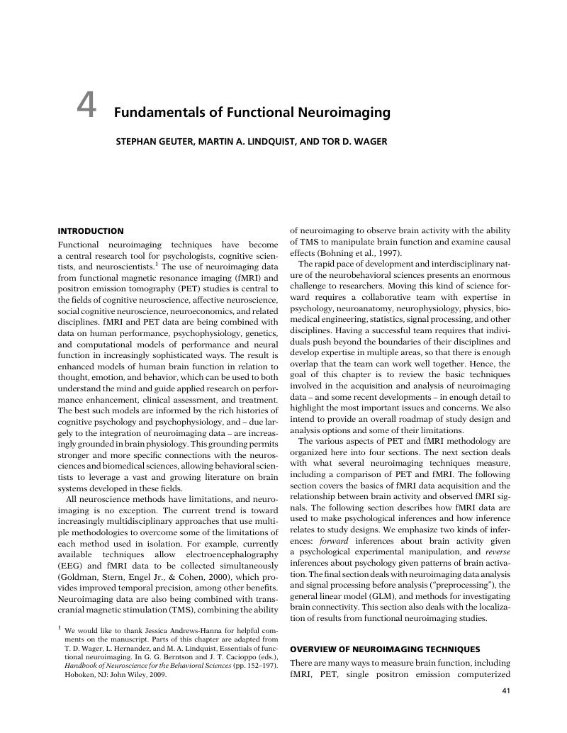
Image of the first page of this content. For PDF version, please use the ‘Save PDF’ preceeding this image.'
Type
Chapter
Information
Publisher: Cambridge University Press
Print publication year: 2016

Access options

Get access to the full version of this content by using one of the access options below. (Log in options will check for institutional or personal access. Content may require purchase if you do not have access.)

References

Aguirre, G. K., Zarahn, E., & D’Esposito, M. (1998). The variability of human, BOLD hemodynamic responses. NeuroImage, 8: 360369.Google Scholar
Amunts, K., Schleicher, A., & Zilles, K. (2007). Cytoarchitecture of the cerebral cortex: more than localization. NeuroImage, 37: 10611065.Google Scholar
Andersson, J. L., Hutton, C., Ashburner, J., Turner, R., & Friston, K. (2001). Modeling geometric deformations in EPI time series. NeuroImage, 13: 903919.CrossRefGoogle ScholarPubMed
Andrews-Hanna, J. R., Reidler, J. S., Huang, C., & Buckner, R. L. (2010). Evidence for the default network’s role in spontaneous cognition. Journal of Neurophysiology, 104: 322335.Google Scholar
Aron, A., Fisher, H., Mashek, D. J., Strong, G., Li, H., & Brown, L. L. (2005). Reward, motivation, and emotion systems associated with early-stage intense romantic love. Journal of Neurophysiology, 94: 327337.Google Scholar
Ashburner, J. (2007). A fast diffeomorphic image registration algorithm. NeuroImage, 38: 95113.Google Scholar
Ashburner, J. & Friston, K. J. (2000). Voxel-based morphometry: the methods. NeuroImage, 11: 805821.Google Scholar
Atlas, L. Y., Lindquist, M. A., Bolger, N., & Wager, T. D. (2014). Brain mediators of the effects of noxious heat on pain. Pain, 155: 16321648.CrossRefGoogle ScholarPubMed
Atlas, L. Y., Whittington, R. A., Lindquist, M. A., Wielgosz, J., Sonty, N., & Wager, T. D. (2012). Dissociable influences of opiates and expectations on pain. Journal of Neuroscience, 32: 80538064.Google Scholar
Bates, D., Maechler, M., Bolker, B., & Walker, S. (2013). LME4: Linear mixed-effects models using Eigen and S4. R package version 1.Google Scholar
Beckmann, C. F., Jenkinson, M., & Smith, S. M. (2003). General multilevel linear modeling for group analysis in FMRI. NeuroImage, 20: 10521063.Google Scholar
Behrens, T. E. J., Berg, H. J., Jbabdi, S., Rushworth, M. F. S., & Woolrich, M. W. (2007). Probabilistic diffusion tractography with multiple fibre orientations: what can we gain? NeuroImage, 34: 144155.Google Scholar
Bendriem, B. & Townsend, D. W. (1998). The Theory and Practice of 3D PET. Boston and Dordrecht: Kluwer.CrossRefGoogle Scholar
Benjamini, Y. & Hochberg, Y. (1995) Controlling the false discovery rate: a practical and powerful approach to multiple testing. Journal of the Royal Statistical Society B, 57: 289300.Google Scholar
Bernstein, M. A., King, K. F., & Zhou, Z. J. (2004). Handbook of MRI Pulse Sequences. Burlington, MA: Elsevier Academic Press.Google Scholar
Binder, J. R., Desai, R. H., Graves, W. W., & Conant, L. L. (2009). Where is the semantic system? A critical review and meta-analysis of 120 functional neuroimaging studies. Cerebral Cortex, 19: 27672796.Google Scholar
Birn, R. M., Saad, Z. S., & Bandettini, P. A. (2001). Spatial heterogeneity of the nonlinear dynamics in the FMRI BOLD response. NeuroImage, 14: 817826.CrossRefGoogle ScholarPubMed
Biswal, B., Yetkin, F. Z., Haughton, V. M., & Hyde, J. S. (1995). Functional connectivity in the motor cortex of resting human brain using echo-planar MRI. Magnetic Resonance in Medicine, 34: 537541.Google Scholar
Bohning, D. E., Pecheny, A. P., Epstein, C. M., Speer, A. M., Vincent, D. J., Dannels, W., & George, M. S. (1997). Mapping transcranial magnetic stimulation (TMS) fields in vivo with MRI. Neuroreport, 8: 25352538.Google Scholar
Bohning, D. E., Shastri, A., McConnell, K. A., Nahas, Z., Lorberbaum, J. P., Roberts, D. R., Teneback, C., Vincent, D. J., & George, M. S. (1999). A combined TMS/fMRI study of intensity-dependent TMS over motor cortex. Biological Psychiatry, 45: 385394.Google Scholar
Bornhovd, K., Quante, M., Glauche, V., Bromm, B., Weiller, C., & Buchel, C. (2002). Painful stimuli evoke different stimulus-response functions in the amygdala, prefrontal, insula and somatosensory cortex: a single-trial fMRI study. Brain, 125: 13261336.CrossRefGoogle ScholarPubMed
Boubela, R. N., Kalcher, K., Huf, W., Seidel, E. M., Derntl, B., Pezawas, L., … & Moser, E. (2015). fMRI measurements of amygdala activation are confounded by stimulus correlated signal fluctuation in nearby veins draining distant brain regions. Scientific Reports, 5: 10499.Google Scholar
Boynton, G. M., Engel, S. A., Glover, G. H., & Heeger, D. J. (1996). Linear systems analysis of functional magnetic resonance imaging in human V1. Journal of Neuroscience, 16: 42074221.Google Scholar
Brett, M., Johnsrude, I. S., & Owen, A. M. (2002). The problem of functional localization in the human brain. Nature Reviews Neuroscience, 3: 243249.Google Scholar
Brooks, J. C., Beckmann, C. F., Miller, K. L., Wise, R. G., Porro, C. A., Tracey, I., & Jenkinson, M. (2008). Physiological noise modelling for spinal functional magnetic resonance imaging studies. NeuroImage, 39: 680692.Google Scholar
Brown, A. K., Fujita, M., Fujimura, Y., Liow, J. S., Stabin, M., Ryu, Y. H., … & Innis, R. B. (2007). Radiation dosimetry and biodistribution in monkey and man of 11C-PBR28: a PET radioligand to image inflammation. Journal of Nuclear Medicine, 48: 20722079.Google Scholar
Buchel, C., Bornhovd, K., Quante, M., Glauche, V., Bromm, B., & Weiller, C. (2002), Dissociable neural responses related to pain intensity, stimulus intensity, and stimulus awareness within the anterior cingulate cortex: a parametric single-trial laser functional magnetic resonance imaging study. Journal of Neuroscience, 22: 970976.Google Scholar
Buckner, R. L., Andrews-Hanna, J. R., & Schacter, D. L. (2008). The brain’s default network. Annals of the New York Academy of Sciences, 1124: 138.CrossRefGoogle ScholarPubMed
Buracas, G. T. & Boynton, G. M. (2002). Efficient design of event-related fMRI experiments using M-sequences. NeuroImage, 16: 801813.Google Scholar
Bush, G., Luu, P., & Posner, M. I. (2000). Cognitive and emotional influences in anterior cingulate cortex. Trends in Cognitive Sciences, 4: 215222.CrossRefGoogle ScholarPubMed
Button, K. S., Ioannidis, J. P., Mokrysz, C., Nosek, B. A., Flint, J., Robinson, E. S., & Munafo, M. R. (2013). Power failure: why small sample size undermines the reliability of neuroscience. Nature Reviews Neuroscience, 14: 365376.Google Scholar
Buxton, R. B. & Frank, L. R. (1997). A model for the coupling between cerebral blood flow and oxygen metabolism during neural stimulation. Journal of Cerebral Blood Flow & Metabolism, 17: 6472.Google Scholar
Buxton, R. B., Frank, L. R., Wong, E. C., Siewert, B., Warach, S., & Edelman, R. R. (1998). A general kinetic model for quantitative perfusion imaging with arterial spin labeling. Magnetic Resonance in Medicine, 40: 383396.Google Scholar
Buxton, R. B., Uludag, K., Dubowitz, D. J., & Liu, T. T. (2004). Modeling the hemodynamic response to brain activation. NeuroImage, 23: S220S233.Google Scholar
Cacioppo, J. T., & Tassinary, L. G. (1990). Inferring psychological significance from physiological signals. American Psychologist, 45: 1628.Google Scholar
Calhoun, V. D., Miller, R., Pearlson, G., & Adali, T. (2014). The chronnectome: time-varying connectivity networks as the next frontier in fMRI data discovery. Neuron, 84: 262274.Google Scholar
Chaimow, D., Yacoub, E., Ugurbil, K., & Shmuel, A. (2011). Modeling and analysis of mechanisms underlying fMRI-based decoding of information conveyed in cortical columns. NeuroImage, 56: 627642.Google Scholar
Cheng, K., Waggoner, R. A., & Tanaka, K. (2001). Human ocular dominance columns as revealed by high-field functional magnetic resonance imaging. Neuron, 32: 359374.Google Scholar
Collins, D. L., Neelin, P., Peters, T. M., & Evans, A. C. (1994). Automatic 3D intersubject registration of MR volumetric data in standardized Talairach space. Journal of Computer Assisted Tomography, 18: 192205.Google Scholar
Coltheart, M. (2006). What has functional neuroimaging told us about the mind (so far)? Cortex, 42: 323331.Google Scholar
Constable, R. T. & Spencer, D. D. (1999). Composite image formation in z-shimmed functional MR imaging. Magnetic Resonance in Medicine, 42: 110117.Google Scholar
Cover, T. M. & Thomas, J. A. (1991). Elements of Information Theory. New York: John Wiley.Google Scholar
Cribben, I., Haraldsdottir, R., Atlas, L. Y., Wager, T. D., & Lindquist, M. A. (2012). Dynamic connectivity regression: determining state-related changes in brain connectivity. NeuroImage, 61: 907920.Google Scholar
de Quervain, D. J., Fischbacher, U., Treyer, V., Schellhammer, M., Schnyder, U., Buck, A., & Fehr, E. (2004). The neural basis of altruistic punishment. Science, 305: 12541258.Google Scholar
Deckers, R. H., van Gelderen, P., Ries, M., Barret, O., Duyn, J. H., Ikonomidou, V. N., … & de Zwart, J. A. (2006). An adaptive filter for suppression of cardiac and respiratory noise in MRI time series data. NeuroImage, 33: 10721081.Google Scholar
Denis Le Bihan, M. D., Mangin, J. F., Poupon, C., Clark, C. A., Pappata, S., Molko, N., & Chabriat, H. (2001). Diffusion tensor imaging: concepts and applications. Journal of Magnetic Resonance Imaging, 13: 534546.Google Scholar
Denny, B. T., Kober, H., Wager, T. D., & Ochsner, K. N. (2012). A meta-analysis of functional neuroimaging studies of self- and other judgments reveals a spatial gradient for mentalizing in medial prefrontal cortex. Journal of Cognitive Neuroscience, 24: 17421752.Google Scholar
Desmond, J. E. & Glover, G. H. (2002). Estimating sample size in functional MRI (fMRI) neuroimaging studies: statistical power analyses. Journal of Neuroscience Methods, 118: 115128.Google Scholar
Detre, J. A., Zhang, W. G., Roberts, D. A., Silva, A. C., Williams, D. S., Grandis, D. J., … & Leigh, J. S. (1994). Tissue-specific perfusion imaging using arterial spin-labeling. NMR in Biomedicine, 7: 7582.Google Scholar
Devlin, J. T. & Poldrack, R. A. (2007). In praise of tedious anatomy. NeuroImage, 37: 10331041.Google Scholar
Disbrow, E. A., Slutsky, D. A., Roberts, T. P., & Krubitzer, L. A. (2000). Functional MRI at 1.5 tesla: a comparison of the blood oxygenation level-dependent signal and electrophysiology. Proceedings of the National Academy of Sciences of the USA, 97: 97189723.Google Scholar
Doucet, G., Naveau, M., Petit, L., Zago, L., Crivello, F., Jobard, G., … & Joliot, M. (2012). Patterns of hemodynamic low-frequency oscillations in the brain are modulated by the nature of free thought during rest. NeuroImage, 59: 31943200.Google Scholar
Duong, T. Q., Yacoub, E., Adriany, G., Hu, X., Ugurbil, K., Vaughan, J. T., … & Kim, S. G. (2002). High-resolution, spin-echo BOLD, and CBF fMRI at 4 and 7 T. Magnetic Resonance in Medicine, 48: 589593.CrossRefGoogle Scholar
Duvernoy, H. M. (2012). The Human Brain Stem and Cerebellum: Surface, Structure, Vascularization, and Three-Dimensional Sectional Anatomy, with MRI. Dordrecht: Springer Science & Business Media.Google Scholar
Eickhoff, S. B., Stephan, K. E., Mohlberg, H., Grefkes, C., Fink, G. R., Amunts, K., & Zilles, K. (2005). A new SPM toolbox for combining probabilistic cytoarchitectonic maps and functional imaging data. NeuroImage, 25: 13251335.CrossRefGoogle ScholarPubMed
Eisenberger, N. I., Lieberman, M. D., & Williams, K. D. (2003). Does rejection hurt? An fMRI study of social exclusion. Science, 302: 290292.Google Scholar
Elster, A. D. (1994). Questions and Answers in Magnetic Resonance Imaging. St. Louis, MO: Mosby.Google Scholar
Ethofer, T., Van De Ville, D., Scherer, K., & Vuilleumier, P. (2009). Decoding of emotional information in voice-sensitive cortices. Current Biology, 19: 10281033.Google Scholar
Feinberg, D. A., Moeller, S., Smith, S. M., Auerbach, E., Ramanna, S., Gunther, M., … & Yacoub, E. (2010). Multiplexed echo planar imaging for sub-second whole brain FMRI and fast diffusion imaging. PLoS One, 5: e15710.CrossRefGoogle ScholarPubMed
Finsterbusch, J., Busch, M. G., & Larson, P. E. Z. (2013). Signal scaling improves the signal-to-noise ratio of measurements with segmented 2D-selective radiofrequency excitations. Magnetic Resonance in Medicine, 70: 14911499.Google Scholar
Fischl, B., Sereno, M. I., & Dale, A. M. (1999). Cortical surface-based analysis. II: Inflation, flattening, and a surface-based coordinate system. NeuroImage, 9: 195207.Google Scholar
Fox, M. D., Snyder, A. Z., Vincent, J. L., Corbetta, M., Van Essen, D. C., & Raichle, M. E. (2005). The human brain is intrinsically organized into dynamic, anticorrelated functional networks. Proceedings of the National Academy of Sciences of the USA, 102: 96739678.Google Scholar
Fox, M. D., Snyder, A. Z., Vincent, J. L., & Raichle, M. E. (2007). Intrinsic fluctuations within cortical systems account for intertrial variability in human behavior. Neuron, 56: 171184.Google Scholar
Frey, K. A. (1999). Positron emission tomography. In Siegel, G. J., Agranoff, B. W., Albers, R. W., Fisher, S. K., & Uhler, M. D. (eds.), Basic Neurochemistry, 6th edn. (pp. 11091131). Philadelphia: Lippincott, Williams, & Wilkins.Google Scholar
Friston, K. J. (2009). Modalities, modes, and models in functional neuroimaging. Science, 326: 399403.Google Scholar
Friston, K. J. (2011). Functional and effective connectivity: a review. Brain Connectivity, 1: 1336.Google Scholar
Friston, K. J. (2012). Ten ironic rules for non-statistical reviewers. NeuroImage, 61: 13001310.Google Scholar
Friston, K. J., Buechel, C., Fink, G. R., Morris, J., Rolls, E., & Dolan, R. J. (1997). Psychophysiological and modulatory interactions in neuroimaging. NeuroImage, 6: 218229.Google Scholar
Friston, K. J., Frith, C. D., Turner, R., & Frackowiak, R. S. (1995). Characterizing evoked hemodynamics with fMRI. NeuroImage, 2: 157165.Google Scholar
Friston, K. J., Harrison, L., & Penny, W. (2003). Dynamic causal modelling. NeuroImage, 19: 12731302.CrossRefGoogle ScholarPubMed
Friston, K. J., Mechelli, A., Turner, R., & Price, C. J. (2000). Nonlinear responses in fMRI: the Balloon model, Volterra kernels, and other hemodynamics. NeuroImage, 12: 466477.Google Scholar
Gianaros, P. J. & Wager, T. D. (2015). Brain–body pathways linking psychological stress and physical health. Current Directions in Psychological Science, 24: 313321.Google Scholar
Glahn, D. C., Paus, T., & Thompson, P. M. (2007a). Imaging genomics: mapping the influence of genetics on brain structure and function. Human Brain Mapping, 28: 461463.Google Scholar
Glahn, D. C., Thompson, P. M., & Blangero, J. (2007b). Neuroimaging endophenotypes: strategies for finding genes influencing brain structure and function. Human Brain Mapping, 28: 488501.Google Scholar
Glover, G. H. & Law, C. S. (2001). Spiral-in/out BOLD fMRI for increased SNR and reduced susceptibility artifacts. Magnetic Resonance in Medicine, 46:515522.Google Scholar
Glover, G. H., Li, T. Q., & Ress, D. (2000). Image-based method for retrospective correction of physiological motion effects in fMRI: RETROICOR. Magnetic Resonance in Medicine, 44: 162167.Google Scholar
Goldman, R. I., Stern, J. M., Engel, J. Jr., & Cohen, M. S. (2000). Acquiring simultaneous EEG and functional MRI. Clinical Neurophysiology, 111: 19741980.Google Scholar
Gonzalez-Castillo, J., Saad, Z. S., Handwerker, D. A., Inati, S. J., Brenowitz, N., & Bandettini, P. A. (2012). Whole-brain, time-locked activation with simple tasks revealed using massive averaging and model-free analysis. Proceedings of the National Academy of Sciences of the USA, 109: 54875492.Google Scholar
Good, C. D., Johnsrude, I. S., Ashburner, J., Henson, R. N. A., Friston, K. J., & Frackowiak, R. S. J. (2001). A voxel-based morphometric study of ageing in 465 normal adult human brains. NeuroImage, 14: 2136.Google Scholar
Grinband, J., Savitskaya, J., Wager, T. D., Teichert, T., Ferrera, V. P., & Hirsch, J. (2011). The dorsal medial frontal cortex is sensitive to time on task, not response conflict or error likelihood. NeuroImage, 57: 303311.Google Scholar
Haacke, E. M. (1999). Magnetic Resonance Imaging: Physical Principles and Sequence Design. New York: John Wiley.Google Scholar
Haines, D. E. (2000). Neuroanatomy: An Atlas of Structures, Sections, and Systems. Philadelphia: Lippincott Williams & Wilkins.Google Scholar
Hare, T. A., Camerer, C. F., & Rangel, A. (2009). Self-control in decision-making involves modulation of the vmPFC valuation system. Science, 324: 646648.Google Scholar
Haxby, J. V., Guntupalli, J. S., Connolly, A. C., Halchenko, Y. O., Conroy, B. R., Gobbini, M. I., … & Ramadge, P. J. (2011). A common, high-dimensional model of the representational space in human ventral temporal cortex. Neuron, 72: 404416.Google Scholar
Haynes, J. D. (2015). A primer on pattern-based approaches to fMRI: principles, pitfalls, and perspectives. Neuron, 87: 257270.Google Scholar
Haynes, J. D., Deichmann, R., & Rees, G. (2005). Eye-specific effects of binocular rivalry in the human lateral geniculate nucleus. Nature, 438: 496499.Google Scholar
Heeger, D. J. & Ress, D. (2002). What does fMRI tell us about neuronal activity? Nature Reviews Neuroscience, 3: 142151.Google Scholar
Henson, R., Shallice, T., & Dolan, R. (2000). Neuroimaging evidence for dissociable forms of repetition priming. Science, 287: 12691272.CrossRefGoogle ScholarPubMed
Honey, C. J., Sporns, O., Cammoun, L., Gigandet, X., Thiran, J. P., Meuli, R., & Hagmann, P. (2009). Predicting human resting-state functional connectivity from structural connectivity. Proceedings of the National Academy of Sciences of the USA, 106: 20352040.CrossRefGoogle ScholarPubMed
Horikawa, T., Tamaki, M., Miyawaki, Y., & Kamitani, Y. (2013). Neural decoding of visual imagery during sleep. Science, 340: 639642.Google Scholar
Huettel, S. A., Song, A. W., & McCarthy, G. (2004). Functional Magnetic Resonance Imaging. Sunderland, MA: Sinauer Associates.Google Scholar
Huth, A. G., Nishimoto, S., Vu, A. T., & Gallant, J. L. (2012). A continuous semantic space describes the representation of thousands of object and action categories across the human brain. Neuron, 76: 12101224.Google Scholar
Johansen-Berg, H. & Behrens, T. E. (2006). Just pretty pictures? What diffusion tractography can add in clinical neuroscience. Current Opinion in Neurology, 19: 379385.Google Scholar
Johansen-Berg, H., Behrens, T. E., Robson, M. D., Drobnjak, I., Rushworth, M. F., Brady, J. M., … & Matthews, P. M. (2004). Changes in connectivity profiles define functionally distinct regions in human medial frontal cortex. Proceedings of the National Academy of Sciences of the USA, 101: 1333513340.Google Scholar
Josephs, O. & Henson, R. N. (1999). Event-related functional magnetic resonance imaging: modelling, inference and optimization. Philosophical Transactions of the Royal Society of London B: Biological Sciences, 354: 12151228.Google Scholar
Kamitani, Y. & Tong, F. (2005). Decoding the visual and subjective contents of the human brain. Nature Neuroscience, 8: 679685.Google Scholar
Kao, M. H., Mandal, A., Lazar, N., & Stufken, J. (2009). Multi-objective optimal experimental designs for event-related fMRI studies. NeuroImage, 44: 849856.Google Scholar
Kastner, S. & Ungerleider, L. G. (2000). Mechanisms of visual attention in the human cortex. Annual Review of Neuroscience, 23: 315341.Google Scholar
Kleinschmidt, A., Buchel, C., Zeki, S., & Frackowiak, R. S. (1998). Human brain activity during spontaneously reversing perception of ambiguous figures. Proceedings of the Royal Society of London B: Biological Sciences, 265: 24272433.Google Scholar
Klunk, W. E., Engler, H., Nordberg, A., Wang, Y., Blomqvist, G., Holt, D. P., … & Långström, B. (2004). Imaging brain amyloid in Alzheimer’s disease with Pittsburgh Compound-B. Annals of Neurology, 55: 306319.Google Scholar
Kober, H., Barrett, L. F., Joseph, J., Bliss-Moreau, E., Lindquist, K., & Wager, T. D. (2008). Functional grouping and cortical–subcortical interactions in emotion: a meta-analysis of neuroimaging studies. NeuroImage, 42: 9981031.Google Scholar
Kong, Y., Jenkinson, M., Andersson, J., Tracey, I., & Brooks, J. C. (2012). Assessment of physiological noise modelling methods for functional imaging of the spinal cord. NeuroImage, 60: 15381549.Google Scholar
Kriegeskorte, N., Lindquist, M. A., Nichols, T. E., Poldrack, R. A., & Vul, E. (2010). Everything you never wanted to know about circular analysis, but were afraid to ask. Journal of Cerebral Blood Flow & Metabolism, 30: 15511557.Google Scholar
Kriegeskorte, N., Simmons, W. K., Bellgowan, P. S., & Baker, C. I. (2009). Circular analysis in systems neuroscience: the dangers of double dipping. Nature Neuroscience, 12: 535540.Google Scholar
Kvitsiani, D., Ranade, S., Hangya, B., Taniguchi, H., Huang, J. Z., & Kepecs, A. (2013). Distinct behavioural and network correlates of two interneuron types in prefrontal cortex. Nature, 498: 363366.Google Scholar
Kwong, K. K., Belliveau, J. W., Chesler, D. A., Goldberg, I. E., Weisskoff, R. M., Poncelet, B. P., … & Turner, R. (1992). Dynamic magnetic resonance imaging of human brain activity during primary sensory stimulation. Proceedings of the National Academy of Sciences of the USA, 89: 56755679.Google Scholar
Laufs, H., Daunizeau, J., Carmichael, D. W., & Kleinschmidt, A. (2008). Recent advances in recording electrophysiological data simultaneously with magnetic resonance imaging. NeuroImage, 40: 515528.Google Scholar
Leitao, J., Thielscher, A., Tunnerhoff, J., & Noppeney, U. (2015). Concurrent TMS-fMRI reveals interactions between dorsal and ventral attentional systems. Journal of Neuroscience, 35: 1144511457.Google Scholar
Lindquist, K. A., Wager, T. D., Kober, H., Bliss-Moreau, E., & Barrett, L. F. (2012a). The brain basis of emotion: a meta-analytic review. Behavioral and Brain Sciences, 35: 121143.Google Scholar
Lindquist, M. A., Caffo, B., & Crainiceanu, C. (2013). Ironing out the statistical wrinkles in “ten ironic rules.” NeuroImage, 81: 499502.Google Scholar
Lindquist, M. A., Spicer, J., Asllani, I., & Wager, T. D. (2012b). Estimating and testing variance components in a multi-level GLM. NeuroImage, 59: 490501.Google Scholar
Lindquist, M. A., Waugh, C., & Wager, T. D. (2007). Modeling state-related fMRI activity using change-point theory. NeuroImage, 35: 11251141.Google Scholar
Lindquist, M. A., Zhang, C. H., Glover, G., & Shepp, L. (2008). Rapid three-dimensional functional magnetic resonance imaging of the initial negative BOLD response. Journal of Magnetic Resonance, 191: 100111.Google Scholar
Liu, T. T. (2004). Efficiency, power, and entropy in event-related fMRI with multiple trial types. Part II: design of experiments. NeuroImage, 21: 401413.Google Scholar
Loggia, M. L., Chonde, D. B., Akeju, O., Arabasz, G., Catana, C., Edwards, R. R., … & Hooker, J. M. (2015). Evidence for brain glial activation in chronic pain patients. Brain, 138: 604615.Google Scholar
Logothetis, N. K. (2008). What we can do and what we cannot do with fMRI. Nature, 453: 869878.Google Scholar
Logothetis, N. K., Pauls, J., Augath, M., Trinath, T., & Oeltermann, A. (2001). Neurophysiological investigation of the basis of the fMRI signal. Nature, 412: 150157.Google Scholar
Maguire, E. A., Gadian, D. G., Johnsrude, I. S., Good, C. D., Ashburner, J., Frackowiak, R. S., & Frith, C. D. (2000). Navigation-related structural change in the hippocampi of taxi drivers. Proceedings of the National Academy of Sciences of the USA, 97: 43984403.Google Scholar
Mai, J. K., Paxinos, G., & Voss, T. (2007). Atlas of the Human Brain, 3rd edn. New York: Academic Press.Google Scholar
Menon, R. S. (2002). Postacquisition suppression of large-vessel BOLD signals in high-resolution fMRI. Magnetic Resonance in Medicine, 47: 19.Google Scholar
Miezin, F. M., Maccotta, L., Ollinger, J. M., Petersen, S. E., & Buckner, R. L. (2000). Characterizing the hemodynamic response: effects of presentation rate, sampling procedure, and the possibility of ordering brain activity based on relative timing. NeuroImage, 11: 735759.Google Scholar
Morawetz, C., Holz, P., Lange, C., Baudewig, J., Weniger, G., Irle, E., & Dechent, P. (2008). Improved functional mapping of the human amygdala using a standard functional magnetic resonance imaging sequence with simple modifications. Magnetic Resonance Imaging, 26: 4553.Google Scholar
Mumford, J. A. & Nichols, T. E. (2008). Power calculation for group fMRI studies accounting for arbitrary design and temporal autocorrelation. NeuroImage, 39: 261268.Google Scholar
Nichols, T. & Hayasaka, S. (2003). Controlling the familywise error rate in functional neuroimaging: a comparative review. Statistical Methods in Medical Research, 12: 419446.Google Scholar
Nichols, T. E. & Holmes, A. P. (2002). Nonparametric permutation tests for functional neuroimaging: a primer with examples. Human Brain Mapping, 15: 125.Google Scholar
Noll, D. C., Fessler, J. A., & Sutton, B. P. (2005). Conjugate phase MRI reconstruction with spatially variant sample density correction. IEEE Transactions on Medical Imaging, 24: 325336.Google Scholar
Norman, K. A., Polyn, S. M., Detre, G. J., & Haxby, J. V. (2006). Beyond mind-reading: multi-voxel pattern analysis of fMRI data. Trends in Cognitive Sciences, 10: 424430.Google Scholar
Northoff, G., Heinzel, A., de Greck, M., Bermpohl, F., Dobrowolny, H., & Panksepp, J. (2006). Self-referential processing in our brain: a meta-analysis of imaging studies on the self. NeuroImage, 31: 440457.Google Scholar
Ogawa, S., Lee, T. M., Kay, A. R., & Tank, D. W. (1990). Brain magnetic resonance imaging with contrast dependent on blood oxygenation. Proceedings of the National Academy of Sciences of the USA, 87: 98689872.Google Scholar
Ogawa, S., Tank, D. W., Menon, R., Ellermann, J. M., Kim, S. G., Merkle, H., & Ugurbil, K. (1992). Intrinsic signal changes accompanying sensory stimulation: functional brain mapping with magnetic resonance imaging. Proceedings of the National Academy of Sciences of the USA, 89: 59515955.Google Scholar
Paton, J. J., Belova, M. A., Morrison, S. E., & Salzman, C. D. (2006). The primate amygdala represents the positive and negative value of visual stimuli during learning. Nature, 439: 865870.Google Scholar
Paus, T. (2001). Primate anterior cingulate cortex: where motor control, drive and cognition interface. Nature Reviews Neuroscience, 2: 417424.Google Scholar
Petrini, K., Pollick, F. E., Dahl, S., McAleer, P., McKay, L. S., Rocchesso, D., … & Puce, A. (2011). Action expertise reduces brain activity for audiovisual matching actions: an fMRI study with expert drummers. NeuroImage, 56: 14801492.Google Scholar
Phillips, C., Rugg, M. D., & Friston, K. J. (2002). Anatomically informed basis functions for EEG source localization: combining functional and anatomical constraints. NeuroImage, 16: 678695.Google Scholar
Poldrack, R. A. (2011). Inferring mental states from neuroimaging data: from reverse inference to large-scale decoding. Neuron, 72: 692697.Google Scholar
Price, C. J., Veltman, D. J., Ashburner, J., Josephs, O., & Friston, K. J. (1999). The critical relationship between the timing of stimulus presentation and data acquisition in blocked designs with fMRI. NeuroImage, 10: 3644.Google Scholar
Raichle, M. E., MacLeod, A. M., Snyder, A. Z., Powers, W. J., Gusnard, D. A., & Shulman, G. L. (2001). A default mode of brain function. Proceedings of the National Academy of Sciences of the USA, 98: 676682.Google Scholar
Rasbash, J. (2002). A User’s Guide to MLwiN. Centre for Multilevel Modelling, University of London.Google Scholar
Raudenbush, S. W. & Bryk, A. S. (2002). Hierarchical Linear Models: Applications and Data Analysis, 2nd edn. Newbury Park, CA: Sage.Google Scholar
Reiman, E. M., Fusselman, M. J., Fox, P. T., & Raichle, M. E. (1989). Neuroanatomical correlates of anticipatory anxiety. Science, 243: 10711074 [erratum published in Science, 256 (1992): 1696].Google Scholar
Rosen, B. R., Buckner, R. L., & Dale, A. M. (1998). Event-related functional MRI: past, present, and future. Proceedings of the National Academy of Sciences of the USA, 95: 773780.Google Scholar
Ruff, C. C., Blankenburg, F., Bjoertomt, O., Bestmann, S., Freeman, E., Haynes, J. D., … & Driver, J. (2006). Concurrent TMS-fMRI and psychophysics reveal frontal influences on human retinotopic visual cortex. Current Biology, 16: 14791488.Google Scholar
Saad, Z. S., Reynolds, R. C., Argall, B., Japee, S., & Cox, R. W. (2004). SUMA: an interface for surface-based intra- and inter-subject analysis with AFNI. IEEE International Symposium on Biomedical Imaging: Nano to Macro, 1512: 15101513.Google Scholar
Sandler, M. P. (2003). Diagnostic Nuclear Medicine. Philadelphia, PA: Lippincott, Williams & Wilkins.Google Scholar
Sarter, M., Berntson, G. G., & Cacioppo, J. T. (1996). Brain imaging and cognitive neuroscience: toward strong inference in attributing function to structure. American Psychologist, 51: 1321.Google Scholar
Schacter, D. L., Buckner, R. L., Koutstaal, W., Dale, A. M., & Rosen, B. R. (1997). Late onset of anterior prefrontal activity during true and false recognition: an event-related fMRI study. NeuroImage, 6: 259269.Google Scholar
Scheibe, C., Ullsperger, M., Sommer, W., & Heekeren, H. R. (2010). Effects of parametrical and trial-to-trial variation in prior probability processing revealed by simultaneous electroencephalogram/functional magnetic resonance imaging. Journal of Neuroscience, 30: 1670916717.Google Scholar
Seeley, W. W., Menon, V., Schatzberg, A. F., Keller, J., Glover, G. H., Kenna, H., … & Greicius, M. D. (2007). Dissociable intrinsic connectivity networks for salience processing and executive control. Journal of Neuroscience, 27: 23492356.Google Scholar
Setsompop, K., Gagoski, B. A., Polimeni, J. R., Witzel, T., Wedeen, V. J., & Wald, L. L. (2012). Blipped-controlled aliasing in parallel imaging for simultaneous multislice echo planar imaging with reduced g-factor penalty. Magnetic Resonance in Medicine, 67: 12101224.Google Scholar
Shulman, R. G. & Rothman, D. L. (1998). Interpreting functional imaging studies in terms of neurotransmitter cycling. Proceedings of the National Academy of Sciences of the USA, 95: 1199311998.Google Scholar
Shulman, R. G., Rothman, D. L., Behar, K. L., & Hyder, F. (2004). Energetic basis of brain activity: implications for neuroimaging. Trends in Neurosciences, 27: 489495.Google Scholar
Sibson, N. R., Dhankhar, A., Mason, G. F., Behar, K. L., Rothman, D. L., & Shulman, R. G. (1997). In vivo 13C NMR measurements of cerebral glutamine synthesis as evidence for glutamate-glutamine cycling. Proceedings of the National Academy of Sciences of the USA, 94: 26992704.Google Scholar
Sinha, R., Lacadie, C., Skudlarski, P., & Wexler, B. E. (2004). Neural circuits underlying emotional distress in humans. Annals of the New York Academy of Sciences, 1032: 254257.Google Scholar
Skudlarski, P., Constable, R. T., & Gore, J. C. (1999). ROC analysis of statistical methods used in functional MRI: individual subjects. NeuroImage, 9: 311329.Google Scholar
Smith, S. M. (2012). The future of FMRI connectivity. NeuroImage, 62: 12571266.Google Scholar
Smith, S. M., Jenkinson, M., Beckmann, C., Miller, K., & Woolrich, M. (2007). Meaningful design and contrast estimability in FMRI. NeuroImage, 34: 127136.Google Scholar
Smith, S. M., Jenkinson, M., Woolrich, M. W., Beckmann, C. F., Behrens, T. E., Johansen-Berg, H., … & Matthews, P. M. (2004). Advances in functional and structural MR image analysis and implementation as FSL. NeuroImage, 23: S208S219.Google Scholar
Sporns, O. (2014). Contributions and challenges for network models in cognitive neuroscience. Nature Neuroscience, 17: 652660.Google Scholar
Stephan, K. E., Penny, W. D., Daunizeau, J., Moran, R. J., & Friston, K. J. (2009). Bayesian model selection for group studies. NeuroImage, 46: 10041017.Google Scholar
Sternberg, S. (2001). Separate modifiability, mental modules, and the use of pure and composite measures to reveal them. Acta Psychologica (Amsterdam), 106: 147246.Google Scholar
Summerfield, C., Greene, M., Wager, T., Egner, T., Hirsch, J., & Mangels, J. (2006). Neocortical connectivity during episodic memory formation. PLoS Biol, 4: e128.Google Scholar
Summerfield, C., Trittschuh, E. H., Monti, J. M., Mesulam, M. M., & Egner, T. (2008). Neural repetition suppression reflects fulfilled perceptual expectations. Nature Neuroscience, 11: 10041006.Google Scholar
Sylvester, C. Y., Wager, T. D., Lacey, S. C., Hernandez, L., Nichols, T. E., Smith, E. E., & Jonides, J. (2003). Switching attention and resolving interference: fMRI measures of executive functions. Neuropsychologia, 41: 357370.Google Scholar
Tagliazucchi, E. & Laufs, H. (2014). Decoding wakefulness levels from typical fMRI resting-state data reveals reliable drifts between wakefulness and sleep. Neuron, 82: 695708.Google Scholar
Talairach, J. & Tournoux, P. (1988). Co-Planar Stereotaxic Atlas of the Human Brain. 3-Dimensional Proportional System: An Approach to Cerebral Imaging. Stuttgart and New York: Thieme.Google Scholar
Taylor, J. E. & Worsley, K. J. (2006). Inference for magnitudes and delays of responses in the FIAC data using BRAINSTAT/FMRISTAT. Human Brain Mapping, 27: 434441.Google Scholar
Thompson, P. M., Schwartz, C., Lin, R. T., Khan, A. A., & Toga, A. W. (1996). Three-dimensional statistical analysis of sulcal variability in the human brain. Journal of Neuroscience, 16: 42614274.Google Scholar
Thompson, P. M., Stein, J. L., Medland, S. E., Hibar, D. P., Vasquez, A. A., Renteria, M. E., … & Drevets, W. (2014). The ENIGMA Consortium: large-scale collaborative analyses of neuroimaging and genetic data. Brain Imaging and Behavior, 8: 153182.Google Scholar
Tye, K. M., Prakash, R., Kim, S.-Y., Fenno, L. E., Grosenick, L., Zarabi, H., … & Deisseroth, K. (2011). Amygdala circuitry mediating reversible and bidirectional control of anxiety. Nature, 471: 358362.Google Scholar
van Ast, V., Spicer, J., Smith, E., Schmer-Galunder, S., Liberzon, I., Abelson, J., & Wager, T. (2014). Brain mechanisms of social threat effects on working memory. Cerebral Cortex (September): bhu206.Google Scholar
Van Essen, D. C. & Dierker, D. L. (2007). Surface-based and probabilistic atlases of primate cerebral cortex. Neuron, 56: 209225.Google Scholar
Van Essen, D. C., Drury, H. A., Dickson, J., Harwell, J., Hanlon, D., & Anderson, C. H. (2001). An integrated software suite for surface-based analyses of cerebral cortex. Journal of the American Medical Informatics Association, 8: 443459.Google Scholar
Vazquez, A. L., Cohen, E. R., Gulani, V., Hernandez-Garcia, L., Zheng, Y., Lee, G. R., … & Noll, D. C. (2006). Vascular dynamics and BOLD fMRI: CBF level effects and analysis considerations. NeuroImage, 32: 16421655.Google Scholar
Vazquez, A. L. & Noll, D. C. (1998). Nonlinear aspects of the BOLD response in functional MRI. NeuroImage, 7: 108118.Google Scholar
Vincent, J. L., Patel, G. H., Fox, M. D., Snyder, A. Z., Baker, J. T., Van Essen, D. C., … & Raichle, M. E. (2007). Intrinsic functional architecture in the anaesthetized monkey brain. Nature, 447: 8386.Google Scholar
Vogt, B. A., Nimchinsky, E. A., Vogt, L. J., & Hof, P. R. (1995). Human cingulate cortex: surface features, flat maps, and cytoarchitecture. Journal of Comparative Neurology, 359: 490506.Google Scholar
Vul, E., Harris, C., Winkielman, P., & Pashler, H. (2009). Puzzlingly high correlations in fMRI studies of emotion, personality, and social cognition. Perspectives on Psychological Science, 4: 274290.Google Scholar
Wager, T. D., Atlas, L. Y., Lindquist, M. A., Roy, M., Woo, C. W., & Kross, E. (2013). An fMRI-based neurologic signature of physical pain. New England Journal of Medicine, 368: 13881397.Google Scholar
Wager, T. D., Jonides, J., & Reading, S. (2004a). Neuroimaging studies of shifting attention: a meta-analysis. NeuroImage, 22: 16791693.Google Scholar
Wager, T. D., Jonides, J., Smith, E. E., & Nichols, T. E. (2005b). Toward a taxonomy of attention shifting: individual differences in fMRI during multiple shift types. Cognitive, Affective, & Behavioral Neuroscience, 5: 127143.Google Scholar
Wager, T. D., Lindquist, M., & Kaplan, L. (2007). Meta-analysis of functional neuroimaging data: Current and future directions. Social Cognitive and Affective Neuroscience, 2: 150158.Google Scholar
Wager, T. D. & Nichols, T. E. (2003). Optimization of experimental design in fMRI: a general framework using a genetic algorithm. NeuroImage, 18: 293309.Google Scholar
Wager, T. D., Reading, S., & Jonides, J. (2004b). Neuroimaging studies of shifting attention: a meta-analysis. NeuroImage, 22: 16791693.Google Scholar
Wager, T. D., Vazquez, A, Hernandez, L, & Noll, D. C. (2005a). Accounting for nonlinear BOLD effects in fMRI: parameter estimates and a model for prediction in rapid event-related studies. NeuroImage, 25: 206218.Google Scholar
Wager, T. D., Waugh, C. E., Lindquist, M., Noll, D. C., Fredrickson, B. L., & Taylor, S. F. (2009). Brain mediators of cardiovascular responses to social threat. Part I: Reciprocal dorsal and ventral sub-regions of the medial prefrontal cortex and heart-rate reactivity. NeuroImage, 47: 821835.Google Scholar
Waugh, C. E., Hamilton, J. P., & Gotlib, I. H. (2010). The neural temporal dynamics of the intensity of emotional experience. NeuroImage, 49: 16991707.Google Scholar
Wiech, K., Jbabdi, S., Lin, C. S., Andersson, J., & Tracey, I. (2014). Differential structural and resting state connectivity between insular subdivisions and other pain-related brain regions. Pain, 155: 20472055.Google Scholar
Wilson, J. L. & Jezzard, P. (2003). Utilization of an intra-oral diamagnetic passive shim in functional MRI of the inferior frontal cortex. Magnetic Resonance in Medicine, 50: 10891094.Google Scholar
Winkler, A. M., Ridgway, G. R., Webster, M. A., Smith, S. M., & Nichols, T. E. (2014). Permutation inference for the general linear model. NeuroImage, 92: 381397.Google Scholar
Wise, R. G., Rogers, R., Painter, D., Bantick, S., Ploghaus, A., Williams, P., … & Tracey, I. (2002). Combining fMRI with a pharmacokinetic model to determine which brain areas activated by painful stimulation are specifically modulated by remifentanil. NeuroImage, 16: 9991014.Google Scholar
Woo, C. W., Koban, L., Kross, E., Lindquist, M. A., Banich, M. T., Ruzic, L., … & Wager, T. D. (2014a). Separate neural representations for physical pain and social rejection. Nature Communications, 5: 5380.Google Scholar
Woo, C. W., Krishnan, A., & Wager, T. D. (2014b). Cluster-extent based thresholding in fMRI analyses: pitfalls and recommendations. NeuroImage, 91: 412419.Google Scholar
Woolrich, M. W., Behrens, T. E., Beckmann, C. F., Jenkinson, M., & Smith, S. M. (2004). Multilevel linear modelling for FMRI group analysis using Bayesian inference. NeuroImage, 21: 17321747.Google Scholar
Worsley, K. J. & Friston, K. J. (1995). Analysis of fMRI time-series revisited – again. NeuroImage, 2: 173181.Google Scholar
Worsley, K. J., Taylor, J. E., Tomaiuolo, F., & Lerch, J. (2004). Unified univariate and multivariate random field theory. NeuroImage, 23: S189S195.Google Scholar
Yacubian, J., Sommer, T., Schroeder, K., Glascher, J., Kalisch, R., Leuenberger, B., … & Buchel, C. (2007). Gene–gene interaction associated with neural reward sensitivity. Proceedings of the National Academy of Sciences of the USA, 104: 81258130.Google Scholar
Yarkoni, T., Poldrack, R. A., Nichols, T. E., Van Essen, D. C., & Wager, T. D. (2011). Large-scale automated synthesis of human functional neuroimaging data. Nature Methods, 8: 665670.Google Scholar
Yeo, B. T., Krienen, F. M., Sepulcre, J., Sabuncu, M. R., Lashkari, D., Hollinshead, M., … & Buckner, R. L. (2011). The organization of the human cerebral cortex estimated by intrinsic functional connectivity. Journal of Neurophysiology, 106: 11251165.Google Scholar
Zarahn, E. & Slifstein, M. (2001). A reference effect approach for power analysis in fMRI. NeuroImage, 14: 768779.Google Scholar

Save book to Kindle

To save this book to your Kindle, first ensure no-reply@cambridge.org is added to your Approved Personal Document E-mail List under your Personal Document Settings on the Manage Your Content and Devices page of your Amazon account. Then enter the ‘name’ part of your Kindle email address below. Find out more about saving to your Kindle.

Note you can select to save to either the @free.kindle.com or @kindle.com variations. ‘@free.kindle.com’ emails are free but can only be saved to your device when it is connected to wi-fi. ‘@kindle.com’ emails can be delivered even when you are not connected to wi-fi, but note that service fees apply.

Find out more about the Kindle Personal Document Service.

Available formats
×

Save book to Dropbox

To save content items to your account, please confirm that you agree to abide by our usage policies. If this is the first time you use this feature, you will be asked to authorise Cambridge Core to connect with your account. Find out more about saving content to Dropbox.

Available formats
×

Save book to Google Drive

To save content items to your account, please confirm that you agree to abide by our usage policies. If this is the first time you use this feature, you will be asked to authorise Cambridge Core to connect with your account. Find out more about saving content to Google Drive.

Available formats
×