There are about 60 million epileptic patients worldwide. Finding a more effective antiepileptic drug with fewer side effects is of continuing importance(Reference Kobow, Auvin and Jensen1). Ganoderma lucidum, a mushroom having been used as Traditional Chinese Medicine, has showed the effect of antiepileptic effect with no side-effects(Reference Wang, Li and Zhou2). The aim of the present study was to assess the mechanisms of antiepileptic effects by Ganoderma lucidum polysaccharides (GLP).
In study 1, effect of GLP on calcium turnover, Ca2+/calmodulin-dependent protein kinase type II α (CaMKII α) and ERK1/2 expression was investigated using primary hippocampal neurons from <1 day old rats. Neurons were cultured with normal medium (Control group), or Mg2+ free medium (Model I group) for 3 hours; neurons were incubated with Mg2+ free medium without GLP (Model II group), or with GLP (0·375 mg/ml) (GLP I group) for 3 hours and then cultured with the normal medium for an extra 3 hours; neurons were incubated with Mg2+ free medium for 3 hours and then cultured with a normal culture medium containing GLP for an extra 3 hours (GLP group II). In study 2, male Wistar rats (n = 60) were randomly divided into A) Control group; B) PBS group; and C) Epilepsy group: rats were given kainic acid (1·5 μg/μl was injected into hippocampus), then they were given GLP (i.p.) once a day for 7 days, GLP doses were D), 25 mg/kg; E), 50 mg/kg), and F, 100 mg/kg, respectively. Rat behavior and EEG were recorded. The number of immunohistochemical staining caveolin-1(Cav-1) and NF-κB positive cells was counted.
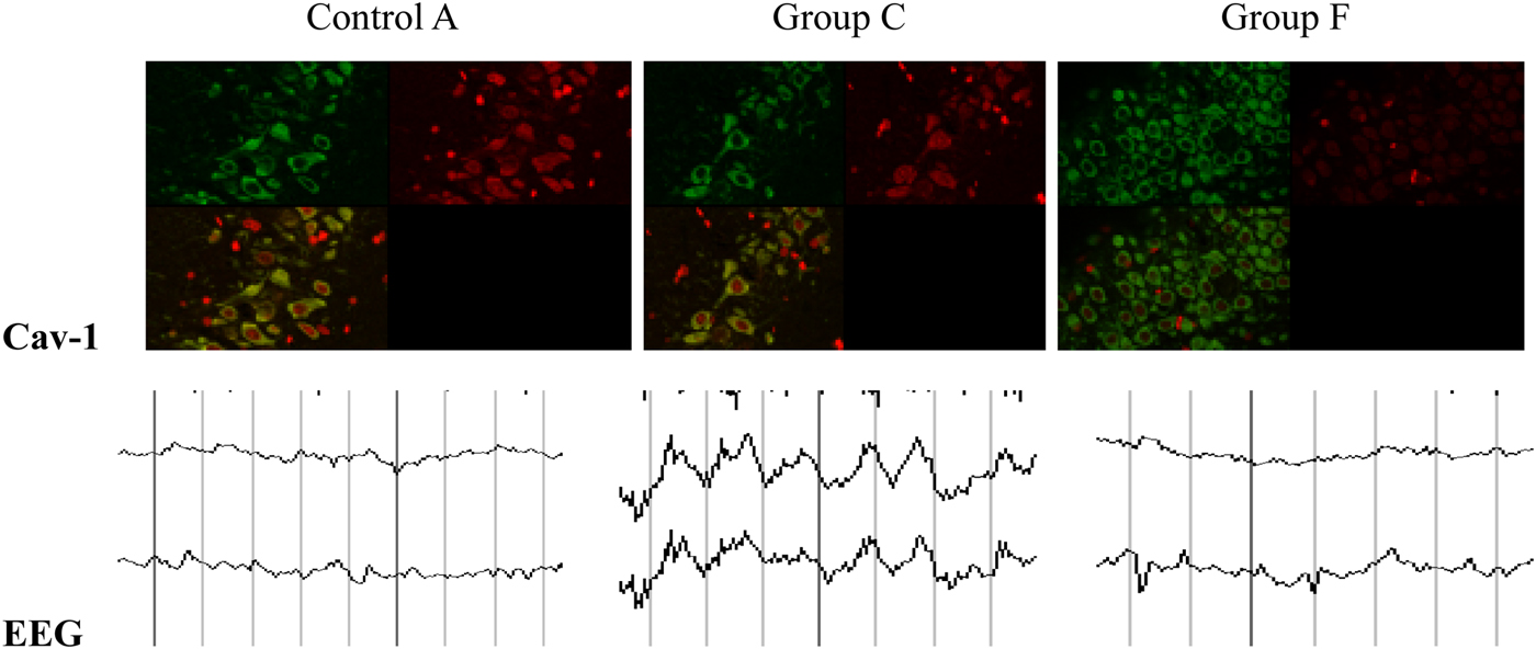
In study 1, GLP treatment increased CaMKIIα expression, and decreased calcium turnover. ERK1/2 had higher expression in cytoplasm and nucleus in Model groups compared with the normal Control groups (P < 0·01). In GLP group, the expression of ERK1/2 (Fig. 1) was inhibited. In study 2. Epileptic symptoms in rats of group C, D, E and F were observed in 7 minutes after kainic acid injection. In day 7, normal EEG, with lower wavelength of α and β wave, was recorded in group A and B; sharp and frequent wavelength was observed in group C, D, E or F (Fig. 2). Even more, compared to group C, more waves with lower wavelength existed in group D, E or F. The frequency of epilepsy discharge from initial 10–15/min reduced to 1/min after GLP treatment. Cav-1 positive cells (Fig. 2) and NF-κB positive cells in group C were significantly higher compared with group A and B. Compared with group C, GLP treatments further increased Cav-1 positive cells, but decreased NF-κB positive cells.

Fig. 1. Control group Model group GLP group (II)

Fig. 2. Control A Group C Group F
In conclusion, GLP improved the epileptic behavior; inhibited calcium overloading and ERK1/2 and NF-KB expression; also stimulated CaMK II α and Cav-1 expression.
This work was supported by the National Natural Science Foundation, China (No. 81241112); Scientific and technological innovation on neurological cell injury and protection (2012TD013, Heilongjiang Province, China); Team focus on fostering technological innovation project funding (04099904, Jiamusi University, China)


