Skip to main content Accessibility help
×
  • Cited by 2
Publisher:
Cambridge University Press
Online publication date:
October 2011
Print publication year:
2011
Online ISBN:
9780511977787

Book description

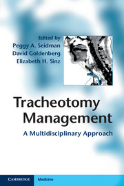
Patients with tracheotomies are managed by a wide variety of healthcare professionals. As a result, information regarding best practice is scattered throughout the medical literature and can be difficult to identify and implement in the clinical setting. Tracheotomy Management: A Multidisciplinary Approach is a practical review of all tracheotomy procedures and acute and chronic tracheotomy care. It combines evidence-based practice and expert opinion to create an invaluable hands-on guide for any healthcare provider managing patients with tracheotomies. Each chapter is authored by at least two different subspecialists, contains case studies with real-life examples of problematic clinical scenarios, and is enhanced by high quality images. The rationale for different approaches is discussed to guide the decision-making process. Written and edited by tracheotomy experts from a wide variety of disciplines, Tracheotomy Management: A Multidisciplinary Approach is essential reading for anesthesiologists, nurse anesthetists and critical care and emergency physicians.

Reviews

"This is an excellent resource for its intended audience and it compares favorably to other books in the field."Doody's Review Services

Refine List

Actions for selected content:

Select all | Deselect all
  • View selected items
  • Export citations
  • Download PDF (zip)
  • Save to Kindle
  • Save to Dropbox
  • Save to Google Drive

Save Search

You can save your searches here and later view and run them again in "My saved searches".

Please provide a title, maximum of 40 characters.
×

Contents

  • Chapter 9 - Intensive care unit tracheotomy care
    pp 117-125
  • View abstract

    Summary

    The platysma muscle together with the subcutaneous tissue comprises the superficial fascia of the head and neck. The deep fascia forms more distinct layers: superficial, pretracheal/middle, prevertebral/ deep, and carotid sheaths. Both the pretracheal and retrovisceral spaces descend into the superior mediastinum acting as important potential conduits of head and neck infections. The thyroid cartilage forms most of the anterior and lateral walls of the larynx. Anterior to the trachea in the neck is the isthmus of the thyroid gland at about the level of the second to fourth tracheal cartilages; below this the inferior thyroid veins, lymph nodes, and sometimes a thyroid ima artery. Lateral to the trachea in the neck are the lobes of the thyroid gland, great vessels, and recurrent laryngeal nerves. A thorough knowledge of anatomy and anatomical variations of the head and neck is essential to avoid or assess complications arising from tracheotomies.
  • Chapter 10 - Complications of tracheotomy
    pp 126-133
  • View abstract

    Summary

    Three main groups of patients that benefit from elective tracheotomy: required prolonged intubation; cannot manage their airway secretions; or have an upper airway obstruction. Tracheotomies can improve the quality of life for patients by allowing return of speech with the use of a Passy-Muir valve or a fenestrated tube, allowing oral intake, improving oral hygiene, and promoting patient mobility. An elective surgical tracheotomy is performed in the operating room under general anesthesia. In cases where difficult intubation is anticipated an elective awake tracheotomy may be performed under local anesthesia. Proper tracheotomy tube selection will minimize discomfort and avoid damage to the tracheal wall. Bleeding intraoperatively or postoperatively is the most common complication from tracheotomies. Inadvertent decannulation is a serious complication that can arise in the early postoperative period. Complications from tracheotomies can be minimized by proper surgical technique and postoperative care.
  • Chapter 11 - Airway manipulation with tracheotomy
    pp 134-145
  • View abstract

    Summary

    Percutaneous tracheotomy (PT) is beneficial in decreasing dead space and reducing airway resistance when compared with intubation. Absolute contraindications include the need for an emergent airway or inability to intubate the patient. All current PT methods are based upon the Seldinger technique of dilators placed over a guidewire. It is recommended that PT be performed under simultaneous video bronchoscopy. Techinques for PT are: Ciaglia method (percutaneous dilating technique), Griggs technique (guidewire dilating forceps (GWDF) technique), Fantoni's technique (translaryngeal approach), and PercTwist (screw-action dilator). Two important issues specific to PT that may arise during the course of the procedure are accidental penetration of the endotracheal tube cuff with the introducer needle during initial puncture of the anterior tracheal wall and possible dislocation of the endotracheal tube. The most common immediate postoperative complication is bleeding. Long-term complications have been reported extensively in a number of studies and metaanalyses.
  • Chapter 12 - Tracheotomy equipment
    pp 146-164
  • View abstract

    Summary

    This chapter presents a case to demonstrate the dire situation of can't ventilate, can't intubate (CVCI) in a patient who cannot breathe. The cricothyroid membrane is composed of fibroelastic tissue bordered by the cricothyroid muscles laterally, thyroid cartilage superiorly, and cricoid ring inferiorly. The fundamental difference between cricothyroidotomy techniques is how the procedure is approached and how the airway lumen is entered: either by needle puncture or blade incision. Needle puncture techniques are based on equipment originally developed for vascular access. The rate and nature of the complications associated with a cricothyroidotomy depend on the choice of technique, skill level of the operator, and patient factors. Surgical cricothyroidotomy is also inadequate for long-term ventilation and is frequently converted to a formal tracheotomy. Simulation is an important component of training and instruction in airway management, especially cricothyroidotomy because it is seldom performed.
  • Chapter 13 - Care of the patient with a tracheotomy
    pp 165-179
  • View abstract

    Summary

    Defective or incomplete separation by the tracheoesophageal septum is one of the most frequent congenital anomalies producing tracheoesophageal fistula (TEF). Anatomic variations that can influence the planning and success of a tracheotomy may be broadly grouped into extrinsic and intrinsic causes. The etiology of the nasal obstruction may include an anterior congenital nasal pyriform aperture stenosis (which is rare), a tumor, or choanal atresia. The most common congenital malformation of the esophagus is esophageal atresia, with or without TEF. Congenital tracheal stenosis may be associated with congenital heart disease, TEF, and skeletal abnormalities. Treatments include tracheoplasty, resection with reanastomosis and stenting. Direct trauma can result in cartilaginous damage and occlusion of the airway lumen by hemorrhage, edema, granulation tissue, scarring, or structural collapse. Congenital tracheal webs are rare presenting with stridor, wheezing, and recurrent respiratory infections. The most common pediatric tracheobronchial tumors include hemangioma, bronchial carcinoid, and papillomatosis.
  • Chapter 14 - Tracheotomy education for home care
    pp 180-194
  • View abstract

    Summary

    The development of advanced neonatal care and anesthesia techniques enabled patients to be intubated for prolonged periods leading to a rise in incidence of acquired subglottic stenosis and the survival of patients with ventilator-dependent respiratory failure. The decision for tracheotomy tube placement should entail a detailed thought process and individualized plan for each patient. When evaluating children for tracheotomy tube placement, it is important to communicate openly with anesthesia staff. Correct size and positioning of the tracheotomy tube can be confirmed with a post-operative chest radiograph and/or passage of a small pediatric flexible endoscope. After tracheotomy, the patient should be closely monitored in the intensive care unit for 5-7 days. Careful dissection and ligature techniques are used for anterior jugular veins and the thyroid isthmus to avoid complications. Patients with tracheotomies have been shown to exhibit difficulties with speech and language development, even after decannulation.

Metrics

Full text views

Total number of HTML views: 0
Total number of PDF views: 0 *
Loading metrics...

Book summary page views

Total views: 0 *
Loading metrics...

* Views captured on Cambridge Core between #date#. This data will be updated every 24 hours.

Usage data cannot currently be displayed.