Introduction
The term platelet ice is used by different authors to mean either the loose platelets underlying a sea-ice sheet, but not yet frozen into place (e.g. Reference DaytonDayton, 1989), or the incorporated layer consisting of platelets and columnar ice formed between the platelets (e.g. Reference Jeffries, Weeks, Shaw and MorrisJeffries and others, 1993; Reference Gow, Ackley, Govoni, Weeks and JeffriesGow and others, 1998). In this paper we will discuss both loose and incorporated platelet ice, and have attempted to make this distinction clear in the terminology used if it is not clear from the context.
There is strong evidence that platelet-ice formation is linked to the presence of ice shelves. Reference MoretskiyMoretskiy (1965) found that loose billows of platelet ice beneath the fast ice off Mirny station, Antarctica, in late November 1958 were thickest adjacent to the ice shelf. Reference Gow, Ackley, Govoni, Weeks and JeffriesGow and others (1998) saw "no systematic variation of [incorporated] platelet thicknesses with location in McMurdo Sound". Reference Crocker and WadhamsCrocker and Wadhams (1989) noted the thickest billows of loose platelets beneath the fast ice of McMurdo Sound in 1986 occurred in regions where Reference Lewis, Perkin and JacobsLewis and Perkin (1985) had observed maximal supercooling in 1982. The generally accepted explanation for the supercooling observed is that sea water near its surface freezing point has been transported by currents beneath the ice shelf and then has been cooled by contact with the ice shelf (Reference Lewis, Perkin and JacobsLewis and Perkin, 1985). When this water rises to the surface along the underside of the ice shelf, it becomes supercooled due to the change in freezing point with depth (Reference Foldvik and KvingeFoldvik and Kvinge, 1974; Reference RobinRobin, 1979; Reference Crocker and WadhamsCrocker and Wadhams, 1989). Of some debate is whether the platelets then form at considerable depths or at the underside of the sea-ice cover. There is evidence for both hypotheses: Reference Dieckmann, Rohardt, Hellmer and KipfstuhlDieckmann and others (1986) observed a large cloud of platelets at 250 m depth in the Weddell Sea, while Reference Gow, Ackley, Govoni, Weeks and JeffriesGow and others (1998) state that the orientation of the platelet crystals beneath McMurdo Sound fast ice indicates that the platelets grow attached to the ice above, rather than forming at depth. It has been acknowledged (Reference Eicken and LangeEicken and Lange, 1989) that different processes may cause platelet-ice formation in different regions of Antarctica. Reference SmetacekSmetacek and others (1992) suggested that platelet ice observed under pack ice could be caused by different processes than those that cause platelet ice observed under fast ice. This possibility was also alluded to by Reference MoretskiyMoretskiy (1965).
In this paper the results of field measurements made in McMurdo Sound in 1999 are presented and discussed in relation to consistency with the conjecture that platelet ice in McMurdo Sound forms in situ.
Locations
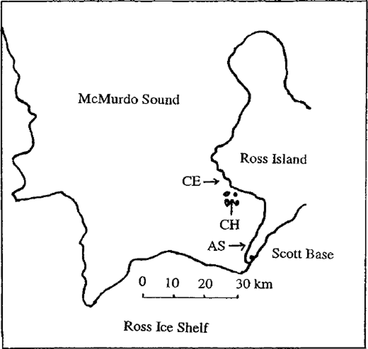
Three sites in McMurdo Sound were studied (see Fig. 1).
-
(1) Site AS (array site) was on the sea ice off Arrival Heights. This site was the location of a thermistor (temperature sensor) array part of an investigation of the thermal conductivity of sea ice by one of us (H.J.T.). The thermistor-array global positioning system (GPS) coordinates were 77°50.197’S, 166°36.764’E. Temperatures were automatically logged simultaneously at each depth every 30 min. Thermistors were spaced 100 mm apart along the 2 m length of the probe. Water salinity and temperature measurements were made and ice cores taken 150 m north of the array. The site was examined twice: once on 23 August 1999, and again on 9 October 1999.
-
(2) Site CH (Camp Haskell) was located between Big Razorback Island and Inaccessible Island on the sea ice between the Dellbridge Islands. The GPS coordinates of the CH site were 77°40.627’ S, 166°27.038’ E.
-
(3) Site CE (Cape Evans) was located on the sea ice approximately 150 m from Scott’s Hut at Cape Evans. (Coordinates of Scott’s Hut at Cape Evans are roughly 77°38’10"S,166°25’10"E.)

Fig. 1. Map of locations in mcmurdo sound.
Measurements at CH and CE were conducted on 17 October 1999.
Ice-Structure Analysis
Ice cores were taken at CE, CH and AS sites for structural analysis. In addition, a slab approximately 1 m by 1 m by 1.93 m was extracted at CH, and vertical thick sections were made from this. When this slab was removed, large platelets approximately 2 mm thick and up to 200 mm in diameter were found attached to the underside of the ice. These were discoloured by brown algae. The base of such a slab is shown in Figure 2. The results of thin- and thick-section analysis of ice cores and slabs from the locations are shown schematically in Figure 3 and summarized in Table 1.
Table 1. Summary of ice structure at the sites


Fig. 2. Base of sea-ice slab taken at ch site, showing attached platelet ice.
From Figure 3 and Table 1, it should be noted that although the CE and CH sites were only approximately 3 km apart, there was no platelet ice observed at CE. A possible explanation for this difference is the existence of a submarine ridge between Cape Evans and Inaccessible Island (Hydrographer of the Navy, 1974; U.S. National Imagery and Mapping Agency, 1997). This ridge could block the flow of ice-shelf water (ISW) to the area off Gape Evans, resulting in an absence of platelet ice in the area. Alternatively, the ridge could block the advection of loose platelet crystals from further south in the Sound.

Fig. 3. Ice-structure diagram for the sites.
The c-axis plot for the horizontal thin section prepared from 2.12 m depth in the AS core from 10 October 1999 (Fig. 4) shows crystals at large angles to each other, which is characteristic of incorporated platelet ice. This is in contrast to the aligned or girdle distributions that would have been obtained for the columnar ice in the study. We have not distinguished between the platelet crystals and the columnar ice between them. Our core was not oriented with respect to a fixed azimuth.

Fig. 4. c-axis plot for horizontal thin section from 2.12 m depth in the as core from 10 october 1999.
The two AS cores were taken just over a month apart. The onset of incorporated platelet ice beneath the columnar ice occurs at the same depth in each core, as would be expected. The increase in the thickness of the incorporated platelet-layer thickness suggests active platelet growth was still occurring between the two core extractions. The layer of loose platelet ice observed on 24 August 1999 was approximately 50 mm thick. This is insufficient to account for the incorporated platelet-ice thickness observed on 10 October 1999 unless platelets were continually advected upwards or were continuously nucleating attached to the ice undersurface. It is possible these platelets were formed at depth and were advected upwards. Reference Gow, Ackley, Buck and GoldenGow and others (1987) observed banded frazil in Weddell Sea pack-ice floes, where plates of ice seemed to have floated up from depth and the resulting c-axis orientations were vertical. In contrast, the observations presented here are more consistent with those of Reference Gow, Ackley, Govoni, Weeks and JeffriesGow and others (1998). In that paper, attached platelets below the incorporated layer of McMurdo Sound fast ice had a near-horizontal c-axis orientation, and the authors hence concluded that those platelets nucleated directly onto the bottom of the sea-ice cover. We did not observe any purely columnar bands within the incorporated platelet-ice layer, as Reference Jeffries, Weeks, Shaw and MorrisJeffries and others (1993) did in their core RS-29. In the absence of such bands, advection theory would require continuous advection of platelets from depth to the site. Otherwise it might be expected that one could observe bands of just columnar ice where all the advected platelets present had been frozen into place and then columnar growth continued.
Isotope Analysis
Oxygen isotope analysis was carried out on sea-ice samples from each site. At each site a core was divided into 100 mm sections for analysis. The samples from AS were taken on 24 August 1999. These were melted and bottled at Scott Base, then transported to New Zealand. Analysis was carried out in February and March 2000. The analysis was referenced to Vienna Standard Mean Ocean Water (VSMOW), with a precision of ±0.04‰. Duplicate subsamples were taken for the analysis. The samples from CH were taken on 19 October 1999 and were melted and bottled at the camp in 100 mL glass bottles with aluminium caps fitted with rubber seals. A duplicate core was sectioned and transported to New Zealand frozen for later alternative preparation and analysis. The samples from GE were also transported frozen back to New Zealand. These samples had the centre of each section removed at room temperature on 10 February 2000, and these were then melted and bottled.
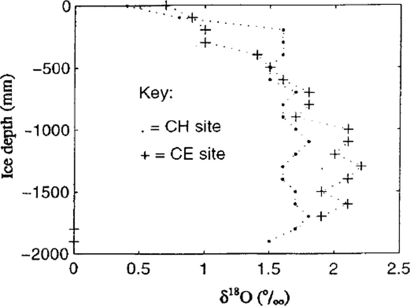
The preliminary isotope results for CH and CE are shown in Figure 5. Although an obvious change from columnar to platelet ice is observed in vertical thin sections of each core, there is no such obvious change in the isotope values at that level. In other words, the isotope values seem to be independent of ice type. This is consistent with the observations of Reference Lange, Schlosser, Ackley, Wadhams and DieckmannLange and others (1990) and Reference Tison, Lorrain, Bouzette, Dini, Bondesan, Stievenard and JeffriesTison and others (1998), although in both those cases the transition in ice types was not columnar to platelet. However, the loose platelets do exhibit a lower δ 18O (≈1.4%‰) than the mixture of platelet and columnar ice above (δ 18O ≈ 1.7%,). This could be due to the presence of sea water in the loose platelets, or it could indicate that the platelets form from fresher water (e.g. from ISW). It is also possible that the platelets and interstitial columnar ice grow at different rates, and the 518O values are therefore an average of the results of the two different growth rates. Below 0.6 m, the CE δ 18O values are consistently higher (+2.0 to +2.3‰) than the CH values (+1.5 to +1.8%,) at the same depths. The salinity profiles (Fig. 6) show the CE ice salinities lower than the CH ice salinities by at least 0.5‰ below 0.6 m. Since no surface water samples were taken for isotopic analysis at these two sites, it cannot be determined whether the same water types were present. If the ice formed from the same water type and desalination was minimal, then Reference Tison, Lorrain, Bouzette, Dini, Bondesan, Stievenard and JeffriesTison and others (1998) state that the isotopic profiles will be determined by the changes in freezing rate at each depth. Reference Tison, Lorrain, Bouzette, Dini, Bondesan, Stievenard and JeffriesTison and others (1998) then state that a lower freezing rate will cause lower bulk salinity and increased δ 18O values in the ice. Hence, if the water at CH and CE were identical, then the δ 18O and salinity values reported here could be explained by lower freezing rates at CE. As discussed below, however, our salinity and temperature measurements indicate that the water was slightly more saline at CE than at CH, which may indicate an absence of ISW at CE. CTD casts at depth are planned for the CE and CH areas in future work, and water samples will be taken for isotope analysis. This will allow determination of the presence or absence of ISW in these areas.

Fig. 5. Oxygen isotope measurements through the ice cores from ceandch.

Fig. 6. Salinity results from the ice cores from ce and ch.
Salinity and Temperature Measurements
Many previous characterizations of the oceanography of the Ross Sea region have been based on ship-based measurements over open water during the austral summer Reference Jacobs, Fairbanks, Horibe and JacobsJacobs and others, 1985). However, Reference Lewis, Perkin and JacobsLewis and Perkin (1985) measured the presence of supercooling beneath the McMurdo Sound fast ice using a Guildline Type 8706 CTD (conductivity- (salinity-), temperature- and depth-measuring device). Due to the presence of the ice cover and the influence of the hole, they relied solely on measurements below 4 m. Their results show supercoolings of up to approximately 0.025°C at the 4 m level.
Our experiments were designed to make similar measurements, but closer to the ice/water interface and away from the influence of the hole.
At the CH, CE and AS sites, a Sea-Bird Electronics (SBE) system was used. The instrument was configured for making measurements beneath the ice and away from the hole. SBE 3plus (temperature) and SBE 4C (conductivity (salinity)) sensors were connected to an SBE 5T pump, and the cables were then connected to a desktop multi-channel counter (SBE 31), driven by a laptop computer running SeaSoft software. This allowed monitoring of the conditions during the experiment, which we found invaluable for detecting any possible icing of the C cell. No depth sensor was used, since all measurements were to be near the ice/water interface. We therefore refer to our equipment as a CT rather than the usual CTD. The cell and pump were mounted on a hinged, alloy arm (Fig. 7) that could be lowered through a hole vertically and then winched into position so that an L-shaped configuration resulted. All experiments were conducted from inside a heated dive hut with a removable floor. This allowed the CT to be kept warmer than the water into which it was being lowered, thus preventing freezing of the cells. The hut exhibited strong temperature gradients (e.g. in the CH experiments the temperature at head height was +13°C, while the floor was −7°C). Because of this, the arm was stored standing upright, with the cells near the roof, until deployment. The CTwas set running in the vertical position to remove any air bubbles in the system, and salinity and temperature were recorded while the cells were put into position. These data were removed from the final analysis. The final positioning of the CTsensors with respect to the underside of the ice was aided by a small black-and-white video camera mounted at the end of the arm (Fig. 8). The depth of the sensors was determined later by measuring along the arm from a mark made to show the ice surface position. The CT was in the water for at least 10min, with data logged 24 times a second. The arm was designed so that the cells were 2 m away from the hole, which ensures the measurements are away from the convective effects of the hole when measuring close to the ice/water interface (Reference ScorerScorer, 1957).

Fig. 7. Photo of ct mounted on arm.

Fig. 8. Diagram ofctarm in deployed position.
Salinities were automatically calculated from the conductivities by the SeaSoft software using the Unesco Practical Salinity Scale 1978 (Unesco, 1981). Following the recommendation of that work, salinities are often given without units (Reference Lewis, Perkin and JacobsLewis and Perkin (1985) followed this convention). However, we have given our results with psu (practical salinity units) following salinity values. This is an alternative convention that has developed in the oceanographic literature (e.g. Reference Jenkins and BomboschJenkins and Bombosch, 1995). It clarifies the distinction between sea-water salinities which are given in psu, and ice salinities, which we have given in parts per thousand.
The freezing points were calculated using the Reference MilleroMillero (1978) formula, with pressure taken to be atmospheric. The errors quoted are based on manufacturers’ calibration before and after the fieldwork.
Near-surface values at the three sites are shown in Table 2.
Table 2. Mean measured s and tvalues at as, ch and ce sites, and supercoolings calculated from these values

The results of the measurements from AS on 9 October 1999 are shown in Figure 9. The upper three lines are the freezing-point temperatures (centre) and error limits, calculated from salinity values. The bottom three lines are the temperature measurements (centre) with error limits. The time-scale is data points, with data recorded 24 times a second.

Fig. 9. Graph showing water temperatures and calculated freezing points from ct measurements at as on 9 october 1999
It can be seen from Figure 9 and Table 2 that at the AS site the water was supercooled by at least 0.006°G on 9 October 1999. At the same site on 23 August 1999, we also measured supercooling of at least 0.006°G (Table 2). However (Table 2), the water was also colder on average by 0.003°C for the October measurements and saltier on average by 0.05 psu. This can be explained as the result of ongoing rejection of salt as the ice continued to thicken in the late winter/ early spring.
The supercooling measured at CH on 17 October 1999 is of a similar magnitude to that observed at the AS site (Table 2). The average salinity of 34.67 psu is higher by 0.02 psu than that observed at AS. In contrast, there is negligible discernible supercooling from the CE site on 17 October 1999 (Table 2). This may be indicative of the absence of ISW in the CE area, but this needs further investigation.
Ice Growth in Supercooled Water
The analysis of measurements from the thermistor array at the AS site yields a result similar to that described in Reference TrodahlTrodahl and others (2000) for a probe at the same site in 1997, namely, that not all the latent heat associated with incorporated platelet-ice formation was conducted to the atmosphere. The experiments described in that paper could not determine whether this was due to platelet formation at the ice/water interface in a supercooled layer or due to advection of platelet crystals that had nucleated at depth, or whether a combination of these processes was occurring. Based on observations of the base of a slab of sea ice taken at the CH site, 10% of the volume of the incorporated platelet ice was estimated to be pure platelet ice. The other 90% of that ice type was interstitial congelation ice that formed between the individual platelets by heat conduction to the air through the ice. The observed thicknesses of incorporated platelet ice at the AS and CH sites were of the order of 0.7 m by mid-October. The pure platelet ice is composed of dendritic crystals, implying growth under supercooled conditions.
Currents in McMurdo Sound have been measured in the Cape Armitage area to be 0.043 ms−1 (Reference Barry and DaytonBarry and Dayton, 1988). If a horizontal current transported supercooled water from beneath an ice shelf and this then interacted with the skeletal interface beneath a growing sea-ice cover, the skeletal layer could experience enhanced growth conditions. This mechanism could explain the predominantly near-horizontal c-axis orientation noted in the platelet ice of McMurdo Sound. The following calculation shows how this forced convection may explain the amounts of platelet ice observed in this study.
The equation for heat flux in the buffer zone, where molecular and eddy transfer are occurring (Reference Kay and NeddermanKay and Nedderman, 1974), is
where q is the heat flux, p and cp are the density and specific heat capacity of sea water, respectively, a is the thermal diffusivity of sea water (conduction term), e is the kinematic eddy viscosity (forced convection term) and dt/dz is the temperature gradient in the buffer zone.
The formation of 0.07 m3 of pure platelet ice (10% of a 0.7 m thick section of incorporated platelet ice in a lm2 area) requires the removal of approximately 21000 kJ of latent heat, q. Assuming the platelet ice began forming in early July (Reference Crocker and WadhamsCrocker and Wadhams, 1989), this gives approximately 100 days for the observed thickness of incorporated platelet ice to form. Thus we can approximate the rate of heat transfer over an area of lm2 as;

Assuming that the water directly in contact with the ice/ water interface is not supercooled, and since the CT cells were 0.5 m below the ice/water interface, we can approximate the temperature gradient as
Using values for sea water of p = 1028 kg m−3 (density), cp = 4.2177 kJ kg−1 K−1 (specific heat), a = 1.3 × 10⁻⁷ m2 s−1 (thermal diffusivity), and solving for the kinematic eddy viscosity gives
Since eddy viscosity tends to zero with decreasing current velocity and decreasing depth below the ice, this value is not inconsistent with values of kinematic eddy viscosity reported beneath first-year land-fast sea ice in the Arctic (Reference Crawford, Padman and McPheeCrawford and others, 1996), where for current velocities of 5−10 cm s−1 and at a depth of 2 m below the ice, the eddy viscosity was reported to be 0.005 m2 s−1.
Therefore the amount of incorporated platelet ice observed at the AS and CH sites could be explained by in situ nucleation of pure platelets due to current-enhanced convection of supercooled water adding to the conduction process, and the interstitial congelation ice being the result of conduction of heat to the air due to the temperature gradient between the air and water.
Conclusions
This study confirms the presence of supercooling in sea water immediately below actively growing platelet ice, and the absence of pronounced supercooling in an area where no platelet ice is present. Combined with the ice-growth calculations in a horizontal current, this result adds weight to the argument that McMurdo Sound fast-ice platelets grow attached to the fast-ice above, rather than being advected from depth. The near-horizontal c-axis orientation of the attached platelets beneath the McMurdo Sound fast ice in August and October 1999 is consistent with the platelets growing at the ice/water interface. If such platelets were advected from depth, it would be expected that the c axes would be more vertically oriented, and that supercooling would already have been relieved, so that no supercooling would be observed next to the ice/water interface. However, it is possible that in other areas of McMurdo Sound, or beneath the Ross Sea pack ice, platelets advecting from depth may be observed, and future work will examine whether different platelet formation processes are present in the wider McMurdo Sound and Ross Sea areas. The focal bathymetry may play a significant role in the presence or absence of platelet ice, with a submarine ridge off Cape Evans either blocking the flow of ISW or blocking the advection of platelets from depth to that area. In future work, it is intended to conduct deeper CTD casts in the CH (Dellbridge Islands) and CE (Cape Evans) areas. The results of these casts will be combined with water-sample isotopic analysis to compare the water masses present in the two areas, and determine if ISW is present in the areas. Isotope and salinity profiles from CH and CE exhibit characteristics that indicate the ice below 600 mm at CE was subject to a lower freezing rate than the ice at similar depths at CH. The detailed analysis of these profiles is ongoing, and a comparison of isotope and salinity profiles with ice-growth rates from AS (sea ice off Arrival Heights) is intended in the future.
Acknowledgements
We wish to thank the Foundation for Research, Science and Technology for their ongoing support of Event K131 research. I.J.S is grateful to Antarctica New Zealand for awarding her the 1999/2000 Antarctica New Zealand Sir Robin Irvine Scholarship and for the research opportunities that resulted from it. Thanks also to Antarctica NZ and the Scott Base staff of the 1998/99 and 1999/2000 seasons for providing essential field and logistical support. We are grateful to the staff of McMurdo station for their assistance, especially W Haals, R. Score of the Crary Laboratory and Jeff and Dennis of the Survey Team.


