Hostname: page-component-78c5997874-lj6df Total loading time: 0 Render date: 2024-11-10T05:29:25.686Z Has data issue: false hasContentIssue false

Persistant Anterior Falcine Sinus: Demonstration by CT Angiography

Published online by Cambridge University Press:  18 February 2016

Charles C. Matouk*
Affiliation:
The University of Toronto Brain Vascular Malformation Study Group, Departments of Surgery and Medical Imaging, The Toronto Hospital, Western Division, Toronto, Ontario, Canada
Daniel M. Mandell
Affiliation:
The University of Toronto Brain Vascular Malformation Study Group, Departments of Surgery and Medical Imaging, The Toronto Hospital, Western Division, Toronto, Ontario, Canada
Timo Krings
Affiliation:
The University of Toronto Brain Vascular Malformation Study Group, Departments of Surgery and Medical Imaging, The Toronto Hospital, Western Division, Toronto, Ontario, Canada
Robert A. Willinsky
Affiliation:
The University of Toronto Brain Vascular Malformation Study Group, Departments of Surgery and Medical Imaging, The Toronto Hospital, Western Division, Toronto, Ontario, Canada
Karel G. terBrugge
Affiliation:
The University of Toronto Brain Vascular Malformation Study Group, Departments of Surgery and Medical Imaging, The Toronto Hospital, Western Division, Toronto, Ontario, Canada
*
The University of Toronto Brain Vascular Malformation Study Group, Departments of Surgery and Medical Imaging, The Toronto Hospital, Western Division, 399 Bathurst Street, Toronto, Ontario, M5T 2S8, Canada
Rights & Permissions [Opens in a new window]

Abstract

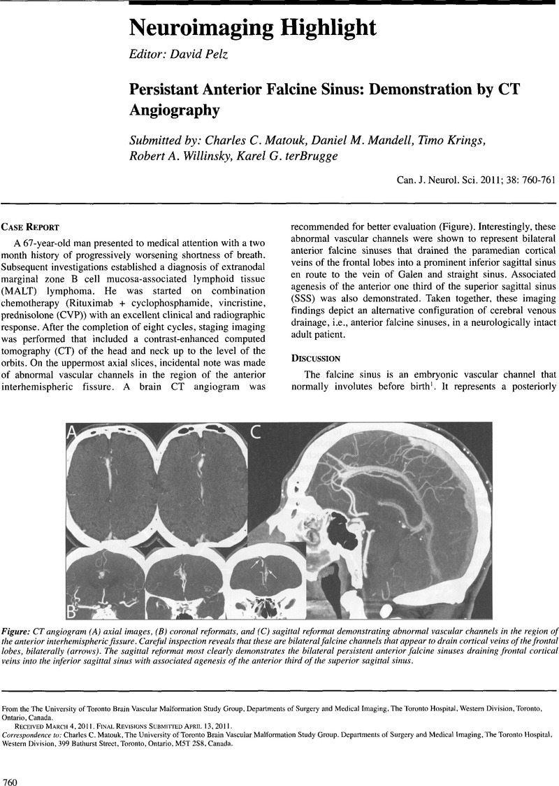
Image of the first page of this content. For PDF version, please use the ‘Save PDF’ preceeding this image.'
Type
Neuroimaging Highlight
Copyright
Copyright © Canadian Neurological Sciences Federation 2011

References

1.Manoj, KS, Krishnamoorthy, T, Thomas, B, Kapilamoorthy, TR. An incidental persistent falcine sinus with dominant straight sinus and hypoplastic distal superior sagittal sinus. Pediatr Radiol. 2006 Jan;36(l):65–7.CrossRefGoogle ScholarPubMed
2.Kesava, PP. Recanalization of the falcine sinus after venous sinus thrombosis. AJNR Am J Neuroradiol. 1996 Oct;17(9):1646–8.Google ScholarPubMed
3.Sener, RN. Association of persistent falcine sinus with different clinicoradiologic conditions: MR imaging and MR angiography. Comput Med Imaging Graph. 2000 Nov-Dec;24(6):343–8.CrossRefGoogle ScholarPubMed
4.Ryu, CW. Persistent falcine sinus: is it really rare? AJNR Am J Neuroradiol. 2010 Feb;31(2):367–9.CrossRefGoogle ScholarPubMed
5.Strub, WM, Leach, JL, Tomsick, TA. Persistent falcine sinus in an adult: demonstration by MR venography. AJNR Am J Neuroradiol. 2005 Apr;26(4):750–1.Google Scholar
6.Streeter, GL. The development of the venous sinuses of the dura mater in the human embryo. Am J Anat. 1915;18:145–78.CrossRefGoogle Scholar
7.Tubbs, RS, Loukas, M, Louis, RG Jr., et al.Anatomy of the falcine venous plexus. J Neurosurg. 2007 Jul;107(l):155–7.CrossRefGoogle ScholarPubMed
8.Raybaud, C. Normal and abnormal embryology and development of the intracranial vascular system. Neurosurg Clin N Am. 2010 Jul;21(3):399426.CrossRefGoogle ScholarPubMed