INTRODUCTION
Legionella pneumophila and other members of the family Legionellaceae are the causative agents of legionellosis. Legionella spp. are aquatic bacteria that can be transmitted to humans by inhalation of water or an aerosol contaminated with the bacteria. The clinical spectrum of legionellosis ranges from asymptomatic infection, through influenza-like disease (Pontiac fever) to Legionnaires' disease (LD), an often severe pneumonia. Legionella infections are underdiagnosed but are nonetheless recognized to be common causes of community-acquired pneumonia [Reference Sopena1, Reference Waterer, Baselski and Wunderink2], in particular in hospitalized patients with exacerbations of chronic obstructive pulmonary disease [Reference Lieberman3].
LD is a notifiable disease in Denmark, and the incidence is about 20 per million per year; about 50–60% are sporadic community-acquired cases [Reference Uldum and Krause4]. The incidence of non-pneumonic legionellosis is unknown. The incidence of community-acquired LD is known to vary geographically, and we have shown that a specific town in Denmark, Randers, has a high incidence of LD. It has not been possible to find the cause of this high incidence or otherwise explain the observation [Reference Rudbeck and Hansen5].
Previous outbreak studies have shown increased antibody levels among individuals exposed to L. pneumophila, although these persons did not develop LD [Reference Boshuizen6–Reference Saravolatz8]. Hence, a possible presence of a continuous environmental infective source would probably result in a high prevalence of antibodies to one or more serogroups of Legionella in the population of Randers. The aim of this study was to: (1) describe the geographical variation in the incidence of LD in towns in Denmark, and (2) determine the seroprevalence of antibodies to Legionella spp. in the general healthy population in a town with a high incidence of LD and compare the seroprevalence with that of a similar town with an average incidence of LD.
METHODS
Incidence study
Cases of Legionella infections were ascertained by reviewing all Legionella laboratory tests analysed at Statens Serum Institut from the two counties of Vejle and Aarhus, between July 1996 and June 2002. In addition, we included cases of LD notified by physicians to the Department of Epidemiology, Statens Serum Institut, during the same period. A few infections (about 3% of all registered infections in the two counties) were diagnosed at the local microbiology departments [Reference Rudbeck and Hansen5] and therefore not confirmed by the reference laboratory at the Statens Serum Institut; these cases were therefore not included in our study.
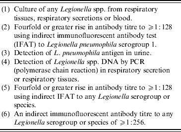
Cases were defined according to the definition of a positive laboratory test by Statens Serum Institut (Table 1). Nosocomial and travel-related notified cases, according to the definitions of the European Working Group for Legionella Infections, were excluded.
Table 1. Laboratory criteria for study inclusion

Criteria (1)–(3) are considered as confirmatory of a current or recent Legionella infection (Legionnaires' disease in a case of pneumonia). Criteria (4)–(6) are considered as presumptive of a current, recent or past Legionella infection (Legionnaires' disease in a case of pneumonia).
By using the Danish Civil Registry number, a unique ID number assigned to all individuals with residence in Denmark, we obtained the addresses of the cases at the time of diagnosis. The addresses were aggregated at the postcode level, and cases were distributed according to seven towns of residence in two neighbouring counties. All towns had a population between 48 000 to 62 000, except Aarhus with a population of about 285 000.
Seroepidemiological study
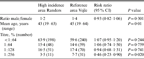
Blood samples were collected from 308 healthy blood donors living in the town of Randers (Aarhus County) and 400 healthy blood donors living in Vejle (Vejle County). Blood donors in Denmark are unpaid healthy volunteers aged between 18 and 65 years. The sampling period was from late February to early June 2004. No difference was found in sampling frequency between the towns. Sampling took place at the sole hospital in each town. The mean age for the blood donors in Randers and Vejle was 43 and 45 years respectively (P=0·01) (Table 2), but there was no difference in the distribution of age groups (<31, 31–40, 41–50, >50 years) between the two towns. Fifty-seven percent were males, and there was no difference in gender distribution between the towns.
Table 2. Seroprevalence of antibodies to Legionella in titresFootnote *

* The titres are based on the highest titre to any antigen in each case.
The blood samples were analysed for antibodies to Legionella spp. by indirect immunofluorescence antibody test (IFAT) with plate-grown and heat-inactivated L. pneumophila serogroup (sg) 1–6 and L. micdadei and L. bozemanii as antigens. The serum samples were titrated from 1:64 and upwards. Antibodies to Legionella spp. were detected with a FITC conjugated rabbit anti-human IgM, A and G antibody (Code F0200, Dako, Glostrup, Denmark). An E. coli blocking fluid was used to block cross-reacting antibodies to Gram-negative bacteria [Reference Bangsborg9]. Samples with an antibody titre of ⩾1:128 were considered as positive for Legionella.
The two towns are located within a distance of about 100 km, and are comparable according to population density, number of citizens in town areas, age composition, and number of unemployed. Each town is served by a single hospital. The incidence of notified LD in Vejle is nearly identical to the national average.
Statistical methods
The home addresses of the blood donors were plotted in a geographical information system (ArcView GIS 3.3, India). Comparisons of proportions were carried out by χ2 tests with a level of statistical significance of P<0·05. The analyses were made in stata version 9.2 (Stata Corp., College Station, TX, USA).
RESULTS
Incidence of community-acquired LD in towns
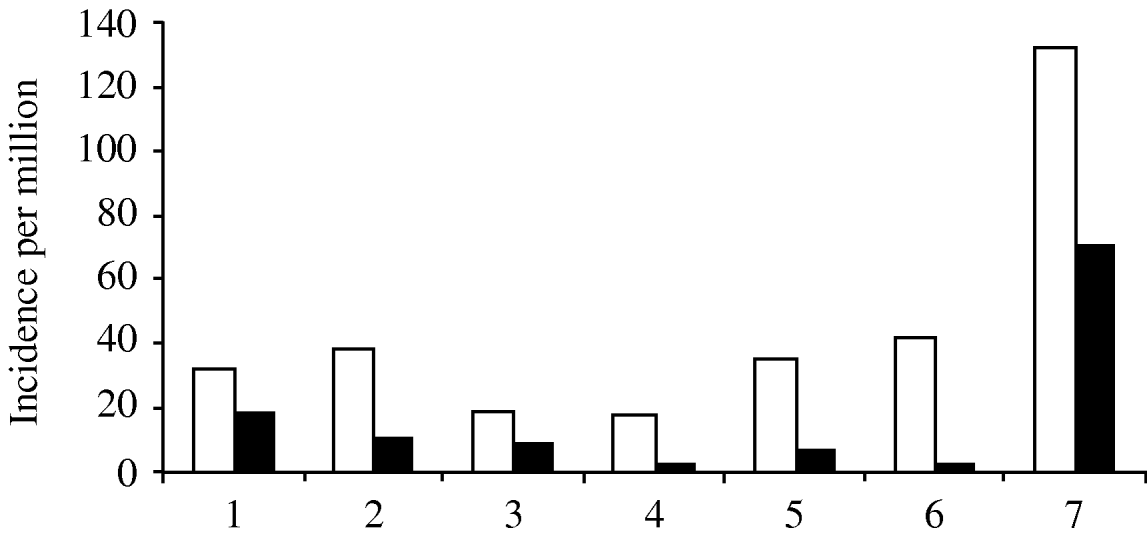
In seven towns Vejle and Aarhus counties in Denmark (Fig. 1), the incidence of notified community-acquired cases varied from 3 to 19 per million, and the incidence of cases with positive laboratory results ranged from 20 to 40 per million. The exception was the hyperendemic area, Randers, where the incidence of notified cases was 70 per million and the incidence of positive laboratory cases was 132 per million (Fig. 2).

Fig. 1. Denmark, study towns. 1, Kolding; 2, Fredericia; 3, Vejle; 4, Horsens; 5, Aarhus; 6, Silkeborg; 7, Randers.

Fig. 2. Incidence of notified cases of Legionnaires' disease (■) and cases with positive Legionella laboratory test (□). 1, Kolding; 2, Fredericia; 3, Vejle; 4, Horsens; 5, Aarhus; 6, Silkeborg; 7, Randers.
Seroprevalence of antibodies to Legionella in healthy blood donors
In Randers, 62 (20·1%) of 308 donors had IFAT titres of ⩾1:128, compared with 101 (25·3%) of 400 donors from Vejle (risk ratio 0·80, 95% CI 0·60–1·05, P=0·109) (Table 2). There was a higher seroprevalence of persons with titres of 1:256 in Vejle (P=0·020) than in Randers (Table 2).
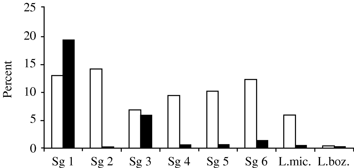
In total there were 61·4% (438) donors with a titre of <1:64, 15·0% (107) with a titre of 1:64, 17·0% (121) with 1:128 and 6·0% (43) with titres of 1:256. The prevalence of positive results (⩾1:128) was highest against sg 2 (14·4%), followed by sg 1 (12·9%), sg 6 (12·3%), sg 5 (10·3%), sg 4 (9·6%), and sg 3 (6·9%) (Fig. 3).

Fig. 3. Legionella seroprevalence in the study population (□) compared to the distribution of species and serogroups isolated from cases of Legionnaires' disease (■) in the Danish population [seroprevalence defined as titres of at least 1:128 in 2004]. Legionnaires' disease 1994–2004 (n=1004), notified to the Department of Epidemiology, Statens Serum Institut, according to the known specified species and serogroups (n=313). L.mic, Legionella micdadei; L.boz, Legionella bozemanii.
There were visually no geographical clusters according to home addresses either in antibody titres or serogroups (not shown).
DISCUSSION
The incidence of community-acquired LD is known to vary geographically. We found a very high variation of from 3 to 70 notified cases per million in different towns. Excluding Randers, case rates ranged from 3 to 19, which is within range of the general incidence in Denmark. The incidence in Randers was as high as 70 per million, and continued to be high in 2004 (data not shown). The incidence of a positive laboratory test is much higher; the true incidence of LD in Denmark may be somewhere between the incidence of notified cases and the incidence of positive laboratory cases. In a previous study in Randers all but one of the individuals with a positive laboratory test were hospitalized, and 91% left the hospital with the diagnosis of either Legionella pneumonia or pneumonia [Reference Rudbeck and Hansen5].
The high incidence of notified community-acquired cases in Randers could be a surveillance artefact, i.e. a result of increased awareness of Legionella among health-care providers of the region. However, a careful analysis could not reveal differences in the frequency of use of diagnostic testing, or outbreaks caused by specific serogroups of Legionella. The incidence in Randers was high in all age groups, being highest among the eldest. In Randers and in the rest of Aarhus County (including the towns of Silkeborg and Aarhus) 0·3% of the population was tested for legionellosis each year. There was no significant difference in sex distribution between the different areas in the county (male/female 1·5:1) [Reference Rudbeck and Hansen5]. A surveillance artefact would be unlikely to explain the hyperendemicity revealed both in the notified cases and in ascertainment of laboratory tests.
One of our main hypotheses was that the hyperendemic situation in Randers could be explained by an ongoing source of Legionella exposure in the community, and that this would result in a high prevalence of antibodies to one or more serogroups. A study conducted in an exposed population in The Netherlands indicates that exposure increases the prevalence of antibodies in the exposed population [Reference Boshuizen10]. Our hypothesis was not confirmed by the present study. Surprisingly, the prevalence of Legionella infection tended to be higher in Vejle, where a greater number of donors had a titre of 1:256.
In the light of the present study, an alternative explanation could be that the hyperendemicity in Randers was due to exposure to one or more virulent subtypes in certain areas or buildings, and that this exposure was not reflected by a general increase in the seroprevalence among healthy blood donors. On the basis of examinations of healthy blood donors, one cannot exclude the existence of subpopulations in Randers with high levels of antibodies. However, the geographical distribution of home addresses did not reveal any obvious clusters, and therefore the existence of high-risk subpopulations remains a speculation.
In addition to the limitations in the selection of the study population, the limitations of seroepidemiology of legionellosis have to be considered. Cross-reactivity has been reported, e.g. to Campylobacter [Reference Boswell, Marshall and Kudesia11]. A Campylobacter-blocking fluid was not used in the present study. However, an E. coli-blocking fluid was used and for ten Campylobacter antibody-positive sera analysed by IFAT, only one had a titre of 1:128, one had a titre of 1:256 and eight were negative, <1:64 (data not shown). These results suggest that there was no general cross-reactivity between Campylobacter-positive sera and the Legionella IFAT antigens. Our results obtained by IFAT were compared with those of other assays. By investigating 200 of the 708 samples we found good correlation with the Focus Legionella IFA kit (Focus Diagnostics Inc., Cypress, CA, USA) and, to a minor degree, the Zeus Scientific L. pneumophila serogroups 1–6 IgG/M/A ELISA (Zeus Scientific Inc., Raritan, NJ, USA). In both kits more positive samples were found than in the in-house assay indicating that cross-reactivity is less of a problem in the in-house assay than in the other assays (P. Elverdal, unpublished data).
We found an overall prevalence for IFAT titres of 1:128 at 17%, and 13% for sg 1. In a neighbouring country, Sweden, the overall prevalence of antibodies to L. pneumophila sg 1, which was the only antigen used, was <1% (IFAT ⩾1:16) [Reference Darelid12]. Even though the results can be difficult to compare because of different assays used, they suggest a higher prevalence in our study population compared with the Swedish population. The Swedish population is otherwise comparable to the Danish population in terms of general health and socioeconomic conditions.
As previously mentioned, the incidence of reported cases of LD is high in Denmark, and in addition, the present study suggests that the prevalence of antibodies to Legionella is high. On the other hand, there have been no major outbreaks. We speculate that a high antibody level reflects a degree of immunity to Legionella in the population that could reduce the risk of disease in events, that otherwise would lead to outbreaks [Reference Boshuizen10, Reference Brydov13].
The prevalence of antibodies to Legionella in Denmark has not previously been published. The prevalence has been described in antibody levels of 1:128 and 1:256; according to the cut-off levels indicating Legionella infection. The distribution of antibodies to Legionella serogroups in healthy blood donors in our study did not reflect the distribution of serogroups isolated from cases of LD (Fig. 3), or the environmental distributions in Denmark [Reference Brydov13, Reference Jeppesen14]. These differences in distributions support the theory that the pathogenicity and virulence of the bacteria varies among different serogroups. The clinical prevalence of Legionella spp. and serogroups also differs from the environmental prevalence in France with L. pneumonia sg 1 accounting for 30%, and serogroups 3 and 6 for more than 20% in the environment, and the incidence of sg 1 in LD of about 85% [Reference Doleans15]. We found that antibodies to L. pneumophila sg 1–6 were almost equally distributed. L. micdadei and L. bozemanii did not appear to be common in Denmark. One limitation of our study is, however, that most positive samples reacted with several serogroup antigens. This is also frequently seen for verified cases of LD, especially later in the course of the disease.
To conclude, the present study did not reveal a higher prevalence of antibodies in blood donors from a hyperendemic area compared with antibodies in blood donors from an area with an average national incidence. In addition, the prevalence of antibodies did not reflect the incidence of Legionella pneumonia in this population. Antibodies to Legionella spp. were common in healthy individuals, suggesting that the bacteria are widely distributed in the environment in general. To date, it is not well known if persons with antibodies have experienced disease or are asymptomatic, and further studies are required to demonstrate if there may be a connection between these antibody levels and disease, socioeconomic factors, or the environment.
ACKNOWLEDGEMENTS
We thank Melina Hansen and Pernille Elverdal for testing of sera using the Focus Legionella IFA kit and the Zeus Scientific L. pneumophila serogroups 1–6 IgG/M/A ELISA kit. We thank Hannah Lewis for language revision.
DECLARATION OF INTEREST
None.


