Published online by Cambridge University Press: 16 April 2020
Social anxiety disorder (SAD) refers to persistent fear of social situations in which the person is exposed to possible scrutiny by others. Anxiety disorders are associated with dysbalanced inhibition within the limbic system towards threatening stimuli (Phillips 2003). It has been shown SAD patients are particularly sensitive towards faces displaying harsh emotions (Phan 2006).
In this study we used fMRI to investigate activation differences between 14 SAD patients and 15 matched healthy controls. The subjects performed a facial emotion discrimination paradigm including a control condition using shape discrimination (modified version of Hariri 2002).
The aim of this study was to investigate emotion task-specific differences in brain activation of patients and healthy controls.
After clinical assessment, 225 whole-brain volumes (TR = 1.8s) were acquired on a Siemens TRIO 3T MR scanner and analyzed using SPM8.
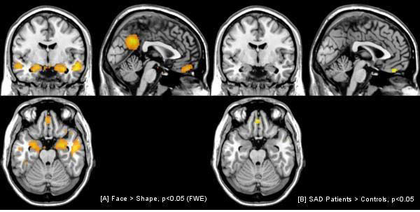
The paradigm activated the amygdalae, the fusiform gyri, the posterior cingulate cortex and the orbitofrontal cortex (OFC) (A). There was a hyperactivation of the OFC in patients compared to healthy controls (B). We found no significant difference in the amygdalae or fusiforme gyrus.

Although not in a social threat situation, SAD patients showed more activity in the orbitofrontal cortex. In contrast to other studies we found no differences in other brain areas indicating an optimized control condition. According to the functional coupling between prefrontal areas and the amygdalae, these data are consistent with an increased effort in down-regulating amygdalar activation by the orbitofrontal cortex.
Comments
No Comments have been published for this article.