Hostname: page-component-cd9895bd7-mkpzs Total loading time: 0 Render date: 2024-12-27T13:29:13.918Z Has data issue: false hasContentIssue false

Heterogeneity in calcium nephrolithiasis: A materials perspective

Published online by Cambridge University Press:  08 May 2017
Benjamin A. Sherer
Affiliation:
Department of Urology, University of California San Francisco, San Francisco, CA 94143
Ling Chen
Affiliation:
Division of Biomaterials and Bioengineering, Department of Preventive and Restorative Dental Sciences, School of Dentistry, University of California San Francisco, San Francisco, CA 94143
Feifei Yang
Affiliation:
Division of Biomaterials and Bioengineering, Department of Preventive and Restorative Dental Sciences, School of Dentistry, University of California San Francisco, San Francisco, CA 94143
Krishna Ramaswamy
Affiliation:
Department of Urology, University of California San Francisco, San Francisco, CA 94143
David W. Killilea
Affiliation:
Children’s Hospital Oakland Research Institute, Oakland, CA 94609
Ryan S. Hsi
Affiliation:
Department of Urologic Surgery, Vanderbilt University Medical Center, Nashville, TN 37232
Marshall L. Stoller
Affiliation:
Department of Urology, University of California San Francisco, San Francisco, CA 94143
Sunita P. Ho*
Affiliation:
Division of Biomaterials and Bioengineering, Department of Preventive and Restorative Dental Sciences, School of Dentistry, University of California San Francisco, San Francisco, CA 94143
*
a) Address all correspondence to this author. e-mail: sunita.ho@ucsf.edu
Get access

Abstract

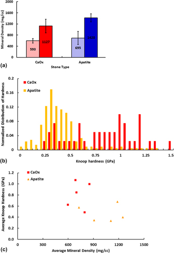
Calcium-based renal calculi demonstrated significant heterogeneity in the structure, density, mineral composition, and material hardness not elucidated by routine clinical testing. Mineral density distributions within calcium oxalate stones revealed differential areas of low (590±80 mg/cc), medium (840±140 mg/cc), and high (1100±200 mg/cc) densities. Apatite stones also contained regions of low (700±200 mg/cc), medium (1100±200 mg/cc), and high (1400±140 mg/cc) densities within layers extending from single or multiple nucleation sites. Despite having lower average mineral density, calcium oxalate (CaOx) stones demonstrated higher material hardness compared to apatite stones, suggesting other chemical components might be involved in determining stone hardness properties. Carbon concentrated sites were identified between morphologic layers in CaOx stones and in stratified layers of apatite stones. Elemental analyses revealed numerous additional trace elements in both stone types. Despite the widespread assumption that stone mineral density is an indicator of susceptibility to lithotripsy, calcium stone mineral density estimates do not directly correlate with actual ex vivo stone hardness. Underlying stone heterogeneity in both structure and mineral density could explain why historical approaches have failed in accurately predicting response of stones to lithotripsy.

Type
Articles
Copyright
Copyright © Materials Research Society 2017 

Access options

Get access to the full version of this content by using one of the access options below. (Log in options will check for institutional or personal access. Content may require purchase if you do not have access.)

Footnotes

Contributing Editor: Adrian B. Mann

References

REFERENCES

Caudarella, R., Tonello, L., Rizzoli, E., and Vescini, F.: Predicting five-year recurrence rates of kidney stones: An artificial neural network model. Arch. Ital. Urol. Androl. 83, 14 (2011).Google ScholarPubMed
Zhong, P., Chuong, C.J., Goolsby, R.D., and Preminger, G.M.: Microhardness measurements of renal calculi: Regional differences and effects of microstructure. J. Biomed. Mater. Res. 26, 1117 (1992).CrossRefGoogle ScholarPubMed
Williams, J.C. Jr., Zarse, C.A., Jackson, M.E., Lingeman, J.E., and McAteer, J.A.: Using helical CT to predict stone fragility in shock wave lithotripsy. In Renal Stone Disease, 1st Annual International Urolithiasis Research Symposium, Vol. 900 (AIP Conf Proc., Indianapolis, IN, 2007); p. 326.Google Scholar
Williams, J.C. Jr., Saw, K.C., Paterson, R.F., Hatt, E.K., McAteer, J.A., and Lingeman, J.E.: Variability of renal stone fragility in shock wave lithotripsy. Urology 61, 1092 (2003).CrossRefGoogle ScholarPubMed
Motley, G., Dalrymple, N., Keesling, C., Fischer, J., and Harmon, W.: Hounsfield unit density in the determination of urinary stone composition. Urology 58, 170 (2001).CrossRefGoogle ScholarPubMed
Khan, S.R. and Kok, D.J.: Modulators of urinary stone formation. Front. Biosci. 9, 1450 (2004).CrossRefGoogle ScholarPubMed
Ramaswamy, K., Killilea, D.W., Kapahi, P., Kahn, A.J., Chi, T., and Stoller, M.L.: The elementome of calcium-based urinary stones and its role in urolithiasis. Nat. Rev. Urol. 12, 543 (2015).CrossRefGoogle ScholarPubMed
Witzmann, F.A., Evan, A.P., Coe, F.L., Worcester, E.M., Lingeman, J.E., and Williams, J.C. Jr.: Label-free proteomic methodology for the analysis of human kidney stone matrix composition. Proteome Sci. 14, 4 (2016).CrossRefGoogle ScholarPubMed
Cloutier, J., Villa, L., Traxer, O., and Daudon, M.: Kidney stone analysis: “Give me your stone, I will tell you who you are!”. World J. Urol. 33, 157 (2015).CrossRefGoogle Scholar
Ruegsegger, P., Koller, B., and Muller, R.: A microtomographic system for the nondestructive evaluation of bone architecture. Calcif. Tissue Int. 58, 24 (1996).CrossRefGoogle ScholarPubMed
Penescu, M., Purcarea, V.L., Sisu, I., and Sisu, E.: Mass spectrometry and renal calculi. J. Med. Life 3, 128 (2010).Google ScholarPubMed
Bak, M., Thomsen, J.K., Jakobsen, H.J., Petersen, S.E., Petersen, T.E., and Nielsen, N.C.: Solid-state 13C and 31P NMR analysis of urinary stones. J. Urol. 164, 856 (2000).CrossRefGoogle ScholarPubMed
Sohnel, O., Grases, F., Garcia-Ferragut, L., and March, J.G.: Study on calcium oxalate monohydrate renal uroliths. III. Composition and density. Scand. J. Urol. Nephrol. 29, 429 (1995).CrossRefGoogle Scholar
Fleisch, H.: Inhibitors and promoters of stone formation. Kidney Int. 13, 361 (1978).CrossRefGoogle ScholarPubMed
Brauer, D.S., Saeki, K., Hilton, J.F., Marshall, G.W., and Marshall, S.J.: Effect of sterilization by gamma radiation on nano-mechanical properties of teeth. Dent. Mater. 24, 1137 (2008).CrossRefGoogle ScholarPubMed
Djomehri, S.I., Candell, S., Case, T., Browning, A., Marshall, G.W., Yun, W., Lau, S.H., Webb, S., and Ho, S.P.: Mineral density volume gradients in normal and diseased human tissues. PLoS One 10, e0121611 (2015).CrossRefGoogle ScholarPubMed
ASTM: ASTM E384-11 Standard Test Method for Knoop and Vickers Hardness of Materials (ASTM International, West Conshohocken, 2011).Google Scholar
Killilea, D.W., Westropp, J.L., Shiraki, R., Mellema, M., Larsen, J., Kahn, A.J., Kapahi, P., Chi, T., and Stoller, M.L.: Elemental content of calcium oxalate stones from a canine model of urinary stone disease. PLoS One 10, e0128374 (2015).CrossRefGoogle ScholarPubMed
Pearle, M.S. and Lotan, Y.: Urinary lithiasis: Etiology, epidemiology, and pathogenesis. In Campbell-walsh Urology, Wein, A.J., Kavoussi, L., Novick, A.C., Partin, A.W., and Peters, C.A., eds. (Saunders Elsevier, Philadelphia, PA, 2012); p. 1257.CrossRefGoogle Scholar
Denstedt, J.D. and Fuller, A.: Epidemiology of stone disease in North America. In Urolithiasis: Basic Science and Clinical Practice, Talati, J., Tiselius, H-G., Albala, D.M., and Ye, Z., eds. (Springer, London, England, 2012); p. 13.CrossRefGoogle Scholar
Echigo, T., Kimata, M., Kyono, A., and Shimizu, M.: Re-investigation of the crystal structure of whewellite [Ca(C2O4)·H2O] and the dehydration mechanism of caoxite [Ca(C2O4)·3H2O]. Mineral. Mag. 69, 77 (2005).CrossRefGoogle Scholar
Conti, C., Casati, M., Colombo, C., Realini, M., Brambilla, L., and Zerbi, G.: Phase transformation of calcium oxalate dihydrate-monohydrate: Effects of relative humidity and new spectroscopic data. Spectrochim. Acta, Part A 128, 413 (2014).CrossRefGoogle ScholarPubMed
Zarse, C.A., McAteer, J.A., Sommer, A.J., Kim, S.C., Hatt, E.K., Lingeman, J.E., Evan, A.P., and Williams, J.C. Jr.: Nondestructive analysis of urinary calculi using micro computed tomography. BMC Urol. 4, 15 (2004).CrossRefGoogle ScholarPubMed
Williams, J.C. Jr., McAteer, J.A., Evan, A.P., and Lingeman, J.E.: Micro-computed tomography for analysis of urinary calculi. Urol. Res. 38, 477 (2010).CrossRefGoogle ScholarPubMed
Williams, J.C. Jr., Lingeman, J.E., Coe, F.L., Worcester, E.M., and Evan, A.P.: Micro-CT imaging of Randall’s plaques. Urolithiasis 43(Suppl 1), 13 (2015).CrossRefGoogle ScholarPubMed
Clarke, B.: Normal bone anatomy and physiology. Clin. J. Am. Soc. Nephrol. 3(Suppl 3), S131 (2008).CrossRefGoogle ScholarPubMed
Fellstrom, B., Lindsjo, M., Danielson, B.G., Karlsson, F.A., and Ljunghall, S.: Binding of glycosaminoglycan inhibitors to calcium oxalate crystals in relation to ionic strength. Clin. Chim. Acta 180, 213 (1989).CrossRefGoogle ScholarPubMed
Walton, R.C., Kavanagh, J.P., and Heywood, B.R.: The density and protein content of calcium oxalate crystals precipitated from human urine: A tool to investigate ultrastructure and the fractional volume occupied by organic matrix. J. Struct. Biol. 143, 14 (2003).CrossRefGoogle ScholarPubMed
McKee, M.D., Nanci, A., and Khan, S.R.: Ultrastructural immunodetection of osteopontin and osteocalcin as major matrix components of renal calculi. J. Bone Miner. Res. 10, 1913 (1995).CrossRefGoogle ScholarPubMed
Perk, H., Serel, T.A., Kosar, A., Deniz, N., and Sayin, A.: Analysis of the trace element contents of inner nucleus and outer crust parts of urinary calculi. Urol. Int. 68, 286 (2002).CrossRefGoogle ScholarPubMed
Chi, T., Kim, M.S., Lang, S., Bose, N., Kahn, A., Flechner, L., Blaschko, S.D., Zee, T., Muteliefu, G., Bond, N., Kolipinski, M., Fakra, S.C., Mandel, N., Miller, J., Ramanathan, A., Killilea, D.W., Bruckner, K., Kapahi, P., and Stoller, M.L.: A drosophila model identifies a critical role for zinc in mineralization for kidney stone disease. PLoS One 10, e0124150 (2015).CrossRefGoogle ScholarPubMed
Turgut, M., Unal, I., Berber, A., Demir, T.A., Mutlu, F., and Aydar, Y.: The concentration of Zn, Mg and Mn in calcium oxalate monohydrate stones appears to interfere with their fragility in ESWL therapy. Urol. Res. 36, 31 (2008).CrossRefGoogle ScholarPubMed
Wiesenthal, J.D., Ghiculete, D., D’A Honey, R.J., and Pace, K.T.: Evaluating the importance of mean stone density and skin-to-stone distance in predicting successful shock wave lithotripsy of renal and ureteric calculi. Urol. Res. 38, 307313 (2010).CrossRefGoogle ScholarPubMed
Ouzaid, I., Al-gahtani, S., Dominique, S., Hupertan, V., Fernandez, P., Hermieu, J., Delmas, V., and Ravery, V.: A 970 hounsfield unit (HU) threshold of kidney stone density on non-contrast computed tomography (NCCT) improves patients’ selection for extracorporeal shockwave lithotripsy (ESWL): Evidence from a prospective study. BJUI 110, E438E442 (2012).Google Scholar
Anastasiadis, A., Onal, B., Modi, P., TUrna, B., Duvdevani, M., Timoney, A., Wolf, J.S. Jr., and De La Rosette, J.: Impact of stone density on outcomes in percutaneous nephrolithotomy (PCNL): An analysis of the clinical research office of the endourological society (CROES) pcnl global study database. Scand. J. Urol. 47, 509514 (2013).CrossRefGoogle ScholarPubMed
Bellin, M.F., Renard-Penna, R., Conort, P., Bissery, A., Meric, J.B., Daudon, M., Mallet, A., Richard, F., and Grenier, P.: Helical CT evaluation of the chemical composition of urinary tract calculi with a discriminant analysis of CT-attenuation values and density. Eur. Radiol. 14, 2134 (2004).CrossRefGoogle ScholarPubMed
Bulakci, M., Tefik, T., Akbulut, F., Ormeci, M.T., Bese, C., Sanli, O., Oktar, T., and Salmaslioglu, A.: The use of non-contrast computed tomography and color Doppler ultrasound in the characterization of urinary stones—Preliminary results. Turk. J. Neurol. 41, 165 (2015).Google Scholar
Zarse, C.A., Hameed, T.A., Jackson, M.E., Pishchalnikov, Y.A., Lingeman, J.E., McAteer, J.A., and Williams, J.C. Jr.: CT visible internal stone structure, but not Hounsfield unit value, of calcium oxalate monohydrate (COM) calculi predicts lithotripsy fragility in vitro. Urol. Res. 35, 201206 (2007).CrossRefGoogle Scholar
Supplementary material: File

Sherer supplementary material

Sherer supplementary material 1

Download Sherer supplementary material(File)
File 16.3 KB
Supplementary material: Image

Sherer supplementary material

Figure S1

Download Sherer supplementary material(Image)
Image 42.2 KB