Hostname: page-component-78c5997874-94fs2 Total loading time: 0 Render date: 2024-11-10T07:44:19.616Z Has data issue: false hasContentIssue false

Antibacterial activity of nanoporous gold against Escherichia coli and Staphylococcus epidermidis

Published online by Cambridge University Press:  16 May 2017

Masataka Hakamada*
Affiliation:
Department of Energy Science and Technology, Graduate School of Energy Science, Kyoto University, Kyoto 606-8501, Japan
Seiji Taniguchi
Affiliation:
Department of Energy Science and Technology, Graduate School of Energy Science, Kyoto University, Kyoto 606-8501, Japan
Mamoru Mabuchi
Affiliation:
Department of Energy Science and Technology, Graduate School of Energy Science, Kyoto University, Kyoto 606-8501, Japan
*
a) Address all correspondence to this author. e-mail: hakamada.masataka.3x@kyoto-u.ac.jp
Get access

Abstract

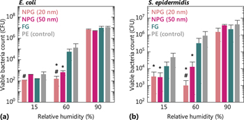
Conventional metallic antibacterial materials release metal ions and reactive oxygen species (ROS) for killing bacteria. Herein, we found that nanoporous gold (NPG) exhibits antibacterial activity (AA) at an intermediate relative humidity (RH) of 60% against Escherichia coli and Staphylococcus epidermidis in contrast to the inert behavior of bulk gold. The dependence of AA on RH, morphological observations of bacteria on NPG, and transcriptomic analyses of NPG-treated Escherichia coli were investigated. These observations collectively suggest that biological processes in cell walls containing peptidoglycan and cell membranes are significantly disrupted by direct contact with NPG. Metal ions and ROS were not detected, and therefore are not responsible for the present antibacterial properties of NPG. The catalytic nature of NPG may be responsible for its AA, probably because of lattice distortion at the surface of nanosized ligaments with large curvature.

Type
Articles
Copyright
Copyright © Materials Research Society 2017 

Access options

Get access to the full version of this content by using one of the access options below. (Log in options will check for institutional or personal access. Content may require purchase if you do not have access.)

Footnotes

b)

Present Address: Sapporo Breweries Ltd., 2 Takasecho, Funahashi, Chiba 273-0014, Japan.

Contributing Editor: Jinju Chen

References

REFERENCES

Zhao, G. and Stevens, S.E. Jr.: Multiple parameters for the comprehensive evaluation of the susceptibility of Escherichia coli to the silver ion. BioMetals 11, 27 (1998).Google Scholar
Galeano, B., Korff, E., and Nicholson, W.L.: Inactivation of vegetative cells, but not spores, of Bacillus anthracis, B. cereus, and B. subtilis on stainless steel surfaces coated with an antimicrobial silver- and zinc-containing zeolite formulation. Appl. Environ. Microbiol. 69, 4329 (2003).Google Scholar
Chatterjee, A.K., Chakraborty, R., and Basu, T.: Mechanism of antibacterial activity of copper nanoparticles. Nanotechnology 25, 135101 (2014).CrossRefGoogle ScholarPubMed
Sadhasivam, S., Shanmugam, P., Veerapandian, M., Subbiah, R., and Yun, K.: Biogenic synthesis of multidimensional gold nanoparticles assisted by Streptomyces hygroscopicus and its electrochemical and antibacterial properties. BioMetals 25, 351 (2012).Google Scholar
Cui, Y., Zhao, Y., Tian, Y., Zhang, W., , X., and Jiang, X.: The molecular mechanism of action of bactericidal gold nanoparticles on Escherichia coli . Biomaterials 33, 2327 (2012).Google Scholar
Zhang, Y., Peng, H., Huang, W., Zhou, Y., and Yan, D.: Facile preparation and characterization of highly antimicrobial colloid Ag or Au nanoparticles. J. Colloid Interface Sci. 325, 371 (2008).CrossRefGoogle ScholarPubMed
Hernández-Sierra, J.F., Ruiz, F., Pena, D.C.C., Martínez-Gutiérrez, F., Martínez, A.E., Guillén, A.de J.P., Tapia-Pérez, H., and Castañón, G.M.: The antimicrobial sensitivity of Streptococcus mutans to nanoparticles of silver, zinc oxide, and gold. Nanomed. Nanotechnol. Biol. Med. 4, 237 (2008).CrossRefGoogle ScholarPubMed
Erlebacher, J., Aziz, M.J., Karma, A., Dimitrov, N., and Sieradzki, K.: Evolution of nanoporosity in dealloying. Nature 410, 450 (2001).CrossRefGoogle ScholarPubMed
Forty, A.J.: Corrosion micromorphology of noble metal alloys and depletion gilding. Nature 282, 597 (1979).Google Scholar
Forty, A.J. and Durkin, P.: A micromorphological study of the dissolution of silver–gold alloys in nitric acid. Philos. Mag. A 42, 295 (1980).Google Scholar
Kameoka, S. and Tsai, A.P.: CO oxidation over a fine porous gold catalyst fabricated by selective leaching from an ordered AuCu3 intermetallic compound. Catal. Lett. 121, 337 (2008).Google Scholar
Zielasek, V., Jürgens, B., Schulz, C., Biener, J., Biener, M.M., Hamza, A.V., and Bäumer, M.: Gold catalysts: Nanoporous gold foams. Angew. Chem., Int. Ed. 45, 8241 (2006).Google Scholar
Xu, C., Su, J., Xu, X., Liu, P., Zhao, H., Tian, F., and Ding, Y.: Low temperature CO oxidation over unsupported nanoporous gold. J. Am. Chem. Soc. 129, 42 (2007).Google Scholar
Wittstock, A., Zielasek, V., Biener, J., Friend, C.M., and Bäumer, M.: Nanoporous gold catalysts for selective gas-phase oxidative coupling of methanol at low temperature. Science 327, 319 (2010).Google Scholar
Hakamada, M., Hirashima, F., and Mabuchi, M.: Catalytic decoloration of methyl orange solution by nanoporous metals. Catal. Sci. Technol. 2, 1814 (2012).CrossRefGoogle Scholar
Seker, E., Berdichevsky, Y., Staley, K.J., and Yarmush, M.L.: Microfabrication-compatible nanoporous gold foams as biomaterials for drug delivery. Adv. Healthcare Mater. 1, 172 (2012).Google Scholar
Tan, Y.H., Terrill, S.E., Paranjape, G.S., Stine, K.J., and Nichols, M.R.: The influence of gold surface texture on microglia morphology and activation. Biomater. Sci. 2, 110 (2014).Google Scholar
Chapman, C.A.R., Chen, H., Stamou, M., Biener, J., Biener, M.M., Lein, P.J., and Seker, E.: Nanoporous gold as a neural interface coating: Effects of topography, surface chemistry, and feature size. ACS Appl. Mater. Interfaces 7, 7093 (2015).Google Scholar
Santos, G.M., de Santi Ferrara, F.I., Zhao, F., Rodrigues, D.F., and Shih, W-C.: Photothermal inactivation of heat-resistant bacteria on nanoporous gold disk arrays. Opt. Mater. Express 6, 1217 (2016).Google Scholar
Anselme, K., Davidson, P., Popa, A.M., Giazzon, M., Liley, M., and Ploux, L.: The interaction of cells and bacteria with surfaces structured at the nanometre scale. Acta Biomater. 6, 3824 (2010).Google Scholar
Rizzello, L., Sorce, B., Sabella, S., Vecchio, G., Galeone, A., Brunetti, V., Cingolani, R., and Pompa, P.P.: Impact of nanoscale topography on genomics and proteomics of adherent bacteria. ACS Nano 5, 1865 (2011).Google Scholar
Japanese Standards Association, Japanese Industrial Standard (JIS) Z 2801 Antibacterial products—Test for antibacterial activity and efficacy.Google Scholar
DAVID Bioinformatics Resources 6.7, http://david.abcc.ncifcrf.gov/ (accessed 27 December 2016).Google Scholar
Møretrø, T. and Langsrud, S.: Effects of materials containing antimicrobial compounds on food hygiene. J. Food Prot. 74, 1200 (2011).Google Scholar
Hirai, Y.: Survival of bacteria under dry conditions; From a viewpoint of nosocomial infection. J. Hosp. Infect. 19, 191 (1991).Google Scholar
Møretrø, T., Høiby-Pettersen, G.S., Halvorsen, C.K., and Langsrud, S.: Antibacterial activity of cutting boards containing silver. Food Control 28, 118 (2012).Google Scholar
Danese, P.N. and Silhavy, T.J.: CpxP, a stress-combative member of the Cpx regulon. J. Bacteriol. 180, 831 (1998).Google Scholar
Thede, G.L., Arthur, D.C., Edwards, R.A., Buelow, D.R., Wong, J.L., Raivio, T.L., and Glover, J.N.M.: Structure of the periplasmic stress response protein CpxP. J. Bacteriol. 193, 2149 (2011).CrossRefGoogle ScholarPubMed
Miot, M. and Betton, M.: Optimization of the inefficient translation initiation region of the cpxP gene from Escherichia coli . Protein Sci. 16, 2445 (2007).Google Scholar
Lima, B.P., Antelmann, H., Gronau, K., Chi, B.K., Becher, D., Brinsmade, S.R., and Wolfe, A.J.: Involvement of protein acetylation in glucose-induced transcription of a stress-responsive promoter. Mol. Microbiol. 81, 1190 (2011).CrossRefGoogle ScholarPubMed
Horler, R.S.P., Müller, A., Williamson, D.C., Potts, J.R., Wilson, K.S., and Thomas, G.H.: Furanose-specific sugar transport characterization of a bacterial galactofuranose-binding protein. J. Biol. Chem. 284, 31156 (2009).Google Scholar
Akiyama, Y.: Quality control of cytoplasmic membrane proteins in Escherichia coli . J. Biochem. 146, 449 (2009).Google Scholar
Coltri, P.P. and Rosato, Y.B.: Regulation of the htpX gene of Xylella fastidiosa and its expression in E. coli . Curr. Microbiol. 48, 391 (2004).Google Scholar
Shimohata, N., Chiba, S., Saikawa, N., Ito, K., and Akiyama, Y.: The Cpx stress response system of Escherichia coli senses plasma membrane proteins and controls HtpX, a membrane protease with a cytosolic active site. Genes Cells 7, 653 (2002).Google Scholar
Nel, A.E., Mädler, L., Velegol, D., Xia, T., Hoek, E.M.V., Somasundaran, P., Klaessig, F., Castranova, V., and Thompson, M.: Understanding biophysicochemical interactions at the nano–bio interface. Nat. Mater. 8, 543 (2009).Google Scholar
Shimada, T., Yamazaki, K., and Ishihama, A.: Novel regulator PgrR for switch control of peptidoglycan recycling in Escherichia coli . Genes Cells 18, 123 (2013).Google Scholar
Castelletto, V., de Santis, E., Alkassem, H., Lamarre, B., Noble, J.E., Ray, S., Bella, A., Burns, J.R., Hoogenboomb, B.W., and Ryadnov, M.G.: Structurally plastic peptide capsules for synthetic antimicrobial viruses. Chem. Sci. 7, 1701 (2016).Google Scholar
Hakamada, M., Takahashi, M., and Mabuchi, M.: Enhanced thermal stability of laccase immobilized on monolayer-modified nanoporous Au. Mater. Lett. 66, 4 (2011).CrossRefGoogle Scholar
Shulga, O.V., Jefferson, K., Khan, A.R., D’Souza, V.T., Liu, J., Demchenko, A.V., and Stine, K.J.: Preparation and characterization of porous gold and its application as a platform for immobilization of acetylcholine esterase. Chem. Mater. 19, 3902 (2007).Google Scholar
Hudson, S., Cooney, J., and Magner, E.: Proteins in mesoporous silicates. Angew. Chem., Int. Ed. 47, 8582 (2008).CrossRefGoogle ScholarPubMed
Supplementary material: PDF

Hakamada supplementary material

Hakamada supplementary material

Download Hakamada supplementary material(PDF)
PDF 431 KB