Hostname: page-component-78c5997874-ndw9j Total loading time: 0 Render date: 2024-11-11T02:33:17.076Z Has data issue: false hasContentIssue false

Photo-crosslinked gelatin methacrylate hydrogels with mesenchymal stem cell and endothelial cell spheroids as soft tissue substitutes

Published online by Cambridge University Press:  26 November 2020

Menekse Ermis*
Affiliation:
BIOMATEN, Middle East Technical University (METU) Center of Excellence in Biomaterials and Tissue Engineering, Ankara, Turkey
*
Get access

Abstract

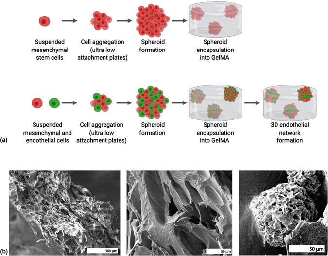
Tumors, trauma, and congenital defects require volume restoration of soft tissues. Tissue engineering provides an alternative source for substituting these defects. Cell encapsulation into hydrogels provides a three-dimensional microenvironment. Spheroids of cells provide close packing and increase cell-to-cell contacts resulting in differentiation. Gelatin is a natural polymer with low immunogenicity and preserved amino acid motifs for cell adhesion and proliferation. In the present study, a soft photo-crosslinked gelatin methacrylate (GelMA) hydrogel with long in vitro lifetime was synthesized. Stem cells (dental pulp derived, DPSC) and endothelial cells (umbilical cord derived, HUVEC) were formed into spheroids to induce prevascular network formation and encapsulated into GelMA (10% weight/volume). Results showed high cell viability, better gel mechanical properties, and longer HUVEC sprouting with spheroids compared to the same combination of cells. Altogether, the photo-crosslinked GelMA hydrogels with DPSC and HUVEC spheroids provided a promising tissue engineering and vascularization strategy in vitro.

Type
Article
Copyright
Copyright © The Author(s), 2020, published on behalf of Materials Research Society by Cambridge University Press

Access options

Get access to the full version of this content by using one of the access options below. (Log in options will check for institutional or personal access. Content may require purchase if you do not have access.)

References

Patrick, C.W.: Tissue engineering strategies for adipose tissue repair. Anat. Rec. 263, 361 (2001).CrossRefGoogle ScholarPubMed
Monfort, A. and Izeta, A.: Strategies for human adipose tissue repair and regeneration. J. Cosmet. Dermatol. Sci. Appl. 02, 93 (2012).Google Scholar
Qi, D., Wu, S., Kuss, M.A., Shi, W., Chung, S., Deegan, P.T., Kamenskiy, A., He, Y., and Duan, B.: Mechanically robust cryogels with injectability and bioprinting supportability for adipose tissue engineering. Acta Biomater. 74, 131 (2018).CrossRefGoogle ScholarPubMed
Huber, B., Borchers, K., Tovar, G.E., and Kluger, P.J.: Methacrylated gelatin and mature adipocytes are promising components for adipose tissue engineering. J. Biomater. Appl. 30, 699 (2016).CrossRefGoogle ScholarPubMed
Kayabolen, A., Keskin, D., Aykan, A., Karslıoglu, Y., Zor, F., and Tezcaner, A.: Native extracellular matrix/fibroin hydrogels for adipose tissue engineering with enhanced vascularization. Biomed. Mater. 12, 035007 (2017).CrossRefGoogle ScholarPubMed
Ibsirlioglu, T., Elçin, A.E., and Elçin, Y.M.: Decellularized biological scaffold and stem cells from autologous human adipose tissue for cartilage tissue engineering. Methods 171, 97 (2020).CrossRefGoogle ScholarPubMed
Luo, L., He, Y., Chang, Q., Xie, G., Zhan, W., Wang, X., Zhou, T., Xing, M., and Lu, F.: Polycaprolactone nanofibrous mesh reduces foreign body reaction and induces adipose flap expansion in tissue engineering chamber. Int. J. Nanomed. 11, 6471 (2016).CrossRefGoogle ScholarPubMed
Sivashanmugam, A., Arun Kumar, R., Vishnu Priya, M., Nair, S.V., and Jayakumar, R.: An overview of injectable polymeric hydrogels for tissue engineering. Eur. Polym. J. 72, 543 (2015).CrossRefGoogle Scholar
Xiao, S., Zhao, T., Wang, J., Wang, C., Du, J., Ying, L., Lin, J., Zhang, C., Hu, W., Wang, L., and Xu, K.: Gelatin methacrylate (GelMA)-based hydrogels for cell transplantation: an effective strategy for tissue engineering. Stem Cell Rev. Rep. 15, 664 (2019).CrossRefGoogle Scholar
Lynn, A.K., Yannas, I.V., and Bonfield, W.: Antigenicity and immunogenicity of collagen. J. Biomed. Mater. Res., Part B 71, 343 (2004).CrossRefGoogle Scholar
Van Den Bulcke, A.I., Bogdanov, B., De Rooze, N., Schacht, E.H., Cornelissen, M., and Berghmans, H.: Structural and rheological properties of methacrylamide modified gelatin hydrogels. Biomacromolecules 1, 31 (2000).CrossRefGoogle ScholarPubMed
Choi, J.R., Yong, K.W., Choi, J.Y., and Cowie, A.C.: Recent advances in photo-crosslinkable hydrogels for biomedical applications. Biotechniques 66, 40 (2019).CrossRefGoogle ScholarPubMed
Eke, G., Mangir, N., Hasirci, N., MacNeil, S., and Hasirci, V.: Development of a UV crosslinked biodegradable hydrogel containing adipose derived stem cells to promote vascularization for skin wounds and tissue engineering. Biomaterials 129, 188 (2017).CrossRefGoogle ScholarPubMed
Tytgat, L., Van Damme, L., Van Hoorick, J., Declercq, H., Thienpont, H., Ottevaere, H., Blondeel, P., Dubruel, P., and Van Vlierberghe, S.: Additive manufacturing of photo-crosslinked gelatin scaffolds for adipose tissue engineering. Acta Biomater. 94, 340 (2019).CrossRefGoogle ScholarPubMed
Tytgat, L., Kollert, M.R., Van Damme, L., Thienpont, H., Ottevaere, H., Duda, G.N., Geissler, S., Dubruel, P., Van Vlierberghe, S., and Qazi, T.H.: Evaluation of 3D printed gelatin-based scaffolds with varying pore size for MSC-based adipose tissue engineering. Macromol. Biosci. 20, 1900364 (2020).CrossRefGoogle ScholarPubMed
Zhao, X., Lang, Q., Yildirimer, L., Lin, Z.Y., Cui, W., Annabi, N., Ng, K.W., Dokmeci, M.R., Ghaemmaghami, A.M., and Khademhosseini, A.: Photocrosslinkable gelatin hydrogel for epidermal tissue engineering. Adv. Healthc. Mater. 5, 108 (2016).CrossRefGoogle ScholarPubMed
Zou, J., Wang, W., Neffe, A.T., Xu, X., Li, Z., Deng, Z., Sun, X., Ma, N., and Lendlein, A.: Adipogenic differentiation of human adipose derived mesenchymal stem cells in 3D architectured gelatin based hydrogels (ArcGel). Clin. Hemorheol. Microcirc. 67, 297 (2017).CrossRefGoogle Scholar
Chen, Y.C., Lin, R.Z., Qi, H., Yang, Y., Bae, H., Melero-Martin, J.M., and Khademhosseini, A.: Functional human vascular network generated in photocrosslinkable gelatin methacrylate hydrogels. Adv. Funct. Mater. 22, 2027 (2012).CrossRefGoogle ScholarPubMed
Lin, R.Z., Chen, Y.C., Moreno-Luna, R., Khademhosseini, A., and Melero-Martin, J.M.: Transdermal regulation of vascular network bioengineering using a photopolymerizable methacrylated gelatin hydrogel. Biomaterials 34, 6785 (2013).CrossRefGoogle ScholarPubMed
Phinney, D.G. and Prockop, D.J.: Concise review: Mesenchymal stem/multipotent stromal cells: The state of transdifferentiation and modes of tissue repair-current views. Stem Cells 25, 2896 (2007).CrossRefGoogle Scholar
Chamberlain, G., Fox, J., Ashton, B., and Middleton, J.: Concise review: Mesenchymal stem cells: Their phenotype, differentiation capacity, immunological features, and potential for homing. Stem Cells 25, 2739 (2007).CrossRefGoogle ScholarPubMed
Harada, H., Kettunen, P., Jung, H.S., Mustonen, T., Wang, Y.A., and Thesleff, I.: Localization of putative stem cells in dental epithelium and their association with Notch and FGF signaling. J. Cell Biol. 147, 105 (1999).CrossRefGoogle ScholarPubMed
Zhang, D., Wei, G., Li, P., Zhou, X., and Zhang, Y.: Urine-derived stem cells: A novel and versatile progenitor source for cell-based therapy and regenerative medicine. Genes Dis. 1, 8 (2014).CrossRefGoogle Scholar
Fuchs, E. and Segre, J.A.: Stem cells: a new lease on life. Cell 100, 143 (2000).CrossRefGoogle Scholar
Wong, C.E., Paratore, C., Dours-Zimmermann, M.T., Rochat, A., Pietri, T., Suter, U., Zimmermann, D.R., Dufour, S., Thiery, J.P., Meijer, D., Beermann, F., Barrandon, Y., and Sommer, L.: Neural crest-derived cells with stem cell features can be traced back to multiple lineages in the adult skin. J. Cell Biol. 175, 1005 (2006).CrossRefGoogle ScholarPubMed
Bianco, P., Riminucci, M., Gronthos, S., and Robey, P.G.: Bone marrow stromal stem cells: Nature, biology, and potential applications. Stem Cells 19, 180 (2001).CrossRefGoogle ScholarPubMed
Charbord, P.. Bone marrow mesenchymal stem cells: historical overview and concepts. Hum. Gene Ther. 21, 1045 (2010).CrossRefGoogle ScholarPubMed
Pittenger, M.F., Mackay, A.M., Beck, S.C., Jaiswal, R.K., Douglas, R., Mosca, J.D., Moorman, M.A., Simonetti, D.W., Craig, S., and Marshak, D.R.: Multilineage potential of adult human mesenchymal stem cells. Science 284, 143 (1999).CrossRefGoogle ScholarPubMed
Zuk, P.A., Zhu, M., Mizuno, H., Huang, J., Futrell, J.W., Katz, A.J., Benhaim, P., Lorenz, H.P., and Hedrick, M.H.: Multilineage cells from human adipose tissue: implications for cell-based therapies. Tissue Eng. 7 (2), 211 (2001).CrossRefGoogle ScholarPubMed
Huang, G.T.J., Gronthos, S., and Shi, S.: Mesenchymal stem cells derived from dental tissues vs. those from other sources: their biology and role in regenerative medicine . J. Dent. Res. 88, 792 (2009).CrossRefGoogle Scholar
Normando, D.: New achievements: from submission to disclosure. Dental Press J. Orthod. 20, 17 (2015).CrossRefGoogle Scholar
Perry, B.C., Zhou, D., Wu, X., Yang, F.C., Byers, M.A., Chu, T.M.G., Hockema, J.J., Woods, E.J., and Goebel, W.S.: Collection, cryopreservation, and characterization of human dental pulp-derived mesenchymal stem cells for banking and clinical use. Tissue Eng., Part C 14, 149 (2008).CrossRefGoogle ScholarPubMed
Gronthos, S., Brahim, J., Li, W., Fisher, L.W., Cherman, N., Boyde, A., DenBesten, P., Robey, P.G., and Shi, S.: Stem cell properties of human dental pulp stem cells. J. Dent. Res. 81, 531 (2002).CrossRefGoogle ScholarPubMed
Kim, D., Kim, J., Hyun, H., Kim, K., and Roh, S.: A nanoscale ridge/groove pattern arrayed surface enhances adipogenic differentiation of human supernumerary tooth-derived dental pulp stem cells in vitro. Arch. Oral Biol. 59, 765 (2014).CrossRefGoogle ScholarPubMed
Bin Lee, Y., Kim, E.M., Byun, H., Kwan Chang, H., Jeong, K., Aman, Z.M., Choi, Y.S., Park, J., and Shin, H.: Engineering spheroids potentiating cell-cell and cell-ECM interactions by self-assembly of stem cell microlayer. Biomaterials 165, 105 (2018).Google Scholar
Moldovan, L., Barnard, A., Gil, C.-H., Lin, Y., Grant, M.B., Yoder, M.C., Prasain, N., and Moldovan, N.I.: iPSC-derived vascular cell spheroids as building blocks for scaffold-free biofabrication. Biotechnol. J. 12, 1700444 (2017).CrossRefGoogle ScholarPubMed
Foty, R.: A simple hanging drop cell culture protocol for generation of 3D spheroids. J. Vis. Exp., 6(51), e2720 (2011).Google Scholar
Liu, T., Chien, C.C., Parkinson, L., and Thierry, B.: Advanced micromachining of concave microwells for long term on-chip culture of multicellular tumor spheroids. ACS Appl. Mater. Interfaces 6, 8090 (2014).CrossRefGoogle ScholarPubMed
Cha, J.M., Park, H., Shin, E.K., Sung, J.H., Kim, O., Jung, W., Bang, O.Y., and Kim, J.: A novel cylindrical microwell featuring inverted-pyramidal opening for efficient cell spheroid formation without cell loss. Biofabrication 9, 035006 (2017).CrossRefGoogle ScholarPubMed
Zuchowska, A., Jastrzebska, E., Chudy, M., Dybko, A., and Brzozka, Z.: 3D lung spheroid cultures for evaluation of photodynamic therapy (PDT) procedures in microfluidic lab-on-a-chip system. Anal. Chim. Acta 990, 110 (2017).Google ScholarPubMed
Moshksayan, K., Kashaninejad, N., Warkiani, M.E., Lock, J.G., Moghadas, H., Firoozabadi, B., Saidi, M.S., and Nguyen, N.T.: Spheroids-on-a-chip: Recent advances and design considerations in microfluidic platforms for spheroid formation and culture. Sens. Actuators, B 263, 151 (2018).CrossRefGoogle Scholar
Ren, T., Steiger, W., Chen, P., Ovsianikov, A., and Demirci, U.: Enhancing cell packing in buckyballs by acoustofluidic activation. Biofabrication 12, 25033 (2020).CrossRefGoogle ScholarPubMed
Laschke, M.W. and Menger, M.D.: Life is 3D: boosting spheroid function for tissue engineering. Trends Biotechnol. 35, 133 (2017).CrossRefGoogle Scholar
Anton, D., Burckel, H., Josset, E., and Noel, G.: Three-dimensional cell culture: a breakthrough in vivo. Int. J. Mol. Sci. 16, 5517 (2015).CrossRefGoogle Scholar
Korff, T. and Augustin, H.G.: Integration of endothelial cells in multicellular spheroids prevents apoptosis and induces differentiation. J. Cell Biol. 143, 1341 (1998).CrossRefGoogle ScholarPubMed
Korff, T., H. G. Augustin: Tensional forces in fibrillar extracellular matrices control directional capillary sprouting. J. Cell Sci. 112(19), 3249 (1999).Google ScholarPubMed
Heiss, M., Hellström, M., Kalén, M., May, T., Weber, H., Hecker, M., Augustin, H.G., and Korff, T.: Endothelial cell spheroids as a versatile tool to study angiogenesis in vitro. FASEB J. 29, 3076 (2015).CrossRefGoogle ScholarPubMed
Rouwkema, J., De Boer, J., and Van Blitterswijk, C.A.: Endothelial cells assemble into a 3-dimensional prevascular network in a bone tissue engineering construct. Tissue Eng. 12, 2685 (2006).CrossRefGoogle Scholar
Liew, A.W.L. and Zhang, Y.: In vitro pre-vascularization strategies for tissue engineered constructs-bioprinting and others. Int. J. Bioprint. 3, 3 (2017).CrossRefGoogle ScholarPubMed
Heo, D.N., Hospodiuk, M., and Ozbolat, I.T.: Synergistic interplay between human MSCs and HUVECs in 3D spheroids laden in collagen/fibrin hydrogels for bone tissue engineering. Acta Biomater. 95, 348 (2019).CrossRefGoogle ScholarPubMed
Alajati, A., Laib, A.M., Weber, H., Boos, A.M., Bartol, A., Ikenberg, K., Korff, T., Zentgraf, H., Obodozie, C., Graeser, R., Christian, S., Finkenzeller, G., Stark, G.B., Héroult, M., and G, H.: Augustin: Spheroid-based engineering of a human vasculature in mice. Nat. Methods 5, 439 (2008).CrossRefGoogle ScholarPubMed
Gutzweiler, L., Kartmann, S., Troendle, K., Benning, L., Finkenzeller, G., Zengerle, R., Koltay, P., Stark, G.B., and Zimmermann, S.: Large scale production and controlled deposition of single HUVEC spheroids for bioprinting applications. Biofabrication 9, 025027 (2017).CrossRefGoogle ScholarPubMed
Inamori, M., Mizumoto, H., and Kajiwara, T.: An approach for formation of vascularized liver tissue by endothelial cell-covered hepatocyte spheroid integration. Tissue Eng., Part A 15, 2029 (2009).CrossRefGoogle ScholarPubMed
Saleh, F., Whyte, M., and Genever, P.: Effects of endothelial cells on human mesenchymal stem cell activity in a three-dimensional in vitro model. Eur. Cells Mater. 22, 242 (2011).CrossRefGoogle Scholar
Dissanayaka, W.L., Zhu, L., Hargreaves, K.M., Jin, L., and Zhang, C.: In vitro analysis of scaffold-free prevascularized microtissue spheroids containing human dental pulp cells and endothelial cells. J. Endod. 41, 663 (2015).CrossRefGoogle ScholarPubMed
Zhu, M., Wang, Y., Ferracci, G., Zheng, J., Cho, N.J., and Lee, B.H.: Gelatin methacryloyl and its hydrogels with an exceptional degree of controllability and batch-to-batch consistency. Sci. Rep. 9, 1 (2019).Google ScholarPubMed
Shirahama, H., Lee, B.H., Tan, L.P., and Cho, N.J.: Precise tuning of facile one-pot gelatin methacryloyl (GelMA) synthesis. Sci. Rep. 6, 1 (2016).CrossRefGoogle ScholarPubMed
Mironi-Harpaz, I., Wang, D.Y., Venkatraman, S., and Seliktar, D.: Photopolymerization of cell-encapsulating hydrogels: Crosslinking efficiency versus cytotoxicity. Acta Biomater. 8, 1838 (2012).CrossRefGoogle ScholarPubMed
Godar, D.E., Gurunathan, C., and Ilev, I.: 3D bioprinting with UVA1 radiation and photoinitiator irgacure 2959: Can the ASTM standard L929 cells predict human stem cell cytotoxicity? Photochem. Photobiol. 95, 581 (2019).CrossRefGoogle ScholarPubMed
Li, X., Chen, S., Li, J., Wang, X., Zhang, J., Kawazoe, N., and Chen, G.: 3D culture of chondrocytes in gelatin hydrogels with different stiffness. Polymers 8, 269 (2016).CrossRefGoogle ScholarPubMed
Hoch, E., Hirth, T., Tovar, G.E.M., and Borchers, K.: Chemical tailoring of gelatin to adjust its chemical and physical properties for functional bioprinting. J. Mater. Chem. B 1, 5675 (2013).CrossRefGoogle ScholarPubMed
Zhou, L., Tan, G., Tan, Y., Wang, H., Liao, J., and Ning, C.: Biomimetic mineralization of anionic gelatin hydrogels: Effect of degree of methacrylation. RSC Adv. 4, 21997 (2014).CrossRefGoogle Scholar
Occhetta, P., Visone, R., Russo, L., Cipolla, L., Moretti, M., and Rasponi, M.: VA-086 methacrylate gelatine photopolymerizable hydrogels: A parametric study for highly biocompatible 3D cell embedding. J. Biomed. Mater. Res., Part A 103, 2109 (2015).CrossRefGoogle ScholarPubMed
Guimarães, C.F., Gasperini, L., Marques, A.P., and Reis, R.L.: The stiffness of living tissues and its implications for tissue engineering. Nat. Rev. Mater. 5, 351 (2020).CrossRefGoogle Scholar
Hasturk, O., Ermis, M., Demirci, U., Hasirci, N., and Hasirci, V.: Square prism micropillars on poly(methyl methacrylate) surfaces modulate the morphology and differentiation of human dental pulp mesenchymal stem cells. Colloids Surf., B 178, 44 (2019).CrossRefGoogle ScholarPubMed
Lv, F.J., Tuan, R.S., Cheung, K.M.C., and Leung, V.Y.L.: Concise review: the surface markers and identity of human mesenchymal stem cells. Stem Cells 32, 1408 (2014).CrossRefGoogle ScholarPubMed
Rodas-Junco, B.A., Canul-Chan, M., Rojas-Herrera, R.A., de-la-Peña, C., and Nic-Can, G.I.: Stem cells from dental pulp: what epigenetics can do with your tooth. Front. Physiol. 8, 999 (2017).CrossRefGoogle Scholar
Iohara, K., Zheng, L., Ito, M., Tomokiyo, A., Matsushita, K., and Nakashima, M.: Side population cells isolated from porcine dental pulp tissue with self-renewal and multipotency for dentinogenesis, chondrogenesis, adipogenesis, and neurogenesis. Stem Cells 24, 2493 (2006).CrossRefGoogle ScholarPubMed
Jo, Y.Y., Lee, H.J., Kook, S.Y., Choung, H.W., Park, J.Y., Chung, J.H., Choung, Y.H., Kim, E.S., Yang, H.C., and Choung, P.H.: Isolation and characterization of postnatal stem cells from human dental tissues. Tissue Eng. 13, 767 (2007).CrossRefGoogle ScholarPubMed
Pettersson, L.F., Kingham, P.J., Wiberg, M., and Kelk, P.: In vitro osteogenic differentiation of human mesenchymal stem cells from jawbone compared with dental tissue. Tissue Eng. Regen. Med. 14, 763 (2017).CrossRefGoogle ScholarPubMed
Struys, T., Moreels, M., Martens, W., Donders, R., Wolfs, E., and Lambrichts, I.: Ultrastructural and immunocytochemical analysis of multilineage differentiated human dental pulp- and umbilical cord-derived mesenchymal stem cells. Cells Tissues Organs 193, 366 (2011).CrossRefGoogle ScholarPubMed
Colle, J., Blondeel, P., De Bruyne, A., Bochar, S., Tytgat, L., Vercruysse, C., Van Vlierberghe, S., Dubruel, P., and Declercq, H.: Bioprinting predifferentiated adipose-derived mesenchymal stem cell spheroids with methacrylated gelatin ink for adipose tissue engineering. J. Mater. Sci. Mater. Med. 31, 1 (2020).CrossRefGoogle ScholarPubMed
Bahcecioglu, G., Hasirci, N., Bilgen, B., and Hasirci, V.: Hydrogels of agarose, and methacrylated gelatin and hyaluronic acid are more supportive for in vitro meniscus regeneration than three dimensional printed polycaprolactone scaffolds. Int. J. Biol. Macromol. 122, 1152 (2019).CrossRefGoogle ScholarPubMed
Kilic Bektas, C. and Hasirci, V.: Cell loaded 3D bioprinted GelMA hydrogels for corneal stroma engineering. Biomater. Sci. 8, 438 (2020).CrossRefGoogle Scholar
Nichol, J.W., Koshy, S.T., Bae, H., Hwang, C.M., Yamanlar, S., and Khademhosseini, A.: Cell-laden microengineered gelatin methacrylate hydrogels. Biomaterials 31, 5536 (2010).CrossRefGoogle ScholarPubMed
Al Rifai, N., Hasan, A., Kobeissy, F., Gazalah, H., and Charara, J.: Culture of PC12 neuronal cells in GelMA hydrogel for brain tissue engineering. In 2015 International Conference on Advances in Biomedical Engineering (ICABME) (IEEE Press, NY, USA, 2015), pp. 254–257.CrossRefGoogle Scholar
Noshadi, I., Hong, S., Sullivan, K.E., Shirzaei Sani, E., Portillo-Lara, R., Tamayol, A., Shin, S.R., Gao, A.E., Stoppel, W.L., Black, L.D., Khademhosseini, A., and Annabi, N.: In vitro and in vivo analysis of visible light crosslinkable gelatin methacryloyl (GelMA) hydrogels. Biomater. Sci. 5, 2093 (2017).CrossRefGoogle ScholarPubMed
Girton, T.S., Oegema, T.R., and Tranquillo, R.T.: Exploiting glycation to stiffen and strengthen tissue equivalents for tissue engineering. J. Biomed. Mater. Res. 46, 87 (1999).3.0.CO;2-K>CrossRefGoogle ScholarPubMed
Girton, T.S., Oegema, T.R., Grassl, E.D., Isenberg, B.C., and Tranquillo, R.T.: Mechanisms of stiffening and strengthening in media-equivalents fabricated using glycation. J. Biomech. Eng. 122, 216 (2000).CrossRefGoogle ScholarPubMed
Jabłońska-Trypuć, A., Matejczyk, M., and Rosochacki, S.: Matrix metalloproteinases (MMPs), the main extracellular matrix (ECM) enzymes in collagen degradation, as a target for anticancer drugs. J. Enzyme Inhib. Med. Chem. 31, 177 (2016).CrossRefGoogle ScholarPubMed
Ries, C., Egea, V., Karow, M., Kolb, H., Jochum, M., and Neth, P.: MMP-2, MT1-MMP, and TIMP-2 are essential for the invasive capacity of human mesenchymal stem cells: Differential regulation by inflammatory cytokines. Blood 109, 4055 (2007).CrossRefGoogle ScholarPubMed
Ho, I.A.W., Chan, K.Y.W., Ng, W.H., Guo, C.M., Hui, K.M., Cheang, P., and Lam, P.Y.P.: Matrix metalloproteinase 1 is necessary for the migration of human bone marrow-derived mesenchymal stem cells toward human glioma. Stem Cells 27, 1366 (2009).CrossRefGoogle ScholarPubMed
Anderson, S.B., Lin, C.C., Kuntzler, D.V., and Anseth, K.S.: The performance of human mesenchymal stem cells encapsulated in cell-degradable polymer-peptide hydrogels. Biomaterials 32, 3564 (2011).CrossRefGoogle ScholarPubMed
Correia, D.M., Padrão, J., Rodrigues, L.R., Dourado, F., Lanceros-Méndez, S., and Sencadas, V.: Thermal and hydrolytic degradation of electrospun fish gelatin membranes. Polym. Test 32, 995 (2013).CrossRefGoogle Scholar
Ahmad, T., Byun, H., Lee, J., Madhurakat Perikamana, S.K., Shin, Y.M., Kim, E.M., and Shin, H.: Stem cell spheroids incorporating fibers coated with adenosine and polydopamine as a modular building blocks for bone tissue engineering. Biomaterials 230, 119652 (2020).CrossRefGoogle ScholarPubMed
Vorwald, C.E., Ho, S.S., Whitehead, J., and Leach, J.K.: High-throughput formation of mesenchymal stem cell spheroids and entrapment in alginate hydrogels in Biomaterials for Tissue Engineering, Humana Press, NY, USA, 2018 139149.Google Scholar
Siltanen, C., Yaghoobi, M., Haque, A., You, J., Lowen, J., Soleimani, M., and Revzin, A.: Microfluidic fabrication of bioactive microgels for rapid formation and enhanced differentiation of stem cell spheroids. Acta Biomater. 34, 125 (2016).CrossRefGoogle ScholarPubMed
Murphy, K.C., Whitehead, J., Zhou, D., Ho, S.S., and Leach, J.K.: Engineering fibrin hydrogels to promote the wound healing potential of mesenchymal stem cell spheroids. Acta Biomater. 64, 176 (2017).CrossRefGoogle ScholarPubMed
Lee, B., Lum, N., Seow, L., Lim, P., and Tan, L.: Synthesis and characterization of types A and B gelatin methacryloyl for bioink applications. Materials 9, 797 (2016).CrossRefGoogle Scholar
Karbanová, J., Soukup, T., Suchánek, J., Pytlík, R., Corbeil, D., and Mokrý, J.: Characterization of dental pulp stem cells from impacted third molars cultured in low serum-containing medium. Cells Tissues Organs 193, 344 (2011).CrossRefGoogle ScholarPubMed
Alge, D.L., Zhou, D., Adams, L.L., Wyss, B.K., Shadday, M.D., Woods, E.J., Chu, T.M.G., and Goebel, W.S.: Donor-matched comparison of dental pulp stem cells and bone marrow-derived mesenchymal stem cells in a rat model. J. Tissue Eng. Regen. Med. 4, 73 (2010).Google ScholarPubMed
Popko, J., Fernandes, A., Brites, D., and Lanier, L.M.: Automated analysis of NeuronJ tracing data. Cytometry, Part A 75, 371 (2009).CrossRefGoogle ScholarPubMed