Hostname: page-component-78c5997874-lj6df Total loading time: 0 Render date: 2024-11-10T21:23:25.679Z Has data issue: false hasContentIssue false

Comment on ‘Imaging tumor hypoxia by magnetic resonance methods’

Published online by Cambridge University Press:  23 November 2011

Syed F. Akber
Affiliation:
Radiological Physicist, Cleveland, Ohio, USA

Abstract

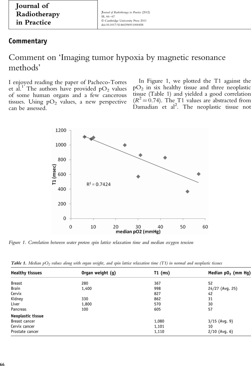
Image of the first page of this content. For PDF version, please use the ‘Save PDF’ preceeding this image.'
Type
Commentary
Copyright
Copyright © Cambridge University Press 2012

Access options

Get access to the full version of this content by using one of the access options below. (Log in options will check for institutional or personal access. Content may require purchase if you do not have access.)

References

Pacheco-Torres, P, Lopez-Larrubia, P, Ballesteros, P, Cerdan, S.Imaging tumor hypoxia by magnetic resonance methods. NMR in Biomedicine 2011; 24:116.CrossRefGoogle ScholarPubMed
Damadian, R, Zaner, K, Hor, D, and DiMaio, T.Human tumors detected by nuclear magnetic resonance. PNAS 1974; 71:14711473.CrossRefGoogle ScholarPubMed
Akber, SF.Correlation between oxygen tension and spin lattice relaxation rate in tumors. Eur J Radiology 1989; 9:5659.Google ScholarPubMed
O’Connor, JPB, Naish, JH, Parker, GJM et al. Preliminary study of oxygen Enhanced longitudinal relaxation in MRI: A potential novel biomarker of oxygen changes in solid tumors. Int J Rad Oncol Biol Phys 2009; 75:12091215.CrossRefGoogle ScholarPubMed
Akber, SF.Organ weight: a new tissue parameter to assess water proton spin lattice relaxation time. Med Hypotheses 1997; 48:101105.Google ScholarPubMed