Coastal sambaquis are shell-mound sites built by fisher-gatherer groups that occupied the Brazilian coast between 8000 and 1000 years BP, although most sambaquis date from 4000 to 2000 years BP. These sites are often found clustered and distributed across southern and southeastern Brazil, as well as part of northeastern Brazil (DeBlasis et al. Reference DeBlasis, Gaspar and Kneip2021; Gaspar et al. Reference Gaspar, DeBlasis, Fish, Fish, Silverman and Isbell2008). After about 2000 years BP, the number of new sambaquis declined until they totally disappeared by 1000 years BP (Gaspar et al. Reference Gaspar, DeBlasis, Fish, Fish, Silverman and Isbell2008). The processes leading to their disappearance are not yet fully understood, but evidence indicates that a myriad of factors are involved, including major sociocultural changes in sambaqui societies, the arrival of ceramist groups from the interior of the country, and environmental changes along the coast occurring between 2000 and 1000 years BP (Angulo et al. Reference Angulo, Lessa and de Souza2006; Barbosa-Guimarães Reference Barbosa-Guimarães2011; DeBlasis et al. Reference DeBlasis, Gaspar and Kneip2021; Gaspar et al. Reference Gaspar, DeBlasis, Fish, Fish, Silverman and Isbell2008; Toso et al. Reference Toso, Hallingstad, McGrath, Fossile, Conlan, Ferreira and Bandeira2021).
In addition to coastal sambaquis, inland shell mounds were built near rivers and are among the oldest sambaquis, reaching back to 10,500 years BP (Figuti and Plens Reference Figuti, Plens, Roksandic, de Souza, Eggers, Burchell and Klokler2014); these sites are known as riverine sambaquis. Most are found in the Ribeira Valley, in São Paulo state, and in the Amazon region (Figuti and Plens Reference Figuti, Plens, Roksandic, de Souza, Eggers, Burchell and Klokler2014; Silveira and Schaan Reference Silveira and Schaan2005).
Sambaquis are usually located near bodies of water such as lagoons and bays, areas that generally yield high biological productivity (Gaspar et al. Reference Gaspar, DeBlasis, Fish, Fish, Silverman and Isbell2008, Reference Gaspar, Klokler, DeBlasis, Roksandic, de Souza, Eggers, Burchell and Klokler2014). In addition to their main matrix components (shellfish, fishbone, sand, and soil), many other cultural elements have been found in sambaquis: stone and bone artifacts used to collect and process food and to perform other daily activities; faunal remains associated with food, tools, and adornments; and human burials (Gaspar et al. Reference Gaspar, DeBlasis, Fish, Fish, Silverman and Isbell2008; Rodrigues-Carvalho Reference Rodrigues-Carvalho2004). Archaeologists consider those human burials to be an important feature of sambaquis because funerary practices played a fundamental role in these groups, influencing the construction of shell mounds, their social lives, and daily activities (Gaspar et al. Reference Gaspar, Klokler, DeBlasis, Roksandic, de Souza, Eggers, Burchell and Klokler2014).
Archaeological studies of sambaqui builders suggest that they had a close connection with the aquatic environment based not only on the location in which their remains are found but also because of these factors: their material culture that is highly associated with fishing activities, the predominance of aquatic species in the faunal assemblage found in these sites, and the occupational stress markers on skeleton remains that are associated with water-related activities. The aquatic environment shaped their daily activities, their food, and their symbolic universe.
There is also evidence that these populations lived in relatively egalitarian heterarchical societies, with a low incidence of violence (DeBlasis et al. Reference DeBlasis, Kneip, Sheel-Ybert, Giannini and Gaspar2007, Reference DeBlasis, Gaspar and Kneip2021; Kneip et al. Reference Kneip, Farias and DeBlasis2018). Additionally, recent research suggests that they had a mixed economy in which horticulture played an important role in daily life alongside fisher-gathering activities. There is compelling evidence that plants were used for food, medicine, ritualistic and religious activities, and the creation of tools (Scheel-Ybert and Boyadjian Reference Scheel-Ybert and Boyadjian2020).
Many researchers have addressed the customs, lifestyle, and health of the sambaqui builders by analyzing their human remains. Early studies of the oral health of these groups suggest that they had homogeneous health conditions and dietary habits; usually, they had high protein intake and low carbohydrate consumption, which led to the identification of shellfish as their main food source (Gaspar Reference Gaspar1996; Souza et al. Reference Souza, Wesolowski, Rodrigues-Carvalho and Oosterbeek2009). However, this perspective changed over the last 30 years as new bioarchaeological and archaeological approaches were employed. For instance, zooarchaeological and isotopic studies have shown that the consumption of fish was predominant and that shellfish were eaten in minor proportions (Bandeira and Fossile Reference Bandeira, Fossile and Zocche2014; Bastos et al. Reference Bastos, Lessa, Rodrigues-Carvalho, Tykot and Santos2014; Colonese et al. Reference Colonese, Collins, Lucquin, Eustace, Hancock, Ponzoni and Mora2014; DeMasi Reference DeMasi2001, Reference DeMasi2009; Ferreira Reference Ferreira, da Rocha Bandeira, Bartz, Fossile and Mayaroka2019; Figuti Reference Figuti1993; Klokler Reference Klokler2008; Pavei Reference Pavei2018). Recent oral health studies point out that these populations had more diversified dietary practices than previously thought (Neves and Wesolowski Reference Neves, Wesolowski, Steckel and Rose2002; Pezo-Lanfranco et al. Reference Pezo-Lanfranco, Eggers, Petronilho, Toso, Bandeira, Von Tersch, dos Santos, Costa, Meyer and Colonese2018; Souza et al. Reference Souza, Wesolowski, Rodrigues-Carvalho and Oosterbeek2009; Wesolowski Reference Wesolowski2007; Wesolowski et al. Reference Wesolowski, de Souza, Reinhard and Ceccantini2010). Current debates regarding Brazilian sambaquis center on the continuity and differences in site construction, funerary and alimentary practices, diet, and subsistence activities (Berredo et al. Reference Berredo, Gaspar, Ramos and Bianchini2020; Gaspar et al. Reference Gaspar, Bianchini, Berredo and Lopes2019, Reference Gaspar, DeBlasis, Fish, Fish, Silverman and Isbell2008, Reference Gaspar, Klokler, DeBlasis, Roksandic, de Souza, Eggers, Burchell and Klokler2014; Mendonça de Souza Reference Mendonça de Souza2018; Scheel-Ybert et al. Reference Scheel-Ybert, Rodrigues-Carvalho, DeBlasis, Gaspar and Klokler2020).
Recent oral health studies on sambaqui groups focus on sites in southern Brazil, and there are few recent publications on oral health in sites found in other regions, such as in Rio de Janeiro state. Oral health analysis has the potential to yield information on sociocultural aspects and continuity and differences between sambaqui societies. This study investigates, using oral health analysis, the differences and similarities in alimentary practices between individuals buried in two sambaquis from the Saquarema city in Rio de Janeiro state.
The Sambaquis from Saquarema
The Saquarema municipality, located in Rio de Janeiro state, has an area of 352,720 km2 and is bounded by the cities of Rio Bonito and Araruama to the north, Araruama to the east, Maricá and Tanguá to the west, and the Atlantic Ocean to the south (Instituto Brasileiro de Geografia e Estatística 2018; Silveira Reference Silveira2001). The area is topographically diverse, encompassing a mountain range, fluvial and marine plains, beach crests, dune remnants, and a complex of four lagoons spread across 215 km2 named Lagoa de Saquarema. Environmentally, the Atlantic Forest biome dominates, comprising mangrove, restinga, and rainforest vegetation and fauna (Silveira Reference Silveira2001).
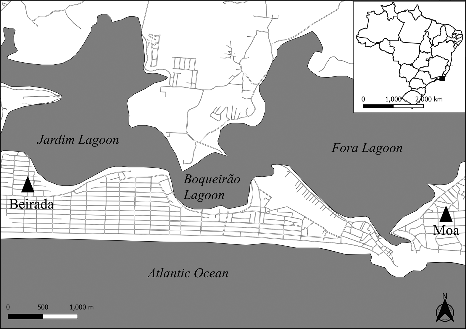
A total of 19 sambaqui sites were found in Saquarema, most of them located in a narrow area between Lagoa de Saquarema and the Atlantic Ocean. According to radiocarbon dates, sambaqui builders occupied the region for approximately 5,000 years from 6631 to 1542 years BP (Guida Reference Guida2019:32). Human skeletal remains were excavated in five of these 19 sites—Beirada, Moa, Saquarema, Pontinha, and Manitiba I (Kneip and Machado Reference Kneip and Machado1993; Kneip et al. Reference Kneip, Pallestrini, Crancio and Machado1991, Reference Kneip, Araujo and Fonseca1995; Machado Reference Machado2001; Silveira, Reference Silveira2001)—totaling 123 individuals; 75% (93/123) of these skeletal remains were excavated from Sambaquis da Beirada and Moa alone. This study analyzes human remains from those two sambaqui sites (Figure 1), both excavated between the decades of the 1980s and 1990s by researchers from the Museu Nacional in Rio de Janeiro, where most of the archaeological materials recovered from those excavations are stored.

Figure 1. Map of Saquarema with the location of Sambaquis do Moa and Beirada archaeological sites. Map created using QGIS software, version 3.2.
Sambaqui da Beirada is located on the southern margin of the Jardim lagoon. The total site area encompassed 2,320 m2, of which 160 m2 were excavated during the 1990s (Kneip and Machado Reference Kneip and Machado1993). This site dates from 5437 to 3440 years cal BP and has four archaeological layers: layer I dates from 4485 to 3440 years cal BP, layer II from 4907 to 3890 years cal BP, layer III from 5196 to 4081 years cal BP, and layer IV from 5437 to 4401 years cal BP (Guida Reference Guida2019:32).
Bone, shell, and stone artifacts were present in all layers of Beirada, with a predominance of the bone and shell. The most abundant artifacts found at the site are needles, fishhooks, whistles, bone points, adornments, and polished stone tools, such as hand axes and mortars (Kneip et al. Reference Kneip, Crancio and Francisco1988, Reference Kneip, Crancio, Pallestrini, Mello, Corrêa, Magalhães and Vogel1994).
The distribution of species of mollusks—used to build this sambaqui—between layers is relatively homogeneous in layers II–IV, with Ostrea sp. and Lucina pectinate being dominant. A shift occurred in layer I, in which Anomalocardia brasiliana became the dominant species (Barbosa-Guimarães Reference Barbosa-Guimarães2011; Kneip et al. Reference Kneip, Pallestrini, Crancio and Machado1991). According to Scheel-Ybert (Reference Scheel-Ybert2000), plant vestiges were found in all layers of this site, mostly in the form of carbonized fruits and charcoal. A total of 39 taxa were identified, most of which were representatives of restinga vegetation.
During the excavation, 29 burials associated with 32 recovered individuals were identified. Twelve individuals were found in layer I, 10 in layer II, 8 in layer III, and 2 in layer IV. When present, grave goods mostly consisted of red sediment and ferruginous concretion over the bodies and, in some cases, tools and adornments made of fishbones (Kneip and Machado Reference Kneip and Machado1993).
Sambaqui do Moa is seated on sandy soil between the sea and Fora lagoon (Kneip and Machado Reference Kneip and Machado1993; Silveira Reference Silveira2001). According to these authors, this site had an area of 2,800 m2, in which a total of 169 m2 was excavated in two campaigns: 49 m2 in 1988 and 120 m2 in 1998. Moa dates from 4770 to 3199 years cal BP and has two layers of excavation: layer I from 4246 to 3199 years cal BP and layer II from 4770 to 3622 years cal BP (Guida Reference Guida2019:32).
The artifact assemblage of Moa is similar to that at Beirada, although a higher amount of chipped stone artifacts was found in Moa (Kneip et al. Reference Kneip, Crancio and Francisco1988, Reference Kneip, Crancio, Pallestrini, Mello, Corrêa, Magalhães and Vogel1994). Pottery fragments associated with a ceramist group known as the Una tradition were found in layer I. However, the presence of these fragments does not indicate contact between this sambaqui and an inland ceramist group because they are from a period more recent than the burials when the sambaqui people were no longer occupying this site (Barbosa-Guimarães Reference Barbosa-Guimarães2011; Kneip et al. Reference Kneip, Crancio, Pallestrini, Mello, Corrêa, Magalhães and Vogel1994).
In contrast to Beirada, shellfish used in site construction in Moa were predominantly Lucina pectinata, followed by Stramonita haemastoma and Perna perna (Barbosa-Guimarães Reference Barbosa-Guimarães2011; Kneip et al. Reference Kneip, Crancio, Pallestrini, Mello, Corrêa, Magalhães and Vogel1994, Reference Kneip, Pallestrini, Crancio and Machado1991). Although plant remains were found at the site, no analyses were performed to identify them (Kneip et al. Reference Kneip, Pallestrini, Crancio and Machado1991). Archaeobotanical studies have only been conducted in two sambaquis in Saquarema: Beirada, as mentioned, and Pontinha (Scheel-Ybert Reference Scheel-Ybert2000).
In Moa, researchers identified 51 burials and recovered 61 individuals from the two campaigns, but information regarding the distribution of burials in the site is available only for the 1988 campaign: 13 were on layer I and 20 on layer II. Common grave goods were red sediment and ferruginous concretion over the bodies, as well as polished stone artifacts. Some burials also contained teeth from small mammals and shell-made tools (Kneip and Machado Reference Kneip and Machado1993).
Oral Health Studies of the Beirada and Moa Populations
Research regarding the oral health and dietary practices of populations from Moa and Beirada is scarce. There is only one oral health study on individuals from these sites, which was published in 1994 by Lilia Machado and Lina Kneip. They analyzed human remains recovered from three sambaquis (Beirada, Moa, and Pontinha) during an excavation campaign in the Saquarema region coordinated by Lina Kneip.
Their results showed that populations from those sites had oral health patterns similar to most sambaqui populations countrywide: intense dental wear, a low incidence of caries, and a high incidence of dental calculus and periodontal lesions. However, given that this study was conducted during the 1990s, its results were interpreted according to the prevailing paradigm that shellfish was the main protein source, a hypothesis since proven to be incorrect. In addition, current research on the oral health of sambaqui builders uses methodologies other than those employed by Machado and Kneip (Reference Machado and Kneip1994), hampering comparison of their results. More importantly, raw data from the 1990s research are no longer available.
This study makes a unique contribution by revisiting the available human remains from Beirada and Moa and employing methodologies that are used in current research on sambaqui builders to assess the oral health of these populations. Thus, it enables the comparison of results of oral health studies in these two sites, as well as with other sambaqui groups from south and southeastern Brazil.
Materials and Methods
We selected for study 35 individuals with a mandible, maxilla, or both that were either fragmented or whole but with examinable teeth. Nineteen were excavated from Beirada and 16 from Moa (Supplemental Table 1). A total of 852 alveoli and 704 teeth were analyzed.
Age-at-Death and Sex Estimation
We followed the methodological standard procedures presented by Buikstra and Ubelaker (Reference Buikstra and Ubelaker1994) to estimate the sex and age-at-death of each individual (Supplemental Table 1): these techniques are used in current bioarchaeological research worldwide, including studies on sambaqui populations. Age-at-death of non-adults was estimated through dental eruption and epiphyseal fusion of the postcranial skeleton. We estimated the age-at-death of adults using pubic symphysis morphology (Brooks and Suchey Reference Brooks and Suchey1990) and cranial suture closure. Sex was estimated only for adults, according to morphological features of the cranium, the subpubic region (Phenice Reference Phenice1969), the greater sciatic notch, and the preauricular sulcus.
Because of poor bone integrity and missing age markers, we adopted broad age categories in our analysis and interpretation: child, up to 12 years old; adolescent, 12–20 years old; and adult, over 20 years old. Individuals for whom age-at-death or sex could not be estimated with a high degree of certainty were registered as “non-observable.”
Oral Health
To assess the oral health of these groups, we investigated dental wear severity, prevalence and frequency of caries, dental calculus, periapical cavities and antemortem teeth loss (AMTL), and prevalence of linear enamel hypoplasia (LEH). Loose teeth that could not be associated with any individual were excluded from oral health analysis.
Dental wear can be defined as a deterioration of the outer surface of the teeth caused by physiological processes such as attrition, abrasion, and corrosion. It is commonly related both to masticatory processes during eating and non-masticatory use, such as parafunctional and idiosyncratic behavior (Burnett Reference Burnett, Irish and Scott2015; Molnar et al. Reference Molnar, Barrett, Brian, Brace, Brose, Dewey and Frisch1972). We used the Smith (Reference Smith1984) scored method to analyze dental wear severity and ranked it in three categories: light wear, scores 1–3; moderate wear, scores 4 and 5; and severe wear, scores 6–8, as used by Wesolowski (Reference Wesolowski2007) in studies on sambaquis in southern Brazil.
Caries is an infectious disease caused by enamel demineralization due to cariogenic microbiota, with the development of carious lesions being significantly influenced by carbohydrate-rich diets and host susceptibility (Keyes and Jordan Reference Keyes, Jordan and Sognnaes1963; Van Houte Reference Van Houte1994). We identified caries as any cavitary lesion with a diameter of 1 mm or more that shows clear signs of demineralization characterized by irregular borders and bottoms (Lukacs Reference Lukacs, Iscan and Kennedy1989, Reference Lukacs1992; Wesolowski Reference Wesolowski2007). The presence of caries was assessed through macroscopic observation on occlusal, interdental, and root surfaces and on the cement-enamel junction of teeth, with the aid of a 10× magnifying glass (Wesolowski Reference Wesolowski2007). Despite not being generally recommended as a means to assess carious lesions (Hillson Reference Hillson2001), we used a dental probe on rare occasions where the presence of carious lesions was not identifiable using a magnifying glass.
Dental calculus is the mineralization of dental plaque (Lieverse Reference Lieverse1999), and its development is influenced by a variety of factors, such as salivary flow, mineral content in water and food, mastication, and genetics (Radini et al. Reference Radini, Nikita, Buckley, Copeland and Hardy2017). We identified dental calculus through macroscopic observation to find mineralized fragments of bacterial plaque.
Linear enamel hypoplasia (LEH) is formed when there is an interruption in the enamel matrix secretion during tooth development; it manifests as a horizontal linear depression around the dental enamel (Goodman and Rose Reference Goodman and Rose1990; Hillson Reference Hillson1996). This defect is usually caused by physiological stress events occurring during infancy, when dental crowns are still forming (Kinaston et al. Reference Kinaston, Roberts, Buckley and Oxenham2016). The presence of LEH was verified macroscopically according to the FIELD method (Brickley and McKinley Reference Brickley and McKinley2004; Buikstra and Ubelaker Reference Buikstra and Ubelaker1994), which uses a 10× magnifying glass and a LED flashlight positioned perpendicularly to teeth occlusal surfaces. Following the guidelines in Buikstra and Ubelaker (Reference Buikstra and Ubelaker1994), only anterior teeth were subjected to this analysis. Cases in which LEH occurred only in one tooth were excluded from further study, because single-tooth LEH can be caused by localized trauma (Hillson Reference Hillson1996).
Periapical lesions are defined as osseous cavities near teeth roots that are caused by infections generated at the level of the dental pulp due to its exposure to bacteria through caries, attrition, or trauma (Dias and Tayles Reference Dias and Tayles1997; Waldron Reference Waldron and Waldron2008). The assessment of periapical cavities was carried out through macroscopic observation of cavities that had signs of osseous reabsorption and were located near teeth roots.
Antemortem tooth loss (AMTL) is caused by a multitude of factors, including caries, attrition, trauma, and periodontal diseases; it is identified by a dental socket with an indication of some degree of bone tissue remodeling (Waldron Reference Waldron and Waldron2008).
To verify whether the differences in the frequencies of oral physiopathological processes found by the intra- and interpopulation comparisons are significant, we carried out statistical analyses using the chi-square test; when this test could not be used because of an insufficient sample size, we applied a Fisher exact test instead. All tests were performed using IBM SPSS statistical software.
Because we did not have archaeological data on burial distribution in Beirada and Moa, we could not determine an adequate correlation between the analyzed individuals and the site layers in which they were found. Consequently, this study approaches both sites as whole structures; that is, the interpretation of the findings does not take into consideration the burial layer. Differences in the period of occupation were considered only when comparing results between the sites.
Results
The poor preservation of most analyzed individuals decreased the accuracy of sex and age estimations and, in some cases, prevented any effective estimation. Most osteological sexual markers were absent in individuals at both sites. Sexual markers in pelvic bones were present in only two individuals, and markers in the skull were absent in five individuals. Hence, sex estimation was not possible in 40% (14/35) of individuals. Our data also indicated a disproportion in sex representation: 76.20% of individuals were classified as females. Considering the disproportion between males and females, any interpretation of results concerning sex differences was made carefully.
The lack of age markers also influenced our analysis, especially in adults. Age intervals of children and adolescents could be inferred through dental eruption and epiphyseal fusion (when postcranial bones were available for the latter). However, estimation of age intervals in all adults, with the exception of one adult in Beirada, was not possible because of the absence of most ossa coxae and the obliteration or absence of age markers on many crania. Therefore, we could not distinguish between young and mature adults. Considering these issues and the underrepresentation of subadults in the sample, a comparison of results between age categories had limited utility.
Oral Analysis
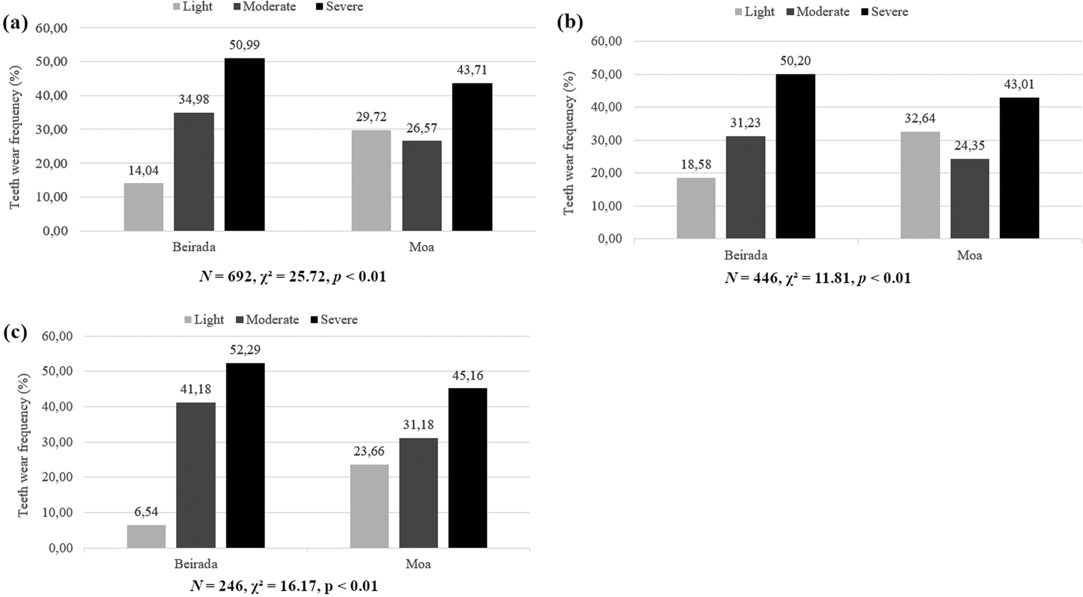
Severe tooth wear was a prominent feature among skeletons at both sites, although the frequency distribution of wear severity differed between them. Whereas Beirada had an increasing frequency of wear severity from light to severe wear, light wear predominated over moderate wear at Moa (p < 0.01; Figure 2a). This scenario was similar in the posterior dentition (p < 0.01; Figure 2b). In the anterior dentition, fewer teeth showed light wear in both sites, and severe wear was predominant (p < 0.01; Figure 2c).

Figure 2. Frequency of dental wear severity in Beirada and Moa individuals: (a) all teeth; (b) posterior dentition; (c) anterior dentition.
Caries analysis showed that none of the 704 teeth observed had any evidence of such infection. Calculus was found in all individuals in Beirada and in 81% (13/16) of Moa individuals (p = 0.086; Table 1). Calculus frequency was 55.58% in Beirada and 49.66% in Moa (p = 0.121). Considering LEH, Beirada presented a higher prevalence than Moa (p = 0.673).
Table 1. Relative Frequency and Prevalence of Oral Physiopathological Processes in Beirada and Moa Individuals.

Only 24 periapical lesions were recorded in this study, 19 in Beirada and five in Moa, which accounted for a frequency of these lesions of 3.97% and 1.34%, respectively (p = 0.022). More than half of the examined individuals from Beirada had periapical lesions, whereas in Moa the prevalence was 18.75% (p = 0.078).
For AMTL, a total of 33 alveoli were found in 15 individuals. Of those, skeletons at Moa exhibited both higher frequency (p = 0.204) and prevalence (p = 0.433) than those at Beirada.
Sex Categories. Wear severity varied greatly between the sexes at Beirada (p < 0.01) and Moa (p < 0.01; Figure 3). For instance, in Beirada, teeth with light wear were not present in males but comprised 11.74% of tooth wear in females (Figure 3a). In addition, males had higher frequencies of severe wear than females. However, in Moa, females presented less light wear and more severe wear than males, who had more light than moderate wear (Figure 3b).

Figure 3. Frequency of dental wear severity according to sex of individuals: (a) Beirada archaeological sites; (b) Moa archaeological sites.
In both sites, females had higher calculus frequency than males (Table 2). Prevalence was higher for males in Moa (p = 1) and equal between sexes in Beirada (p = 1). The proportions of LEH in males and females differed in the two sites: Beirada recorded a higher prevalence in males (p = 1), whereas females in Moa had more LEH (p = 0.5). Regarding periapical lesions, males had higher prevalence (p = 1) and frequency (p = 0.735) in Beirada, whereas in Moa females had higher prevalence (p = 1) and frequency (p = 0.307). AMTL was recorded only in female individuals in Beirada (p = 0.491); in Moa, female individuals showed higher prevalence (p = 1) and frequency (p = 0.154) than males.
Table 2. Relative Frequency and Prevalence of Oral Physiopathological Processes Regarding Sex in Beirada and Moa Individuals.

Age Categories. Regarding tooth wear severity differences between adults and subadults, adults exhibited more intense tooth wear, and greater wear severity was also correlated with older age categories (Figure 4). Children's teeth in Beirada (Figure 4a) presented moderate wear, whereas the entire dentition in children in Moa (Figure 4b) had relatively light wear.

Figure 4. Frequency of dental wear severity according to age categories of individuals: (a) Beirada archaeological site; (b) Moa archaeological site.
As seen in Table 3, calculus frequency increased with age in Beirada (p = 0.555). In Moa, adolescents had the highest frequencies, followed by adults (p = 0.006). Prevalence in Moa also increased with age (p = 0.245).
Table 3. Relative Frequency and Prevalence of Oral Physiopathological Processes Regarding Age Categories in Beirada and Moa Individuals.

Only adults were affected by periapical lesions and AMTL. The frequency and prevalence of the former were almost twice as great at Beirada than at Moa (Table 3). The prevalence and frequency of AMTL in Beirada were 43.75% (p = 1) and 3.52% (p = 0.800), respectively. For Moa, prevalence was 75% (p = 0.037), and frequency was 6.81% (p = 0.019).
Discussion
As previously stated, sambaqui builders had a long temporal and wide spatial distribution, occupying mainly the southern and southeastern Brazilian coast. Funerary practices played a central role in their mound construction and daily lifestyle. The relationships between funerary practices, site construction, and lifestyle in these populations have been the subject of many years of research from a number of sambaqui sites (Berredo et al. Reference Berredo, Gaspar, Ramos and Bianchini2020; Gaspar et al. Reference Gaspar, Bianchini, Berredo and Lopes2019, Reference Gaspar, DeBlasis, Fish, Fish, Silverman and Isbell2008, Reference Gaspar, Klokler, DeBlasis, Roksandic, de Souza, Eggers, Burchell and Klokler2014; Scheel-Ybert et al. Reference Scheel-Ybert, Rodrigues-Carvalho, DeBlasis, Gaspar and Klokler2020).
Archaeological and bioarchaeological studies from the last three decades revealed evidence of a diverse diet that included fish, mollusks, and plants, with marine fish being the primary source of food (Bastos et al. Reference Bastos, Lessa, Rodrigues-Carvalho, Tykot and Santos2014; Boyadjian and Eggers Reference Boyadjian, Eggers, Roksandic, de Souza, Eggers, Burchell and Klokler2014; DeMasi Reference DeMasi2001; Oppitz Reference Oppitz2015; Scheel-Ybert and Boyadjian Reference Scheel-Ybert and Boyadjian2020; Wesolowski et al. Reference Wesolowski, de Souza, Reinhard and Ceccantini2010). Some results indicate a great variation in health indices among populations from different regions of the country, which could relate to distinctive alimentary practices and lifestyles (Mendonça de Souza Reference Mendonça de Souza2018; Souza et al. Reference Souza, Wesolowski, Rodrigues-Carvalho and Oosterbeek2009).
Oral Health of Sambaqui Builders from Saquarema
The predominance of severe tooth wear encountered is expected: this pattern is found in some other hunter-gatherer groups as an indication of intense use of the buccal apparatus in consuming tough, abrasive, or fibrous diets (Deter Reference Deter2009; Hillson Reference Hillson1996; Smith Reference Smith1984). External elements embedded in food, such as shells, earth, and sand, could also have influenced the abrasion intensity, given these groups’ occupation of a region with a great availability of marine resources (Kneip Reference Kneip1998; Kneip et al. Reference Kneip, Araujo and Fonseca1995). Tooth wear is also correlated to age progression, so that mature individuals tend to have a higher degree of wear than young ones (Hillson Reference Hillson1996); this was also the case in this study. However, moderate to severe wear found in young individuals, particularly in a child in Beirada, reinforces the idea that these individuals used the buccal apparatus intensively from an early age.
Differences found in the degree of teeth wear between sexes can assist in the investigation of sexual divisions in alimentary and cultural practices where teeth are used as a third hand. Despite the preliminary evidence provided in this study, more research regarding sexual division in sociocultural traditions of sambaqui societies is necessary to make more statistically reliable suppositions.
The absence of caries in both sites can be the result of a low intake of plants rich in carbohydrates, a pattern typically found in hunter-gatherer groups (Larsen Reference Larsen1997), including sambaqui groups from other regions in Brazil (Mendonça de Souza Reference Mendonça de Souza2018). However, this does not imply that these groups did not have access to plants for consumption. Archaeobotanical and stable isotope studies of sambaqui groups showed that plants were part of their diet (Boyadjian and Eggers Reference Boyadjian, Eggers, Roksandic, de Souza, Eggers, Burchell and Klokler2014; Pezo-Lanfranco et al. Reference Pezo-Lanfranco, Eggers, Petronilho, Toso, Bandeira, Von Tersch, dos Santos, Costa, Meyer and Colonese2018; Scheel-Ybert and Boyadjian Reference Scheel-Ybert and Boyadjian2020; Toso et al. Reference Toso, Hallingstad, McGrath, Fossile, Conlan, Ferreira and Bandeira2021). Moreover, different rates of prevalence of carious lesions were reported for Brazilian sambaqui populations and other hunter-gatherer groups, ranging from 0% to 62.96% (Da-Gloria and Larsen Reference Da-Gloria and Larsen2014; Mendonça de Souza Reference Mendonça de Souza2018; Neves and Wesolowski Reference Neves, Wesolowski, Steckel and Rose2002; Okumura and Eggers Reference Okumura, Eggers, Collard, Morris and Perego2012; Pezo-Lanfranco et al. Reference Pezo-Lanfranco, Eggers, Petronilho, Toso, Bandeira, Von Tersch, dos Santos, Costa, Meyer and Colonese2018; Souza et al. Reference Souza, Wesolowski, Rodrigues-Carvalho and Oosterbeek2009; Wesolowski Reference Wesolowski2007), indicating that plants rich in carbohydrate played an important role in the diverse alimentary practices of Brazilian hunter-gatherer societies.
Results from dental calculus analysis are consistent with our expectations for Brazilian sambaqui builders (Souza et al. Reference Souza, Wesolowski, Rodrigues-Carvalho and Oosterbeek2009). Dental calculus formation is associated with a myriad of factors, including salivary flow, food and water mineral content, genetic factors, and diet (Radini et al. Reference Radini, Nikita, Buckley, Copeland and Hardy2017). Its high prevalence and frequency could be linked to severe tooth wear because intensive mastication can lead to increased salivary flow (Radini et al. Reference Radini, Nikita, Buckley, Copeland and Hardy2017). Another factor is the mineral content in water and food consumed by these populations. Both archaeological sites are situated in an Atlantic Forest region between the sea and a lagoon complex, the latter being fed by seawater and water derived from a mountain range (Silveira Reference Silveira2001); the rich faunal remains found in both sites point to a regular practice of subsistence and cultural activities involving eating and chewing animals (Machado and Kneip Reference Machado and Kneip1994; Silveira Reference Silveira2001). That could mean that the amount and mineral composition of animals and water consumed (and processed) by these human groups could also relate to the high amount of calculus found in this study. The diet of sambaqui groups predominantly comprised animal protein as well, especially marine fish (Bandeira and Fossile Reference Bandeira, Fossile and Zocche2014; Colonese et al. Reference Colonese, Collins, Lucquin, Eustace, Hancock, Ponzoni and Mora2014; DeMasi Reference DeMasi2001, Reference DeMasi2009; Ferreira Reference Ferreira, da Rocha Bandeira, Bartz, Fossile and Mayaroka2019; Figuti Reference Figuti1993; Klokler Reference Klokler2008; Oppitz Reference Oppitz2015; Pavei Reference Pavei2018). A high intake of protein, which increases the alkalinity of the oral environment, is indirectly associated with the development of dental calculus (Lieverse Reference Lieverse1999). Even though diet plays a minor role in calculus formation (Lieverse Reference Lieverse1999; Radini et al. Reference Radini, Nikita, Buckley, Copeland and Hardy2017), it could have been an added factor in the frequent and prevalent formation of high calculus in both populations.
The incidence of LEH varies greatly among sambaqui populations, ranging from 10% to 100% of individuals with this marker (Fischer Reference Fischer2012; Giusto Reference Giusto2018; Neves and Wesolowski Reference Neves, Wesolowski, Steckel and Rose2002; Souza et al. Reference Souza, Wesolowski, Rodrigues-Carvalho and Oosterbeek2009). The prevalence found for both sites in this study falls in the lowest part of this range. Hypotheses about the occurrence of LEH among these sambaqui groups center on the possible lack of important macronutrients in their diet, infections occurring during infancy, and alimentary changes during the weaning period (Guatelli-Steinberg Reference Guatelli-Steinberg, Irish and Scott2016; Mendonça de Souza Reference Mendonça de Souza2018).
Periapical lesions and AMTL are associated with caries, trauma, and periodontal diseases (Dias and Tayles Reference Dias and Tayles1997; Hillson Reference Hillson, Katzenberg and Saunders2008; Waldron Reference Waldron and Waldron2008). These physiopathological processes were mostly found in the posterior section of the dental arch, a region prone to suffer from these processes (Hillson Reference Hillson1996; Nelson Reference Nelson, Irish and Scott2016). Considering that caries were absent in both sites and that these populations manipulated shells and bones with the mouth and presented severe tooth wear, it is probable that periapical lesions and AMTL are related to trauma caused by the intense use of the buccal apparatus for processing food and for paramasticatory activities. Periodontal disease, not analyzed in this study, remains another possible explanation for these patterns.
Differences between Moa and Beirada
The oral health results were compatible with those from other populations of sambaqui builders in Brazil: all the indices calculated in this study fell within the ranges of variation known for such groups. However, differences between both sites point to distinct strategies in the exploitation of environmental resources and alimentary practices.
First, individuals from Beirada seem to have used the buccal apparatus more intensely than those from Moa, as inferred from the higher severity of teeth wear, higher prevalence and frequency of dental calculus, and higher prevalence of periapical lesions. These oral physiopathologies could be related to the resources that constituted their diet, how food was processed and consumed, and the types of artifacts and how they were fashioned, given that the mouth could be used to make them. Thus, differences in oral physiopathologies can provide valuable information on diverse aspects of funerary rituals and subsistence activities of sambaqui groups.
In addition, the LEH results indicate that individuals from Beirada suffered more physiological stress during childhood than did those from Moa. According to current hypotheses on LEH in sambaqui groups, Beirada residents experienced more health problems, malnutrition, and dietary shifts during infancy than those from Moa (Mendonça de Souza Reference Mendonça de Souza2018), which can reflect differences in the availability of environmental resources, alimentary practices, and subsistence activities.
Other forms of evidence can help elucidate these results. For instance, even though the availability and composition of natural resources in the region were the same for both sites, the richness, quantity, and diversity of malacological species varied greatly between them (Barbosa-Guimarães Reference Barbosa-Guimarães2011; Kneip et al. Reference Kneip, Crancio, Pallestrini, Mello, Corrêa, Magalhães and Vogel1994). According to previous studies, there was a greater quantity of mollusks in Beirada than in Moa. In the former settlement, there was also a shift in the predominance of mollusk species through time, whereas in Moa a few species remained predominant in all layers of excavation. Additionally, more than twice the number of specimens from the two predominant ichthyological species were recovered in Moa than in Beirada (Kneip et al. Reference Kneip, Crancio, Pallestrini, Mello, Corrêa, Magalhães and Vogel1994). The position of these faunal remains could relate to sambaqui funerary practices (Gaspar et al. Reference Gaspar, Klokler, DeBlasis, Roksandic, de Souza, Eggers, Burchell and Klokler2014), alimentary practices (Bandeira and Fossile Reference Bandeira, Fossile and Zocche2014; Colonese et al. Reference Colonese, Collins, Lucquin, Eustace, Hancock, Ponzoni and Mora2014; Ferreira Reference Ferreira, da Rocha Bandeira, Bartz, Fossile and Mayaroka2019; Figuti Reference Figuti1993; Klokler Reference Klokler2008), and, in the case of mollusks, site construction (Gaspar et al. Reference Gaspar, DeBlasis, Fish, Fish, Silverman and Isbell2008).
Lithic artifacts also vary by site. According to Kneip and collaborators (Reference Kneip, Crancio and Francisco1988, Reference Kneip, Crancio, Pallestrini, Mello, Corrêa, Magalhães and Vogel1994), most artifacts in Beirada were made of polished stone, whereas the lithic industry in Moa was dominated by chipped stone artifacts. According to these authors the greater presence of chipped stone artifacts in Moa suggests that individuals buried in this site placed more emphasis on fishing activities than ones from Beirada.
Unfortunately, direct evidence for dietary components is absent for the Beirada population. The osteological collection of the Museu Nacional was severely damaged by a 2018 fire before stable isotope analyses of δ13C and δ15N could be performed for the Beirada population. However, these data are available for the Moa population. A study by Bastos and collaborators (Reference Bastos, Guida, Rodrigues-Carvalho, Toso, Santos and Colonese2022) using Bayesian stable isotope mixing models indicates that marine fish was the primary source of protein intake, which is expected for sambaqui populations. It also finds that C3 plants contributed significantly to their diet, strengthening the current view that sambaqui people were also managing and cultivating plants (Scheel-Ybert and Boyadjian Reference Scheel-Ybert and Boyadjian2020).
Another issue to contemplate is the accuracy of the dating of both sites. The original dating had standard deviations close to ± 200 years (Kneip et al. Reference Kneip, Crancio, Pallestrini, Mello, Corrêa, Magalhães and Vogel1994), which could imply that Beirada and Moa were contemporaneous for some time. A greater temporal difference could help explain the disparities in oral health and other archaeological evidence found in these sambaquis. To answer this question, excavation campaigns in Saquarema must be resumed.
In addition, the difficulty in associating the analyzed skeletons with available data on the burial distribution of both sites should be taken into account. Because there is no proper stratigraphical control regarding the human remains, it is not possible to analyze intraspecific differences in the oral health of the individuals buried in each site through time, which could be used to infer sociocultural changes within these groups.
Faunal remains, lithic artifacts, and oral health analysis suggest that Beirada and Moa populations had variations in cultural and alimentary practices. The findings of this study highlight these variations and add to recent debates about continuities and differences in sambaqui societies (Berredo et al. Reference Berredo, Gaspar, Ramos and Bianchini2020; Gaspar et al. Reference Gaspar, Klokler, Scheel-Ybert and Bianchini2013, Reference Gaspar, Bianchini, Berredo and Lopes2019; Scheel-Ybert et al. Reference Scheel-Ybert, Rodrigues-Carvalho, DeBlasis, Gaspar and Klokler2020). These prehistoric societies were socioculturally diverse and complex, engaging in alimentary and funerary practices and exploration of the environment.
Conclusions
The growing number of studies on the oral health of sambaqui groups have enhanced our understanding of their past health and diseases, alimentary practices, and subsistence activities (Souza et al. Reference Souza, Wesolowski, Rodrigues-Carvalho and Oosterbeek2009). Most of these studies have been conducted in southern Brazilian sites, such as those located in Santa Catarina state; there are few studies in other Brazilian regions (Guida Reference Guida2019:84–86).
Our oral health analyses showed that indices from two spatially and temporally similar sites in Rio de Janeiro state were in accord with such analyses from other Brazilian sambaquis. However, there were variations between the two sites—Beirada and Moa—indicating differences in food processing and consumption.
New excavation campaigns in Saquarema municipality are needed to recover additional archaeological evidence regarding sambaqui societies. Future excavations and research should resolve some of the current problems hindering our oral health analyses of these sites, such as the unavailability of dietary isotopic information, the potentially inaccurate dating of the sites, and the partial destruction of the osteological collection at the Museu Nacional.
Acknowledgments
We thank Dr. Michael B. C. Rivera for his feedback and help with the English editing of this manuscript.
Funding Statement
This study was financed in part by the Coordenação de Aperfeiçoamento de Pessoal de Nível Superior— Brasil (CAPES), Finance Code 001.
Data Availability Statement
All raw data from oral health analysis are held by the authors and are available for researchers on request to the lead author. All skeletal material analyzed in this article was severely damaged or destroyed by a fire that occurred in the Museu Nacional on September 2, 2018. Therefore, replicability of these results is not possible for these human remains.
Competing Interests
The authors declare none.
Supplemental Material
For supplemental material accompanying this article, visit https://doi.org/10.1017/laq.2022.98.
Supplemental Table 1. Discrimination of Sex, Age, Number of Teeth, Alveoli, and Postmortem Tooth Loss from Beirada and Moa Individuals.


