In schizophrenia, impairments in social cognition as well as metacognition result in poor social functioning Reference Fett, Viechtbauer, Dominguez, Penn, van Os and Krabbendam1 and act as a mediator between neurocognition and real-world functioning. Reference Sergi, Rassovsky, Widmark, Reist, Erhart and Braff2,Reference Lysaker, Shea, Buck, Dimaggio, Nicolò and Procacci3 Empathy, the ability to infer and share another's internal emotional states, plays a pivotal role in social interaction Reference Kohut4,Reference Preston and de Waal5 and is a multidimensional phenomenon; Decety & Jackson Reference Decety and Jackson6 postulated that at least three core components can be identified: (a) recognition of emotions in oneself and others; (b) an affective component, i.e. the ability to experience and share similar emotions as others (affective responsiveness, AR); and (c) a cognitive component, i.e. the ability to take the perspective of another person and infer their feelings (emotional perspective-taking, EPT). Persons with schizophrenia show substantial deficits in nearly every domain also in empathy, with impairments in emotion recognition (ER) being investigated most frequently (for meta-analysis Reference Kohler, Walker, Martin, Healey and Moberg7 ). However, a thorough investigation of all three components demonstrated that individuals with schizophrenia suffer from a significant deficit in every single domain, Reference Derntl, Finkelmeyer, Toygar, Hülsmann, Schneider and Falkenberg8 thus indicating a much broader emotional deficit. Neuroimaging research extended the behavioural findings by highlighting functional abnormalities during empathy-related tasks in individuals with schizophrenia, Reference Derntl, Finkelmeyer, Voss, Eickhoff, Kellermann and Schneider9,Reference Harvey, Zaki, Lee, Ochsner and Green10 which importantly were further associated with social community functioning. Reference Smith, Schroeder, Abram, Goldman, Parrish and Wang11 Compared with the plethora of studies addressing socio-emotional processes in schizophrenia, relatively little is known about empathy in people at risk for psychosis. Few studies investigated emotional abilities in persons genetically at risk for schizophrenia and observed significant behavioural deficits; Reference Eack, Mermon, Montrose, Miewald, Gur and Gur12 however, mixed results were reported regarding neural activation compared with controls (amygdala hypofunction; Reference Habel, Klein, Shah, Toni, Zilles and Falkai13 neural hyperactivation; Reference Van Buuren, Vink, Rapcencu and Kahn14 no difference Reference Rasetti, Mattay, Wiedholz, Kolachana, Hariri and Callicott15 ). Even less is known in patients at clinical high risk for psychosis. Behavioural studies in such patients addressing facial or vocal affect processing reported mixed findings: ranging from a rather general impairment Reference Addington, Penn, Woods, Addington and Perkins16 to emotion-specific deficits Reference Amminger, Schäfer, Papageorgiou, Klier, Schlögelhofer and Mossaheb17,Reference Kohler, Richard, Brensinger, Borgmann-Winter, Conroy and Moberg18 or no difference. Reference Addington, Piskulic, Perkins, Woods, Liu and Penn19 Seiferth et al Reference Seiferth, Pauly, Habel, Kellermann, Shah and Ruhrmann20 investigated neural activation during ER in individuals at clinical high risk of psychosis and observed frontolimbic hyperactivation particularly during neutral face-processing. Notably, there is a lack of studies investigating cognitive and affective empathy in this group of patients. This seems particularly surprising, given the evidence of a significant association between empathy and social functioning in individuals with schizophrenia Reference Smith, Schroeder, Abram, Goldman, Parrish and Wang11,Reference Smith, Horan, Karpouzian, Abram, Cobia and Csernansky21 and consistent findings on social interaction difficulties acting as a precursor of schizophrenia. Reference Cannon22
The aim of the present study therefore was to investigate the behavioural and neural substrates of empathy in individuals at clinical high risk of psychosis and matched controls, enabling more detailed analyses of emotional competencies, their interactions and possible neural dysfunctions in this preclinical group. In addition to group differences, we also analysed potential associations between behavioural performance, neural activation and clinical parameters. Based on inconsistent results regarding ER and previous neuroimaging data in individuals at clinical high risk of psychosis, Reference Seiferth, Pauly, Habel, Kellermann, Shah and Ruhrmann20 we expected unimpaired recognition accuracy but dysfunctional neural activation. Additionally, to further examine differences and similarities in empathy between individuals at clinical high risk of psychosis and patients with borderline personality disorder, behavioural and neural data of individuals at clinical high risk of psychosis in affective and cognitive empathy was compared with previously published data on individuals with schizophrenia. Reference Derntl, Finkelmeyer, Voss, Eickhoff, Kellermann and Schneider9
Method
Sample
A total of 15 patients at clinical high risk of psychosis (CHR group) and 15 healthy controls (CON group) were included. For comparison, we relied on data of 15 individuals with schizophrenia (SZP group) that have been published previously. Reference Derntl, Finkelmeyer, Voss, Eickhoff, Kellermann and Schneider9 Due to excessive head movement, functional data of two CHR group participants were excluded from functional magnetic resonance imaging (fMRI) data analysis. All patients were White, native German-speaking and were matched for age, parental education and neurocognitive function.
The CHR group were recruited through clinical services at the Department of Psychiatry, Psychotherapy and Psychosomatics (DPPP), RWTH Aachen University. Experienced psychiatrists identified clinical risk for psychosis and diagnostic inclusion criteria were assessed by the Structured Interview for Prodromal Syndromes and the Scale of Prodromal Symptoms (SIPS/SOPS); Reference McGlashan, Miller and Woods23 positive and negative symptoms were also assessed with the Positive and Negative Syndrome Scale (PANSS). Reference Kay, Fiszbein and Opler24 Based on a two-phase approach, Reference Schultze-Lutter, Ruhrmann, Hoyer, Klosterkötter and Leweke25 participants had to fulfil the criteria for a late initial prodromal state. Attenuated psychotic symptoms (APS) and the Brief Limited Intermittent Psychotic Symptoms (BLIPS) were applied as criteria; thus symptoms of clinical high risk of psychosis were below criteria for a full-blown manifest psychotic episode. Social and occupational functioning (Social and Occupational Functioning Assessment Scale, SOFAS Reference Goldman, Skodol and Lave26 ) and depressive symptoms (Montgomery–Åsberg Depression Rating Scale, MADRS Reference Montgomery and Åsberg27 ) were assessed in the CHR group. Four CHR participants had a familial risk for psychosis besides clinical symptoms. Three CHR patients were taking atypical agents (1 aripiprazole, 1 quetiapine and 1 risperidone) and two were on antidepressive medication (selective serotonin reuptake inhibitors).
The SZP group was recruited from in- and out-patient units of the DPPP, received atypical antipsychotics and no other medication. Symptom severity was assessed with standardised scales (PANSS Reference Kay, Fiszbein and Opler24 ). Exclusion criteria for all participants included substance abuse within the last 6 months, left-handedness and any other psychiatric or neurological illness based on the Structured Clinical Interview for DSM-IV Disorders. Reference Wittchen, Zaudig and Fydrich28
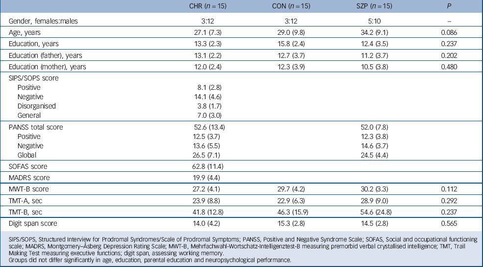
Moreover, to assess self-reported empathic abilities the German version of the Interpersonal Reactivity Index Reference Paulus29 was administered and participants were asked to answer 16 items on a 5-point Likert scale ranging from ‘Does not describe me well’ to ‘Describes me very well’. Additionally, all participants completed neuropsychological tests, tapping premorbid crystallised verbal intelligence (Mehrfachwahl-Wortschatz-Intelligenztest, MWT-B), Reference Lehrl30 information processing speed and executive functions (Trail Making Tests, versions A and B) Reference Reitan31 as well as working memory (digit spans from WAIS-IV). Reference Petermann32 Demographic, clinical and neuropsychological characteristics are shown in Table 1.
TABLE 1 Mean and standard deviation (in parentheses) of sociodemographic and neuropsychological characteristics of participants with clinical high risk for psychosis (CHR), healthy controls (CON), and patients with schizophrenia (SZP)

| CHR (n = 15) | CON (n = 15) | SZP (n = 15) | P | |
|---|---|---|---|---|
| Gender, females:males | 3:12 | 3:12 | 5:10 | – |
| Age, years | 27.1 (7.3) | 29.0 (9.8) | 34.2 (9.1) | 0.086 |
| Education, years | 13.3 (2.3) | 15.8 (2.4) | 12.4 (3.5) | 0.237 |
| Education (father), years | 13.1 (2.2) | 12.7 (3.7) | 11.2 (3.7) | 0.202 |
| Education (mother), years | 12.0 (2.4) | 12.3 (3.9) | 10.5 (3.8) | 0.480 |
| SIPS/SOPS score | ||||
| Positive | 8.1 (2.8) | |||
| Negative | 14.1 (4.6) | |||
| Disorganised | 3.8 (1.7) | |||
| General | 7.0 (3.0) | |||
| PANSS total score | 52.6 (13.4) | 52.0 (7.8) | ||
| Positive | 12.5 (3.7) | 12.3 (3.8) | ||
| Negative | 13.6 (5.5) | 14.6 (3.7) | ||
| Global | 26.5 (7.1) | 24.5 (4.4) | ||
| SOFAS score | 62.8 (11.4) | |||
| MADRS score | 19.9 (4.4) | |||
| MWT-B score | 27.2 (4.1) | 29.7 (4.2) | 30.2 (3.3) | 0.112 |
| TMT-A, sec | 23.9 (8.8) | 22.9 (6.3) | 28.9 (9.0) | 0.292 |
| TMT-B, sec | 41.8 (12.8) | 46.3 (15.9) | 54.6 (24.8) | 0.237 |
| Digit span score | 14.0 (4.2) | 15.3 (2.8) | 14.5 (2.8) | 0.565 |
SIPS/SOPS, Structured Interview for Prodromal Syndromes/Scale of Prodromal Symptoms; PANSS, Positive and Negative Syndrome Scale; SOFAS, Social and occupational functioning scale; MADRS, Montgomery–Åsberg Depression Rating Scale; MWT-B, Mehrfachwahl-Wortschatz-Intelligenztest-B measuring premorbid verbal crystallised intelligence; TMT, Trail Making Test measuring executive functions; digit span, assessing working memory.
Groups did not differ significantly in age, education, parental education and neuropsychological performance.
After complete description of the study to the participants, written informed consent was obtained. All participants were paid for their participation. The ethics committee of RWTH Aachen University approved the study.
Functional tasks
We used three tasks tapping each empathy component separately, which have been used before in neuroimaging studies. Reference Derntl, Finkelmeyer, Voss, Eickhoff, Kellermann and Schneider9 Every response was recorded and the sum of correct responses added up to an accuracy score for each task that was used for statistical analyses. Analysis of internal consistency across the whole sample revealed Cronbach's alpha of 0.649 (ER), 0.839 (EPT) and 0.882 (AR), indicating acceptable to good reliability.
Emotion recognition
Thirty photographs of White faces depicting five basic emotions (happiness, sadness, anger, fear and disgust) and neutral expressions were presented. Stimuli were selected from a standardised stimulus set. Reference Gur, Sara, Hagendoorn, Marom, Hughett and Macy33 Participants were instructed to choose the correct emotion from two possibilities via button press. Stimuli were presented maximally for 5 s with a randomised, variable interstimulus interval ranging from 8 s to 12 s.
Emotional perspective-taking
Participants viewed 35 scenes showing two White people involved in social interaction thereby portraying the emotions described above. The face of one person was masked, and participants were asked to infer the emotional expression of the masked face. Stimuli were shown for 4 s and participants responded immediately afterwards by selecting between two different facial expressions that were taken from the stimulus pool described above.
Affective responsiveness
We presented 35 short written sentences describing real-life situations inducing one of the emotions described above. Participants were asked to imagine how they would feel if they were experiencing those situations. Sentences were presented for 4 s and response format was the same as for EPT.
All stimuli were presented by goggles (VisuaStimDigital, Resonance Technology Inc, Los Angeles, California, USA). Presentation of images, recording of responses and synchronisation with the scanner was achieved using Presentation (Neurobehavioral Systems, Inc, Albany, California, USA).
Behavioural data analysis
Statistical analyses were performed by SPSS 20.0 and level of significance was set at P = 0.05. Correct responses of each empathy task acquired during scanning were analysed by mixed effects analysis of variance (ANOVAs) with task as within-participant factor and group as between-participant factor. Bonferroni corrected P-values are reported for all post hoc comparisons.
Group differences in self-report data were assessed by univariate ANOVAs. For the self-report empathy scale we used sums of the four subscales (each made up of four different items) for further analyses. Regarding the statistical analysis of neuropsychological tests, we relied on the sum of correctly marked words in the MWT-B. For the TMT-A and TMT-B we used reaction time to finish the tasks and regarding digit span we relied on the sum of the correct forward and backward trials.
Correlations between accuracy measures and self-report scores were computed using Pearson correlations, except for AR where performance scores were not normally distributed (here Spearman's rank correlations were applied). For the CHR group, additional analyses investigating potential associations between clinical parameters and behavioural measures as well as self-report scores were performed by Pearson correlations, again with the exception of analyses involving AR scores. All correlation significance tests were two-tailed and corrected for multiple testing.
fMRI acquisition parameters and data processing
All participants were examined with a 3T whole-body scanner (Philips, Best, The Netherlands) at the Medical Faculty of RWTH Aachen University. Functional imaging was performed by gradient-recalled EPI; 35 oblique axial slices were acquired by asymmetric k-space sampling (repetition time = 2200 ms, echo time = 31 ms).
Functional data were pre-processed by SPM8 (www.fil.ion.ucl.ac.uk/spm/spm8.html). Images were slice-timing corrected, realigned and normalised into the standardised stereotactic space. Functional data-sets were spatially smoothed by an isotropic Gaussian kernel with a full-width-at-half- maximum of 8 mm. Prior to pre-processing, a signal artefact correction algorithm was employed to reduce large, slice-wise signal fluctuations in the fMRI time-series as previously reported. Reference Derntl, Finkelmeyer, Voss, Eickhoff, Kellermann and Schneider9
fMRI data analysis
For this event-related design, each stimulus type was modelled with a separate regressor by a delta function convolved with the hemodynamic response function. For EPT and AR the initial task stimulus (scene/sentence) and the response option were modelled with separate regressors. First-level models included motion parameters as regressors of no interest.
To detect group differences, contrast images from all participants for each task were included in a second-level random effects analysis. We performed a 3 (group)×3 (task) mixed effects ANOVA and significant F-contrasts were explored by subsequent post hoc t-contrasts. For EPT and AR only activation related to the initial task stimulus was analysed at the second level.
For whole-brain analyses we applied a correction for multiple comparisons at the cluster-level based on Monte-Carlo simulations by AlphaSim. Reference Cox34 According to 1000 simulations based on a cluster-defining threshold of P<0.001 (uncorrected) and the spatial properties of the residual image an extent threshold of 55 contiguous voxels sufficed to comply with a family-wise error of P<0.05. All results were depicted at this threshold.
Region of interest (ROI) analysis
We performed an ROI analysis for the amygdala region due to its role in emotion processing and previous studies observing functional abnormalities of the amygdala in individuals at clinical high risk for psychosis Reference Seiferth, Pauly, Habel, Kellermann, Shah and Ruhrmann20 and those with schizophrenia. Reference Derntl, Finkelmeyer, Voss, Eickhoff, Kellermann and Schneider9 Based on previous results, Reference Amunts, Kedo, Kindler, Pieperhoff, Mohlberg and Shah35 values for both amygdala ROIs were extracted drawing two spheres (10 mm) centred at [x,y,z: ±20, 0, −20]. A three-way ANOVA was applied with group as between-participant factor, and task and laterality as repeated factors.
Corollary analyses
Pearson correlations were performed with accuracy data from ER and EPT and mean amygdala parameter estimates (taken from the ROI analysis). Spearman's rank correlation was used for correlation analyses between amygdala activation and AR scores. For the CHR group, additional Pearson correlations between amygdala activation and clinical parameters (SIPS/SOPS, PANSS, MADRS, SOFAS) were conducted.
Results
Behavioural performance
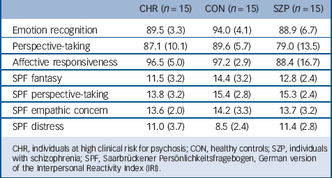
Analysis of percent correct demonstrated a significant task effect (F(2,40) = 17.141, P<0.001) and a significant group effect (F(2,40) = 4.156, P = 0.025), but no task×group interaction (F(2,40) = 1.568, P = 0.194). Bonferroni corrected post hoc tests of the task effect revealed significantly worse performance in EPT compared with AR (P<0.001) and ER (P = 0.003), whereas performance in ER did not differ significantly from AR (P = 0.069). Regarding the group effect, the CON group outperformed the SZP group (P = 0.025), whereas no difference emerged between the other groups (CON-CHR: P = 1.000; CHR-SZP: P = 0.200).
TABLE 2 Performance accuracy of the empathy tasks (percent correct) and self-reported empathic abilities for each group (means and standard deviations)

| CHR (n = 15) | CON (n = 15) | SZP (n = 15) | |
|---|---|---|---|
| Emotion recognition | 89.5 (3.3) | 94.0 (4.1) | 88.9 (6.7) |
| Perspective-taking | 87.1 (10.1) | 89.6 (5.7) | 79.0 (13.5) |
| Affective responsiveness | 96.5 (5.0) | 97.2 (2.9) | 88.4 (16.7) |
| SPF fantasy | 11.5 (3.2) | 14.4 (3.2) | 12.8 (2.4) |
| SPF perspective-taking | 13.8 (3.2) | 15.4 (2.8) | 15.3 (2.4) |
| SPF empathic concern | 13.6 (2.0) | 14.2 (3.3) | 13.7 (3.2) |
| SPF distress | 11.0 (3.7) | 8.5 (2.4) | 11.4 (2.8) |
CHR, individuals at high clinical risk for psychosis; CON, healthy controls; SZP, individuals with schizophrenia; SPF, Saarbrückener Persönlichkeitsfragebogen, German version of the Interpersonal Reactivity Index (IRI).
Figure DS1 in an online data supplement to this paper illustrates performance on the empathy tasks across groups, and Table 2 gives details on performance accuracy.
Empathy questionnaire
Direct comparison of self-report scores demonstrated significant group effects for the fantasy scale (F(2,40) = 3.340, P = 0.047) and the distress scale (F(2,40) = 3.525, P = 0.040), whereas no other significant difference emerged (Ps>0.269). Post hoc analyses showed that the CHR group had significantly lower fantasy scores than the CON group (P = 0.043) but did not differ from the SZP group (P = 0.825) who also did not differ from the CON group (P = 0.569). Regarding the distress scale, post hoc tests revealed no significant difference between the groups (Ps>0.071). Please see Table 2 for means and standard deviations for each group.
Functional data
Separate group analyses for the CHR, SZP and CON groups showed activation of a widespread cortical-subcortical network including frontal regions, orbitofrontal cortex, temporal gyri, fusiform gyri, inferior parietal cortex and cingulate cortex across all tasks. Please note that differences between the SZP and CON groups will not be reported since they were not the focus of the current paper and resemble findings from a previously published study. Reference Derntl, Finkelmeyer, Voss, Eickhoff, Kellermann and Schneider9
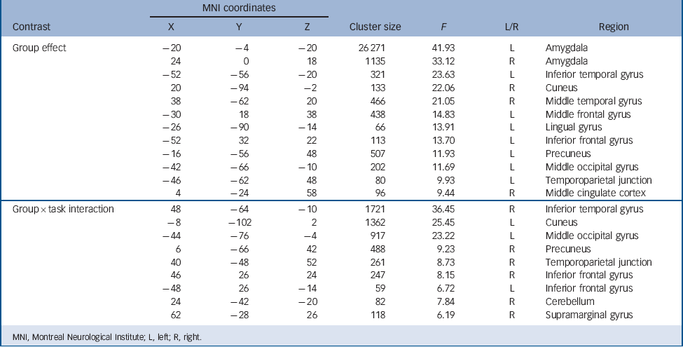
The mixed effects ANOVA revealed significant group differences across all empathy tasks in several regions, for example bilateral amygdala, left inferior and middle frontal gyrus, middle cingulate cortex, and left temporoparietal junction (online Fig. DS2(a) and Table 3).
TABLE 3 Results from the mixed effects ANOVA with group and task as factors, showing a significant main effect of group (threshold: F = 7.32, P<0.05 cluster level corrected) and a significant group×task interaction (threshold: F = 4.95, P<0.05 cluster level corrected)

| MNI coordinates | |||||||
|---|---|---|---|---|---|---|---|
| Contrast | X | Y | Z | Cluster size | F | L/R | Region |
| Group effect | −20 | −4 | −20 | 26 271 | 41.93 | L | Amygdala |
| 24 | 0 | 18 | 1135 | 33.12 | R | Amygdala | |
| −52 | −56 | −20 | 321 | 23.63 | L | Inferior temporal gyrus | |
| 20 | −94 | −2 | 133 | 22.06 | R | Cuneus | |
| 38 | −62 | 20 | 466 | 21.05 | R | Middle temporal gyrus | |
| −30 | 18 | 38 | 438 | 14.83 | L | Middle frontal gyrus | |
| −26 | −90 | −14 | 66 | 13.91 | L | Lingual gyrus | |
| −52 | 32 | 22 | 113 | 13.70 | L | Inferior frontal gyrus | |
| −16 | −56 | 48 | 507 | 11.93 | L | Precuneus | |
| −42 | −66 | −10 | 202 | 11.69 | L | Middle occipital gyrus | |
| −46 | −62 | 48 | 80 | 9.93 | L | Temporoparietal junction | |
| 4 | −24 | 58 | 96 | 9.44 | R | Middle cingulate cortex | |
| Group × task interaction | 48 | −64 | −10 | 1721 | 36.45 | R | Inferior temporal gyrus |
| −8 | −102 | 2 | 1362 | 25.45 | L | Cuneus | |
| −44 | −76 | −4 | 917 | 23.22 | L | Middle occipital gyrus | |
| 6 | −66 | 42 | 488 | 9.23 | R | Precuneus | |
| 40 | −48 | 52 | 261 | 8.73 | R | Temporoparietal junction | |
| 46 | 26 | 24 | 247 | 8.15 | R | Inferior frontal gyrus | |
| −48 | 26 | −14 | 59 | 6.72 | L | Inferior frontal gyrus | |
| 24 | −42 | −20 | 82 | 7.84 | R | Cerebellum | |
| 62 | −28 | 26 | 118 | 6.19 | R | Supramarginal gyrus | |
MNI, Montreal Neurological Institute; L, left; R, right.
Moreover, a significant task effect and a significant task×group interaction emerged. Post hoc t-tests revealed several significant findings (online Fig. DS2(b) and Table DS1).
For ER, the CHR group showed stronger activation compared with the CON group in a widespread network, including bilateral amygdala, right inferior frontal gyrus (IFG), right middle temporal gyrus (MTG), left parahippocampal gyrus, right middle cingulate gyrus and left thalamus. Comparing the CHR group with the SZP group, stronger recruitment of left amygdala, left thalamus, and inferior frontal gyri bilaterally was apparent in the CHR group. The CON and SZP groups showed no stronger activation than the CHR group.
For EPT, the CHR group showed a stronger neural response in the right IFG, left superior medial frontal gyrus, right middle and left inferior temporal gyrus as well as left parahippocampal gyrus than the CON group. The CHR group also exhibited stronger activation than the SZP group of the amygdala bilaterally, left superior medial frontal gyrus, bilateral parahippocampal gyrus, left putamen, bilateral middle frontal gyrus and right lingual gyrus. Again, the CON and SZP groups demonstrated no stronger activation than the CHR group.
Regarding AR, group comparisons revealed stronger activation of the CHR group than the CON group only in the left MTG, whereas the CON group recruited bilateral superior temporal gyrus and right temporoparietal junction more strongly. In comparison with the SZP group, the CHR group showed stronger neural response of the amygdala bilaterally, right IFG, bilateral superior frontal gyrus and thalamus. Again, the SZP group exhibited no stronger activation than the CHR group.
ROI analysis
The ANOVA demonstrated a significant task effect (F(2,80) = 17.675, P50.001), a significant group effect (F(2,40) = 5.104, P = 0.011), and a significant task×laterality interaction (F(2,80) = 6.304, P = 0.003). Moreover, a trend towards a task×group interaction occurred (F(2,80) = 2.482, P = 0.051), but no significant laterality effect (F(2,40) = 2.367, P = 0.133) and no other interaction reached significance (Ps>0.068). Post hoc analysis of the task effect showed that amygdala activation was stronger during ER compared with both other tasks (Ps<0.001), whereas activation did not differ between EPT and AR (P = 1.000). Regarding the group effect, post hoc tests demonstrated significantly less amygdala activation in the SZP group compared with the CON group (P = 0.024) and the CHR group (P = 0.030), whereas the CHR group did not differ from the CON group (P = 1.000). The significant task×laterality interaction revealed a laterality effect only for AR, where the left amygdala showed a significantly stronger response than the right (P = 0.008). Both other tasks showed no laterality difference (Ps>0.069).
Correlation analyses
Behavioural data
Positive symptoms as assessed via SIPS/SOPS showed a significant correlation with accuracy in EPT (r = −0.704, P= 0.007). No other correlation regarding symptom severity (SIPS/SOPS, PANSS) or depression scores (MADRS) reached significance (Ps>0.197). SOFAS scores correlated with performance in ER (r = 0.793, P = 0.001) and EPT (r = 0.625, P = 0.022), but not with AR (P = 0.234).
Functional data
Correlation analyses between performance accuracy and task-specific amygdala activation failed to show significant associations in any group (Ps>0.184).
Regarding clinical parameters in the CHR group, positive symptoms as well as depression scores correlated significantly with amygdala activation during EPT (SIPS_P: r = 0.580, P = 0.038; MADRS: r = −0.563, P = 0.045). A trend for a similar correlation emerged for depression scores and amygdala activation during AR (ρ = −0.572, P = 0.052), whereas all other associations did not reach significance (Ps>0.062).
Discussion
The aim of the current study was to explore behavioural and neural correlates of the core components of empathy, namely ER, EPT and AR in persons at clinical high risk of psychosis compared with healthy controls and individuals with schizophrenia. To the best of our knowledge, no neuroimaging study has addressed a broader range of emotional competencies in the CHR group.
The CHR group showed similar behavioural performance compared with the CON group, thereby supporting findings on unimpaired ER/differentiation in individuals at clinical high risk of psychosis Reference Addington, Piskulic, Perkins, Woods, Liu and Penn19 and extending such evidence to further empathy components. In line with previous work, Reference Seiferth, Pauly, Habel, Kellermann, Shah and Ruhrmann20 the CHR group differed markedly in their neural activation from the CON group. Comparing neural activation of the CHR and SZP groups demonstrated significant differences across all tasks, with the CHR group showing hyperactivation mainly in emotion-related areas. Additionally, significant correlations between performance, neural activation, clinical parameters and social functioning emerged.
Abnormalities in the neural empathy network in the CHR group
Compared with the CON group, neural activation of the CHR group was mainly characterised by hyperactivation of the IFG, the middle cingulate cortex and the parahippocampal gyrus. Interestingly, the MTG showed hyperactivation during all tasks. Elevated MTG activation was recently reported during application of an emotion regulation strategy (distancing) in healthy participants. Reference Koenigsberg, Fan, Ochsner, Liu, Guise and Pizzarello36 Here, the CHR group showed stronger activation of the MTG during all tasks but particularly during AR, the only self-centred task where we asked participants to imagine going through a certain situation and how this would make them feel. The stronger activation of the MTG might indicate that the CHR group was actively using this strategy to prevent them from reacting too emotionally. Since the MTG is only one node in the neural network underlying emotion regulation Reference Koenigsberg, Fan, Ochsner, Liu, Guise and Pizzarello36 and due to multiple functions of each brain region as well as the limited data available in the CHR group, this interpretation needs additional support from the literature. Alternatively, the MTG was proposed to play a major role in mentalising and understanding other people's intentions and emotions. Reference Amodio and Frith37 Stronger activation of the CHR group might then reflect compensatory mechanisms, probably the reason for their unimpaired behavioural performance.
Activation of the IFG was repeatedly observed during various emotional processes including empathy. Reference Derntl, Finkelmeyer, Voss, Eickhoff, Kellermann and Schneider9,Reference Harvey, Zaki, Lee, Ochsner and Green10 In our data, the CHR group demonstrated hyperactivation of this region particularly during ER and EPT compared with the CON group and during ER and AR compared with the SZP group. Seiferth et al Reference Seiferth, Pauly, Habel, Kellermann, Shah and Ruhrmann20 reported stronger activation of the IFG during processing of neutral faces, probably reflecting aberrant attributions of motivational/emotional salience to neutral stimuli, which were also proposed to characterise individuals with schizophrenia. Reference Kapur38 However, direct comparison indicated that, although the SZP group showed a significant reduction in IFG activation across all three tasks, Reference Derntl, Finkelmeyer, Voss, Eickhoff, Kellermann and Schneider9 the CHR group mainly demonstrated hyperactivation of this region. These diverging activation patterns might point to neural changes before illness onset. Given the functionality of sub-regions of the IFG for action observation, imitation, mentalising and emotion processing, Reference Nishitani, Schürmann, Amunts and Hari39 we speculate that the hyperactivation in the CHR group might reflect compensation mechanisms that enabled the CHR group to perform similar to the CON group, whereas the hypoactivation in the SZP group indicates the consequences of illness onset and denotes one important substrate of an impaired capacity to spontaneously simulate another person's subjective world.
Another region that showed elevated activation in the CHR group compared with the CON group as well as the SZP group was the parahippocampal gyrus. Recently, grey matter volume reductions in individuals at ultra-high risk for psychosis were investigated and persons who later developed a psychotic disorder showed distinct reductions of parahippocampal cortex volume. Reference Mechelli, Riecher-Rössler, Meisenzahl, Tognin, Wood and Borgwardt40 Hence, volume alterations of this region may be crucial to the expression of illness.
Notably, we observed greater activation of the amygdala bilaterally in the CHR group compared with the CON group during ER and compared with the SZP group across all tasks. This exaggerated activation might be due to disturbed frontolimbic connectivity in the CHR group, which was recently shown in children and adolescents with familial risk for psychosis. Reference Diwadkar, Wadehra, Pruitt, Keshavan, Rajan and Zajac-Benitez41 This dysfunctional connectivity could lead to elevated emotional arousal, hypersensitivity for emotional stimuli, particularly facial expressions of emotions, causing heightened negative affect and stress. The assumption of amygdala involvement in the pathophysiology of psychosis stems from studies demonstrating amygdala dysfunction in the SZP group and patients with bipolar disorder with psychotic history Reference Delvecchio, Sugranyes and Frangou42 and findings of elevated amygdala activation in psychosis-prone individuals, Reference Modinos, Pettersson-Yeo, Allen, McGuire, Aleman and Mechelli43 which was also accompanied by less prefrontal-amygdala coupling. Interestingly, in our study positive symptoms were associated with less accuracy and higher amygdala activation during perspective-taking, perhaps also reflecting less prefrontal amygdala coupling during this cognitive empathy task.
Mechanism of empathy abnormalities in individuals at clinical high risk of psychosis
There is very limited evidence on neural correlates of emotional abilities in individuals at clinical high risk of psychosis. Seiferth et al Reference Seiferth, Pauly, Habel, Kellermann, Shah and Ruhrmann20 investigated ER performance and observed stronger activation in visual areas in participants at clinical high risk of psychosis compared with controls for all stimuli, whereas neutral facial expressions prompted marked hyperactivation in frontolimbic regions. They suggested that this hyperactivation might reflect hypersensitivity to affectively irrelevant stimuli in brain areas relevant for attributing affective salience.
Greater activation in people at clinical high risk of psychosis in regions known to be essential for emotion and sociocognitive processing were also reported in studies addressing theory of mind. Reference Marjoram, Job, Whalley, Gountouna, McIntosh and Simonotto44,Reference Brüne, Ozgürdal, Ansorge, van Reventlow, Peters and Nicolas45 Brüne et al Reference Brüne, Ozgürdal, Ansorge, van Reventlow, Peters and Nicolas45 concluded that their findings indicate that individuals at clinical high risk of psychosis were emotionally more aroused during task performance and may show compensatory hyperactivation, suggesting a ‘graded’ activation pattern, with people at clinical high risk of psychosis showing the highest activation and individuals with schizophrenia the least activation. Similar to our study, none of these three studies reported behavioural impairments in individuals at clinical high risk of psychosis.
Taken together, previous studies exploring emotional or social cognitive abilities and their neural underpinnings mostly reported hyperactivation in individuals at clinical high risk of psychosis compared with controls and individuals with schizophrenia. The possible mechanism underlying these findings is currently unclear, however, some suggestions have been made: Kapur Reference Kapur38 proposed that the development of psychosis is characterised by a stage of ‘heightened awareness and emotionality’, possibly caused by changes in cerebral dopamine metabolism. Phillips & Seidman Reference Phillips and Seidman46 speculated that an overactive amygdala – as apparent in the present study – might not only lead to stronger arousal but may also signal a threat where it does not exist causing increased negative affect and stress. Frontolimbic connectivity was shown to be disturbed in persons with first-episode and chronic schizophrenia Reference Anticevic, Tang, Cho, Repovs, Cole and Savic47 but also in children and adolescents at familial risk for schizophrenia, Reference Diwadkar, Wadehra, Pruitt, Keshavan, Rajan and Zajac-Benitez41 further supporting the notion that neural abnormalities in emotion processing and emotion regulation might be apparent prior to illness onset and before detectable behavioural deficits arise.
Our finding of hyperactivation in emotion-related brain regions corroborates data from neuroimaging studies in unaffected siblings, i.e. persons genetically at risk for psychosis. Reference Van Buuren, Vink, Rapcencu and Kahn14 However, several other studies in these individuals observed hypoactivation of the neural circuitry of emotion Reference Habel, Klein, Shah, Toni, Zilles and Falkai13 indicating that clinical symptoms, as apparent in individuals at clinical high risk of psychosis, lead to partly divergent neural activation patterns and behavioural performance than just the genetic risk.
Limitations
The CHR group was relatively small even for neuroimaging studies and it remained unknown how many persons were about to develop a manifest illness. Thus, we are unable to firmly draw the conclusion that greater activation of frontolimbic regions in the CHR group is in any way linked to a disease process. This aspect can only be addressed in longitudinal studies, testing non-converters and converters prior and after manifestation of psychosis. Moreover, not only empathic but also metacognitive abilities might be of interest as previous studies in individuals at clinical high risk of psychosis, first-episode psychosis and chronic schizophrenia indicated developmental changes throughout the course of the disorder, although findings are not always consistent. Reference Barbato, Penn, Perkins, Woods, Liu and Addington48–Reference Vohs, Lysaker, Francis, Hamm, Buck and Olesek50 Additionally, besides the one third that may convert to develop psychosis, Addington et al Reference Addington, Cornblatt, Cadenhead, Cannon, McGlashan and Perkins51 pointed out that the other two-thirds will most likely be further characterised by low functional outcome. Hence, they also might benefit from treatment interventions addressing empathic, social cognitive and metacognitive abilities Reference Kurtz and Richardson52,Reference Lysaker and Dimaggio53 and thus better characterisation of their resources and deficits are needed.
Our sample was heterogeneous with respect to gender and medication. Gender differences in empathy were reported previously in healthy individuals. Reference Schulte-Rüther, Markowitsch, Shah, Fink and Piefke54 Thus, future studies should explore whether female and male individuals at clinical high risk of psychosis are also characterised by distinct neural responses. Three individuals in the CHR group were taking antipsychotic medication. Re-running all analyses without these individuals revealed similar results, with the main findings remaining significant. Moreover, in our previous neuroimaging study on empathy in schizophrenia we observed similar results when controlling for medication as covariate, Reference Derntl, Finkelmeyer, Voss, Eickhoff, Kellermann and Schneider9 which was in line with non-significant findings from a meta-analysis. Reference Kohler, Walker, Martin, Healey and Moberg7 Previous neuroimaging studies were very inconsistent in whether to exclude or include medicated participants. As it is unclear how antipsychotic medication actually affects symptomatology, behaviour and neural activation in individuals at clinical high risk of psychosis, future research should be dedicated to further investigate this aspect.
In comparison to matched healthy controls and individuals with schizophrenia, individuals clinically at risk for psychosis showed frontotemporolimbic hyperactivation during empathy despite unimpaired behavioural performance. Our results are consistent with findings from previous fMRI studies addressing social cognitive aspects in this group, which also reported neural hyperactivation. Possible mechanisms driving this greater activation besides compensatory overactivation might be hypersensitivity to emotional stimuli, elevated negative affect as well as dysfunctional emotion regulation. Further investigations will have to clarify the role of the described neural alterations for the development and exacerbation of psychotic symptoms. Particularly, specificity and prognostic value of these brain activation differences in psychosisprone patients need to be examined, thereby gaining information on early diagnosis and treatment possibilities.
Funding
This study was supported by the German Research Foundation (DFG, IRTG 1328 and PREVENT – Secondary Prevention of Schizophrenia). B.D. and U.H. were further supported by the DFG (Ha3202/7) and the Austrian Science Fund (FWF P23533).


eLetters
No eLetters have been published for this article.