Compelling evidence from epidemiological and clinical studies has established a positive correlation between the consumption of green and black tea and protection against atherosclerosis and CVDReference Hertog, Feskens, Hollman, Katan and Kromhout1–Reference Sesso, Gaziano, Buring and Hennekens4. In addition to the antioxidative, anti-inflammatory, anti-proliferative and anti-thrombotic properties of polyphenols contained in tea, favourable effects on endothelial function are the main underlying mechanisms suggested as being involved in the prevention of CHD by tea consumptionReference Sano, Inami, Seimiya, Ohba, Sakai, Takano and Mizuno5, Reference Nakachi, Matsuyama, Miyake, Suganuma and Imai6. Evidence is accumulating that catechins, the main polyphenolic compounds in green tea, are the substances responsible for these beneficial effects. Accordingly, we and others have found that catechins, particularly epigallocatechin-3-gallate (EGCG), evoke endothelial-dependent vasodilation via rapid activation of endothelial NO synthase (eNOS)Reference Huang, Chan, Lau, Yao, Chan and Chen7, Reference Lorenz, Wessler, Follmann, Michaelis, Dusterhoft, Baumann, Stangl and Stangl8.
Both green and black teas contain catechins. In black tea, however, catechin concentrations are significantly lower than in green tea. This is ascribed to the manufacturing process, which either prevents or allows tea polyphenols in the leaves to be oxidised. Whereas in green tea the intention is to avoid oxidation of polyphenols, black tea is manufactured by promoting enzymic oxidation (fermentation). During this process, catechins are oxidised to theaflavins and thearubigins. As a result, catechins constitute about 80–90 % of total flavonoids in green tea, whereas catechin content in black tea is only 20–50 % or even lowerReference Balentine, Wiseman and Bouwens9. Tea is the major beverage after water consumed in the world. Since green tea is mainly consumed in East Asia and black tea is preferred in the Western world, a question of much interest is whether green tea is superior to black tea regarding beneficial effects on endothelial functionReference Robertson, Willson and Clifford10. The aim of the present study was therefore to compare the effects of green and black tea on the production of NO in endothelial cells, on vasorelaxation in rat aortic ring preparations, and on flow-mediated dilation (FMD) in human subjects.
Methods
Measurement of endothelial nitric oxide synthase activity and phosphorylation
Bovine aortic endothelial cells were maintained and incubated as recently describedReference Lorenz, Jochmann, Krosigk, Martus, Baumann, Stangl and Stangl11. Subsequently, bovine aortic endothelial cells were treated with the indicated doses of green or black tea for 15 min. eNOS activity was assessed by the formation of l-[3H]citrulline from l-[3H]arginine after separation of the amino acids by cation exchange chromatography as described previouslyReference Lorenz, Wessler, Follmann, Michaelis, Dusterhoft, Baumann, Stangl and Stangl8. Briefly, stimulation was initiated by addition of the tea preparations, 10 μm-cold l-arginine, and l-[3H]arginine (111 GBq (3 μCi)/ml). After 15 min, the reaction was terminated with ice-cold stop solution containing 5 mm-l-arginine and 4 mm-EDTA. Cells were denatured with 96 % ethanol and, after evaporation, the soluble cellular components were extracted with 20 mm-HEPES-Na (pH 5·5). l-[3H]citrulline was separated from l-[3H]arginine by Dowex chromatography, and l-[3H]citrulline formation was quantified by liquid scintillation counting. For Western blots, cells were treated with the two types of tea, or water as control, washed twice with PBS, and lysed in buffer containing (mm): HEPES (pH 7·9), 20; NaCl, 100; Na3VO4, 1; sodium pyrophosphate, 4; EDTA, 10; phenylmethylsulfonyl fluoride, 1; NaF, 10; okadaic acid, 0·1; Triton X-100, 1 %. Total protein (15 μg per lane) was subjected to SDS-PAGE, and membranes were probed with anti-eNOS from BD Transduction Laboratories (Lexington, KY, USA) and with anti-phospho-eNOS (Ser1179) from Cell Signaling Technologies (Beverly, MA, USA). Bands were visualised by using either BCIP (5-bromo-4-chloro-3-indolyl phosphate, p-toluidine salt) and nitro blue tetrazolium (Sigma, Deisenhofen, Germany), or the enhanced chemiluminescence detection system (Amersham, Freiburg, Germany).
Vasorelaxation studies
Thoracic aortas from male Wistar rats were rapidly excised, cleaned of connective tissue, and cut into rings 2 to 3 mm in length for organ-chamber experiments. The rings were prepared as recently describedReference Lorenz, Wessler, Follmann, Michaelis, Dusterhoft, Baumann, Stangl and Stangl8. Following equilibration and submaximal precontraction with phenylephrine (0·05 μm), relaxation to cumulative doses (5–50 μl) of black and green tea was performed. Selected studies were conducted in rings treated with the NOS inhibitor N-nitro-l-arginine methyl ester (1 mm) before phenylephrine exposure. Vasorelaxation is expressed as percentage of precontraction with phenylephrine.
Endothelial function in human subjects
Endothelial function was measured by high-resolution vascular ultrasound (Sonoline Antares; Siemens, Erlangen, Germany) as recently describedReference Lorenz, Jochmann, Krosigk, Martus, Baumann, Stangl and Stangl11. Briefly, endothelium-dependent FMD was assessed by measuring the change in brachial artery diameter after reactive hyperaemia for 2 min, according to established guidelinesReference Corretti, Anderson and Benjamin12, Reference Pyke and Tschakovsky13. Endothelium-independent nitro-mediated dilation (NMD) was measured after sublingual application of nitroglycerine spray (0·4 mg) for 6 min. FMD and NMD were defined as the maximum percentage change in brachial artery diameter compared with baseline measurement. Analyses of diameter changes were conducted offline (Tom Tec Imaging Systems, Unterschleissheim, Germany) by two different investigators blinded to subject treatment.
Study design
As part of our research, we recently showed that addition of milk to black tea inhibits the vascular effects of black tea aloneReference Lorenz, Jochmann, Krosigk, Martus, Baumann, Stangl and Stangl11. In the present analysis we investigated the effects of green and black tea on FMD in human subjects in a direct comparison of the two types of tea. Study subjects were recruited by press advertisements. None of the participants had taken medication for at least 3 months before entering the study. Subjects with cardiovascular risk factors such as high blood cholesterol, diabetes, arterial hypertension and obesity were excluded. The participants were asked not to drink tea 4 weeks before and during the study. Study subjects were required to make three clinical visits, at least 3 d apart, and at the same time of the day after fasting overnight. Each subject consumed either 500 ml boiled water, freshly brewed black tea or green tea in a cross-over study design. A quantity of 5 g tea leaves (Darjeeling black or Darjeeling green tea; King's Teagarden, Berlin, Germany) were brewed for 3 min with 500 ml boiled water. FMD and NMD were measured before and 2 h after (i.e. 2 h of FMD and NMD) consumption of the beverages. The participants had a standardised breakfast during consumption of the beverage. The study was approved by the Charité University Hospital Ethics Committee, and participants provided their written informed consent.
Analysis of tea components
Tea was prepared as described in the Study design. The concentrations of individual tea substances in brewed tea were determined as described, with slight modifications14. In brief, tea samples were diluted with 10 % acetonitrile containing EDTA (500 μg/ml) and ascorbic acid. The samples were analysed by HPLC. The HPLC detection system consisted of an Agilent 1100 (Agilent Technology, San Diego, CA, USA) with a binary pump, a thermostated autosampler, a column oven, a photodiode array detector, and a data system with Agilent 1100 ChemStation software. The column was eluted at 35°C with a binary gradient of 100 % solution A (9 % acetonitrile, 2 % acetic acid, containing EDTA (20 μg/ml)) for 10 min, 68 % solution A and 32 % of solution B (80 % acetonitrile, 2 % acetic acid, containing EDTA (20 μg/ml)) for 10 min at a flow rate of 1·0 ml/min. The eluent was monitored at 278 nm. The signals were verified by using UV spectra (diode array detector) and comparisons of the retention times with reference compounds. Quantification was carried out using the relative response factor concept of ISO 14 505-214.
Statistical analysis
Goodness of fit for normal distribution was examined by determining skewness and kurtosis. A general linear model was applied to compare the three beverages (water, black tea, and green tea). Baseline was included as a continuous covariate, and the beverages and time (as a nuisance factor) were coded by dummy variables. In cases of overall significance, pairwise tests were performed without further adjustment for multiple testing (closed testing procedure)Reference Hochberg and Tamhane15. Adjustment of the correlation of measurements from the same subjects took place by application of generalised estimating equationsReference Liang and Zeger16. No time effects or time v. treatment interactions were found. All statistical tests were two-sided (level of significance = 0·05). We performed statistical analysis by using SPSS (release 12.0.1; SPSS Inc., Chicago, IL, USA), except for generalised estimating equations analysis, in which we applied SAS (release 9.1; SAS Institute Inc., Cary, NC, USA).
Results
Analysis of tea components
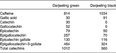
We measured the concentrations of various tea compounds, including the catechins, in both green tea and black tea preparations. The concentrations of single tea compounds are shown in Table 1. The overall catechin concentration in black tea was about half that in green tea. The concentrations of EGCG – one of the most potent catechins – were 30 % lower in black tea than in green tea.
Table 1 Concentrations of individual tea components (μm)

Measurement of endothelial nitric oxide synthase activity in endothelial cells
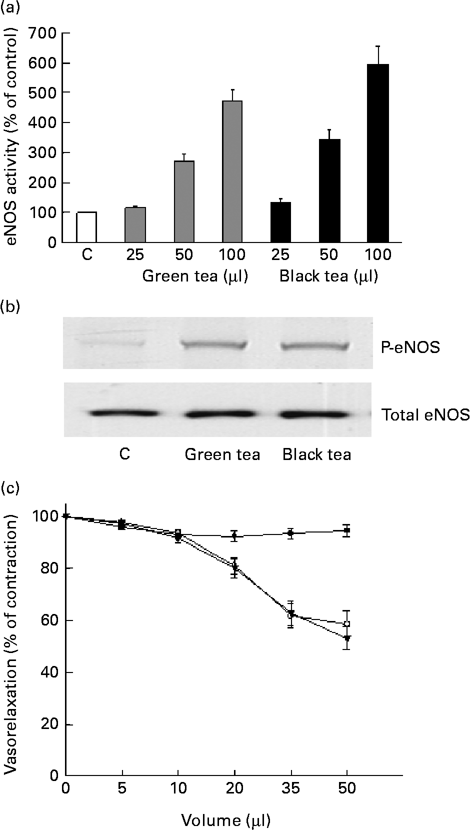
To clarify whether eNOS activation is one of the underlying mechanisms by which both teas enhance endothelial function, we incubated bovine aortic endothelial cells with increasing concentrations of green or black tea, and measured eNOS activity in intact cells. As shown in Fig. 1(a), incubation of cells with varying amounts of either green or black tea activated eNOS dose dependently. There was no difference in the magnitude of the increase in eNOS activity after treatment of cells with green or black tea. The final concentration of total catechins after addition of 100 μl of green and black tea to cells was 63·2 and 35 μm, respectively. Likewise, both green and black tea preparations induced comparable levels of eNOS phosphorylation (Fig. 1(b)).

Fig. 1 Impact of green and black tea preparations on NO production in endothelial cells and on vasorelaxation in aortic rings. (a) Cells were treated for 15 min with the indicated amounts of green or black tea, or with water (control; C), and endothelial NO synthase (eNOS) activity was measured. Values are means (n 5), with standard errors represented by vertical bars. (b) Endothelial cells were treated for 15 min with 100 μl of the indicated tea preparations. The Western blot showing the phosphorylation of eNOS (P-eNOS) was performed with an antibody specific to the eNOS Ser1179 phosphorylation site. Total eNOS served as the control for equal loading. The Western blot is representative of three independent experiments. (c) Precontracted rat aortic rings were treated with cumulative doses of either water (control; –●–; n 16), green tea (–○–; n 18) or black tea (–▾–; n 18), and vasorelaxation was determined. Vasorelaxation is expressed as percentage of contraction. Values are means, with standard errors represented by vertical bars.
Tea-induced vasorelaxation of rat aortic rings
To compare the effects of green and black tea on vasoreactivity, we exposed phenylephrine-precontracted rat aortic rings to cumulative doses of the respective tea preparation. Both green and black tea (n 18) induced pronounced, dose-dependent vasorelaxation in the aortic rings (Fig. 1(c)). In aortic rings, the final concentration of total catechins after addition of 10 μl of green and black tea was 1 and 0·56 μm, respectively. The degree of vasodilation evoked by green and black tea was virtually identical. Pretreatment of rat aortic rings with the NOS-inhibitor N-nitro-l-arginine methyl ester completely prevented tea-induced vasodilation, indicating that relaxations of rat aortic rings induced by green or black tea are due to generation of NO, and are hence endothelial dependent. The vasodilating effect of tea occurred rapidly, within minutes.
Endothelial function in human subjects
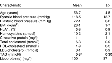
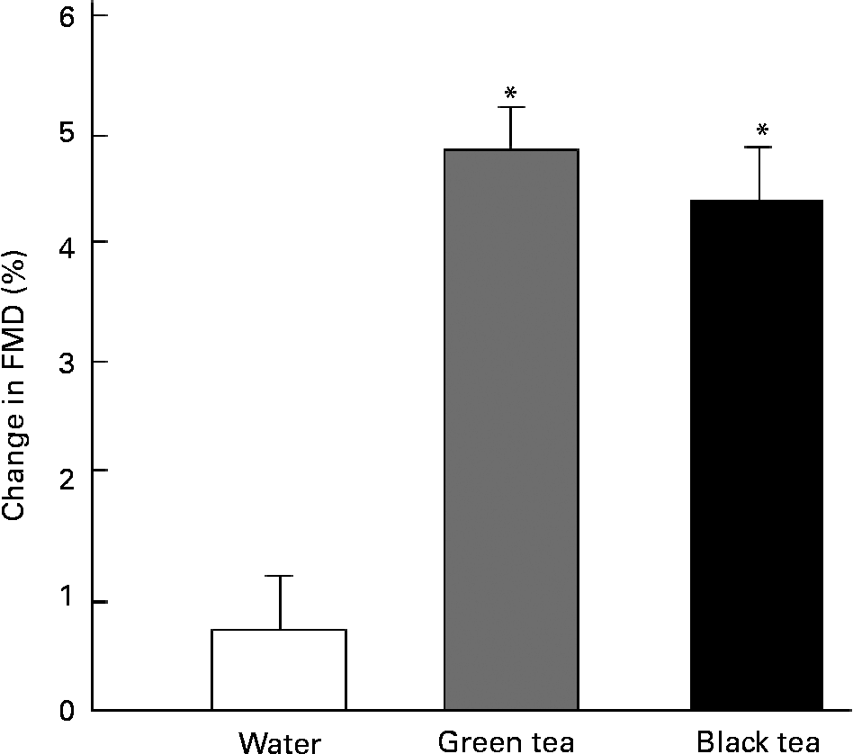
A total of twenty-one healthy postmenopausal women completed the study. Baseline characteristics of the study group are shown in Table 2. Endothelial function was assessed by measuring FMD of the forearm brachial artery, before and 2 h after ingestion either of 500 ml water (control), black tea, or green tea in a cross-over study. All results of the intervention study are presented in detail in Tables 3 and 4. Briefly, ingestion of green and black tea led to significant increase of FMD: from 5·4 (sd 2·3) to 10·2 (sd 3) % (baseline-adjusted difference for 2 h of FMD, green tea v. water: 5·0 (95 % CI 3·0, 7·0) %; P < 0·001), and from 5 (sd 2·6) to 9·1 (sd 3·6) % (baseline-adjusted difference for 2 h of FMD, black tea v. water: 4·4 (95 % CI 2·3, 6·5) %; P < 0·001), respectively (Fig. 2). The increase in FMD was not significantly different between the two tea preparations (baseline-adjusted difference for 2 h of FMD, green tea v. black tea: 0·66 (95 % CI − 0·76, 2·09) %; P = 0·36). Water showed no effect on FMD. Endothelial-independent NMD was not significantly different after consumption of the three beverages.
Table 2 Baseline characteristics of the study population
(Mean values and standard deviations for twenty-one subjects)

Table 3 Flow-mediated dilation (FMD) and nitro-mediated dilation (NMD) before and 2 h after consumption of water, green tea and black tea
(Mean values and standard deviations for twenty-one subjects)

FMDbase, diameter of brachial artery before hyperaemic stimulus; FMDmax, maximum dilation of brachial artery after hyperaemic stimulus, shown in mm and as maximum percentage change in brachial artery diameter compared with FMDbase; NMDbase, diameter of brachial artery before application of 0·4 mg nitroglycerine; NMDmax, maximum dilation of brachial artery after application of 0·4 mg nitroglycerine, shown in mm and as maximum percentage change in brachial artery diameter compared with NMDbase.
Table 4 Baseline-adjusted differences for 2 h of flow-mediated dilation (FMD) 2 h after consumption of green tea and black tea
(Percentages and 95 % confidence intervals for twenty-one subjects)

* P < 0·001.

Fig. 2 Changes in flow-mediated dilation (FMD) (%) after consumption of tea preparations. Volunteers consumed either 500 ml of water, green tea, or black tea, and changes in FMD were measured. Values are means for twenty-one subjects, with standard errors represented by vertical bars. * Mean value was significantly higher than that for water (P < 0·001).
Discussion
The present study is the first to compare the effects of green and black tea on endothelial function in vitro and in vivo. In both rat aortic rings and in healthy women, green and black tea significantly enhanced endothelial function in a comparable manner. The amount of tea-induced increase of eNOS activity in endothelial cells was furthermore not different after incubation of the cells with green or black tea. In the present study, we used freshly brewed tea (rather than tea extracts) for our in vivo as well as in vitro studies, to mimic real-life conditions. Although, as expected, the catechin content was much lower in black tea (Table 1), the efficacy of this fermented tea preparation in terms of amelioration of endothelial function was identical to green tea.
Several clinical studies have described beneficial effects of both green and black tea on endothelial functionReference Lorenz, Jochmann, Krosigk, Martus, Baumann, Stangl and Stangl11, Reference Duffy, Keaney, Holbrook, Gokce, Swerdloff, Frei and Vita17–Reference Hodgson, Puddey, Burke, Watts and Beilin19. We recently showed that addition of milk to black tea completely prevented the improvement of endothelial function by black tea alone in human subjects. In addition to these in vivo results, we were able to demonstrate a selective decrease in concentrations of catechins in tea with milk compared with tea without milk – findings that suggest an important role of catechins in black tea for amelioration of endothelial functionReference Lorenz, Jochmann, Krosigk, Martus, Baumann, Stangl and Stangl11.
The underlying molecular mechanisms for favourable tea-induced effects on endothelial function are multifactorial; they involve direct effects on the NO system, as well as antioxidative effects associated with an increase in NO availability. Although information is limited concerning the biological activities of theaflavins, and especially of thearubigins – the heterogeneous oxidation products that are the main polyphenols in black tea – there is increasing evidence that they are effective antioxidantsReference Yoshida, Ishikawa and Hosoai20, Reference Menet, Sang, Yang, Ho and Rosen21. However, data are not uniform with regard to the antioxidative capacity of theaflavins as compared with catechins. Lee et al. reported that the antioxidative capacity per serving of green tea is much higher than that of black tea in commercial tea productsReference Lee, Lee and Lee22. Some authors found that the antioxidant effects of theaflavins in black tea are equal to that of catechins in green teaReference Leung, Su, Chen, Zhang, Huang and Chen23. Yoshida et al. have shown that theaflavin digallate had a higher inhibitory effect on LDL oxidation in macrophages in vitro when compared with EGCG. The authors postulated that the higher antioxidative capacity of theaflavins is due to their greater number of hydroxyl (OH) groups, since theaflavins represent polymers of catechinsReference Yoshida, Ishikawa and Hosoai20. It is assumed that the degree of hydroxylation is positively correlated with their antioxidant efficiencyReference Rice Evans, Miller and Paganga24. Antioxidative activities of black tea thearubigins on lipid peroxidation have likewise been reportedReference Yoshino, Hara, Sano and Tomita25. In conclusion, these data suggest that the antioxidative effects of green and black tea may differ depending on the polyphenol composition.
Beyond the antioxidant properties of catechins and their oxidised products theaflavins and thearubigins, direct effects on the NO system may also contribute to improvement of endothelial function after green and black tea consumption. We have recently shown that EGCG, the main catechin in green tea, acts as a natural eNOS activator by directly enhancing eNOS activityReference Lorenz, Wessler, Follmann, Michaelis, Dusterhoft, Baumann, Stangl and Stangl8. In a similar manner, black tea polyphenols stimulate eNOS activity through activation of oestrogen receptor α in vascular endothelial cellsReference Anter, Chen, Shapira, Karas and Keaney26. In corroboration of these data, we found a comparable increase in eNOS activity by green and black tea in the present study.
At present, there are few data directly comparing the efficacy of green and black tea on cardiovascular effects. Here we show for the first time that green and black teas are equally effective in ameliorating endothelial function. Accordingly, drinking black tea may result in beneficial cardiovascular health effects similar to those of green tea. These results suggest that other polyphenolic compounds, such as theaflavins and thearubigins generated during oxidation of catechins in the process of black tea manufacture, may compensate for the reduced catechin content in black tea.
Acknowledgements
We would like to thank Angelika Vietzke, Thomas Düsterhöft and Wanda Michaelis for their excellent technical assistance.


