EDITOR:
One-lung ventilation is desirable for open thoracotomy or video-assisted thoracoscopic surgery to facilitate lung exposure for the surgical procedure by collapsing the lung. Double-lumen endotracheal tubes are commonly used for this purpose. The univent single-lumen tube with an endobronchial blocker, has some advantages over the double-lumen tube: easier insertion in patients with difficult airways [Reference Garcia-Aguado, Mateo, Onrubia and Bolinches1] and no need for tube exchange when postoperative mechanical ventilation is required. Fibreoptic bronchoscopy has been considered necessary to verify the position of the univent tube blocker [Reference Campos2,Reference Campos3]. This study was designed to evaluate whether correct position of the endobronchial blocker could be achieved without using a fibreoptic bronchoscope in right lung surgery patients.
The study was approved by our hospital review board. Written, informed consent was obtained from all patients. Sixty patients (18–75 yr old), undergoing thoracic surgery for which one-lung ventilation was required, were enrolled. In Group 1 (n = 30) the endobronchial blocker was advanced blindly as described below, and in Group 2 (n = 30) fibreoptic bronchoscopy was used.
The cuffs of the univent tube (Fuji Systems Corp, Tokyo, Japan) and bronchial blocker were tested for leaks before intubation. The bronchial blocker was lubricated with 10% lidocaine spray. The tube size was adapted to sex, height and weight of the patients (6.5 or 7.0 mm for females and 7.0 or 7.5 mm for males). Anaesthesia was induced with lidocaine 40 mg, propofol 2.5 mg kg−1 and rocuronium 0.6 mg kg−1 intravenously. The univent tube was inserted under direct laryngoscopy. In Group 1, once the tube cuff had passed the vocal cords, the tube was rotated 90° towards the right. The bronchial blocker was advanced sufficiently, and 4 mL of air was injected into its cuff. Breath sounds were auscultated to confirm whether the blocker was in the right bronchus (the case was considered a failure if it was in the left bronchus). The lumen at the distal end of the bronchial blocker was connected to a capnograph for analysis of end-tidal CO2 (etCO2) wave forms. If necessary, 1 mL at a time was added to the endobronchial cuff until the etCO2 wave form ceased, indicating complete blocking of the bronchus. Then, the bronchial blocker was slowly withdrawn until etCO2 reappeared. The scale mark on the blocker was noted and it was advanced 2.5 cm into the right bronchus again until etCO2 ceased. If breath sounds could be heard over the right upper lung field due to an unobstructed right upper lobe bronchus, the bronchial blocker was withdrawn 0.5 cm at a time until the sounds disappeared. At this stage, the position was checked by an independent observer using a fibreoptic bronchoscope (Olympus LF-GP, Tokyo, Japan). If the upper part of the endobronchial cuff was located just below or at the level of the carina, adequately blocking the right bronchus, the position was considered ideal.
At the end of the operation, when one-lung ventilation was no longer necessary, the deflated blocker was pulled back. Using a fibreoptic bronchoscope, the bronchial mucosa was carefully examined for signs of injury in the two groups.
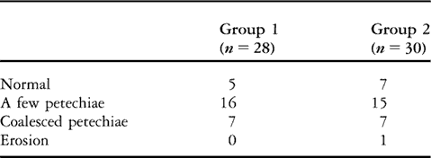
In Group 1, the blocker was inserted in the right bronchus at first attempt in all patients. In 28 of the 30 patients, it was in an ideal position. In this group, the blocker became dislocated during the operation in five cases. Reinsertion to the initial depth resulted in satisfactory lung collapse in all cases. In Group 2, the blocker was placed in an ideal position in all cases and there were five cases of dislodgement during the operation, and the blocker was reinserted under fibreoptic bronchoscopic control. There was no significant difference in bronchial injury between the two techniques of bronchial blocker placement (Table 1).
Table 1 Degree of mucosal injury at the site of the bronchial blocker evaluated by fibreoptic bronchoscopy.

Group 1: insertion without fibreoptic bronchoscope, Group 2: insertion under fibreoptic bronchoscopic control.
In this study, univent tubes with internal diameters of 6.5–7.5 mm were used. The length of the bronchial blocker cuff is equal (24 mm) in all these tube sizes. Therefore, we inserted the blocker in the right bronchus to the same depth initially in all cases and then made adjustments during auscultation of breath sounds. The insertion depth of the bronchial blocker has to be modified for ID 8.0 mm univent tubes because its cuff length is longer, 28 mm. Because of anatomical reasons, where the right mainstem bronchus continues at a more acute angle than the left one, the bronchial blocker had a tendency to enter into the right bronchus. In our study, blind insertion into the right bronchus was successful in all cases.
The bronchial blocker cuff is a low volume–high pressure device [Reference Hannallah4]. The manufacturer has recommended that 4–8 mL of air be used to seal the bronchus. The smallest volume possible should be used to prevent over-inflation, which might result in pressure damage to the bronchial mucosa [Reference Hannallah and Benumof5]. We inflated the blocker cuff gradually, starting at 4 mL. Once the wave form of etCO2 ceased without oscillation, we considered that the blocker balloon had sealed the entrance of the bronchus completely. In most cases, adequate lung collapse was achieved with 4 mL but in a few cases larger volumes were necessary.
A properly positioned blocker can dislodge either when the patient is turned to the lateral decubitus position or during surgical manipulation. This occurred in several cases in our study. There was no difference between the groups. In all cases, the blocker was repositioned to its previous depth and surgery was finished uneventfully.
We found that our technique to position the bronchial blocker of the univent tube, without the use of fibreoptic bronchoscopy for right lung surgery, resulted in satisfactory lung collapse in 28 out of 30 cases. There was no more bronchial injury than in patients in whom fibreoptic bronchoscopy was utilized. We do not propose the routine use of our method, but it can be used as an alternative when fibreoptic bronchoscopy is not available. A limitation of this technique is that it is not applicable for a selective lobar blockade or an anomalous bronchus such as a tracheal bronchus.
Acknowledgement
This work was supported by the St Vincent Hospital, College of Medicine, The Catholic University of Korea.


