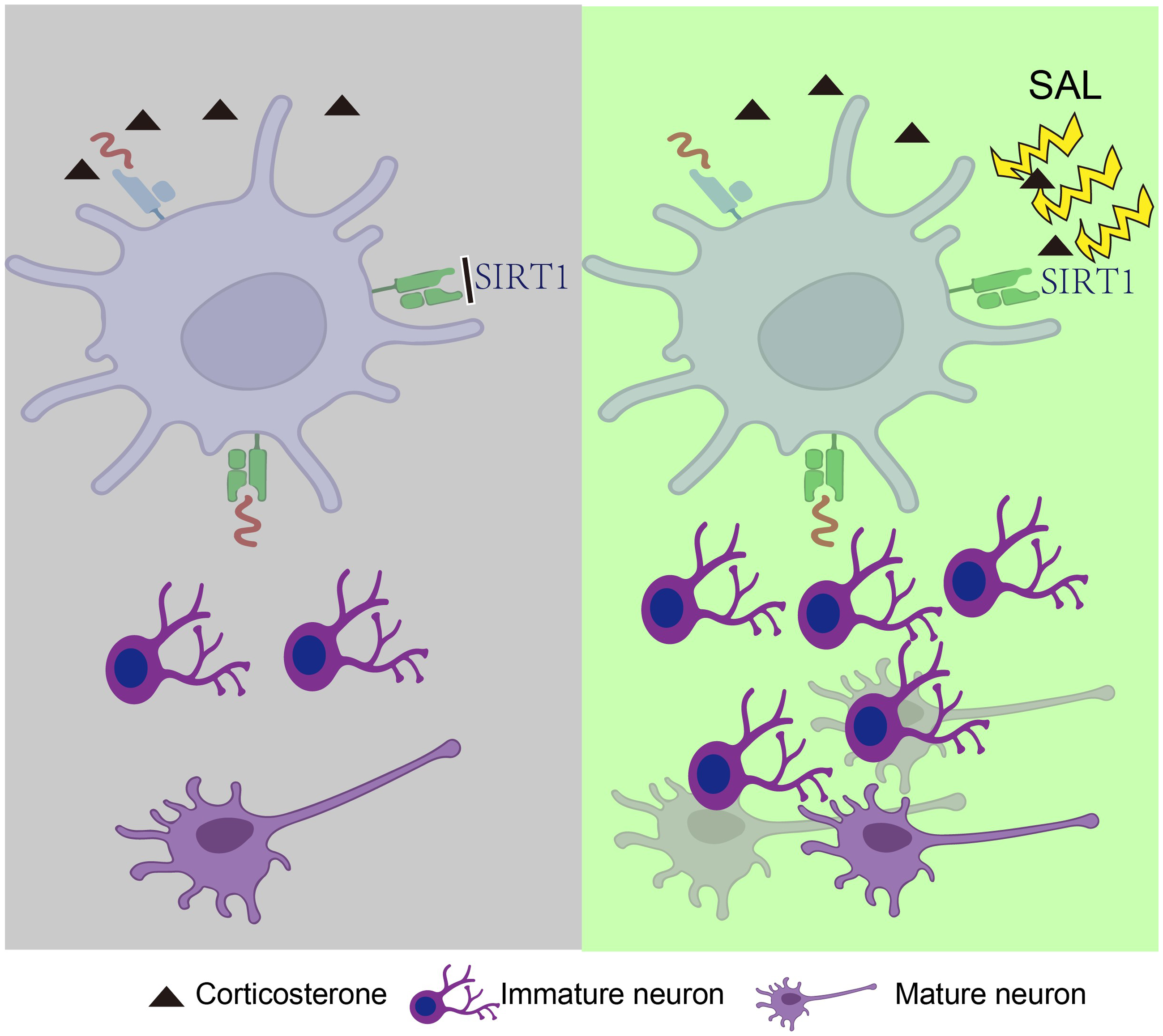
Significant outcomes
-
Salidroside could rapidly relieve depression-like behaviour in a depression model induced by CORT.
-
Salidroside might be partially attributed to an increase in cell proliferation in the hippocampal DG area.
-
Salidroside exerted antidepressant-like effects by promoting hippocampal neurogenesis through the SIRT1/PGC-1α signalling pathway.
Limitations
-
Our study of the antidepressant effects of SAL has been only validated in CORT depressed mice, and more animal models of depression are needed.
-
A single dose of SAL cannot significantly increase the expression of SIRT1, but relies on SIRT1 signalling to produce antidepressant effects, which may suggest that the effect of SAL is delayed, or that SAL can regulate an upstream signal of SIRT1 signalling to produce a cascade effect, which is still to be further explored.
Introduction
Depression is a frequent mental illness that is also a primary cause of disability. Some of the persistent symptoms of this condition include feelings of sadness, loss of interest in activities, thoughts of self-harm, slowed or restless movements, alterations in appetite and sleep patterns, decreased energy or heightened fatigue, and difficulty focusing (Lei et al., Reference Lei, Wang, Wang, Li, Liu, Fang, You, Guo and Lu2020). Depressive disorders have a highly complicated pathophysiology. Currently, there exist numerous hypotheses concerning the development and mechanisms of depression, including the hypothalamus-pituitary-adrenal axis in addition to clubhouse, monoamine neurotransmitter, immune and inflammatory, nerve regeneration, epigenetic, and glutamate system hypotheses (Jellinger, Reference Jellinger2023). The enhancement of synaptic protein expression, axon remodelling, neurotrophic, glial density and hippocampal pyramidal neurons are the microcosmic basis of antidepressant effects (Hassamal, Reference Hassamal2023). Hence, it is crucial to explore novel and powerful treatment methods to avert this catastrophic illness. In the past few years, there has been an increasing amount of research dedicated to the study of antidepressants by extracting bioactive components from plants found in nature. The overall aim is to develop medications that have notable effectiveness and minimal adverse reactions, ultimately benefiting individuals suffering from depression depression (Monchaux De Oliveira et al., Reference Monchaux De Oliveira, Pourtau, Vancassel, Pouchieu, Capuron, Gaudout and Castanon2021).
Salidroside (SAL), a phenylpropanoid glycoside discovered in the herb Rhodiola rosea L., possesses diverse pharmacological effects such as anti-inflammatory (Li et al., Reference Li, Fu, Zhang, Su, Liu, Guo, Li, Liang, Liu, Zhang, Cao, Zhang and Yang2013), anti-oxidative (Pan et al., Reference Pan, Zhu, Li, Zhang and Xu2023) and anti-fatigue (Wang et al., Reference Wang, Qian, Meng, Wang, Cheng, Zhou, Zhu and Liu2023), and regulation of cognitive functions. According to recent research, SAL has been found to demonstrate anti-stress properties and act as an antidepressant in models of immobilisation stress and behavioural despair respectively (Panossian et al., Reference Panossian, Hambardzumyan, Hovhanissyan and Wikman2007, Reference Panossian, Nikoyan, Ohanyan, Hovhannisyan, Abrahamyan, Gabrielyan and Wikman2008; Chai et al., Reference Chai, Cai, Fu, Wang, Zhang, Zhang, Zhu, Miao and Yan2022a). In the meantime, SAL has the ability to demonstrate the promotion of neurogenesis in the hippocampus and exerts potent antidepressant-like impacts in chronic stress-depressed or despair animals (Vasileva et al., Reference Vasileva, Saracheva, Ivanovska, Petrova, Marchev, Georgiev, Murdjeva and Getova2018). SAL has protective effects on various diseases such as Alzheimer’s disease (Chen et al., Reference Chen, Liu, Qu, Guo, Zhang, Kong, Yu, Liu, Zang, Li and Li2023). The objective of this study is to investigate the mechanisms underlying depression and the potential antidepressant properties of SAL, along with its corresponding mechanism of action.
Sirtuin 1 (SIRT1) is a NAD-dependent deacetylase that plays an important role in cellular metabolism (Lei et al., Reference Lei, Wang, Wang, Li, Liu, Fang, You, Guo and Lu2020). Previous researches suggest that SIRT1 play critical roles in the pathogenesis and therapy of depression (Abe-Higuchi et al., Reference Abe-Higuchi, Uchida, Yamagata, Higuchi, Hobara, Hara, Kobayashi and Watanabe2016; Kishi et al., Reference Kishi, Yoshimura, Kitajima, Okochi, Okumura, Tsunoka, Yamanouchi, Kinoshita, Kawashima, Fukuo, Naitoh, Umene-Nakano, Inada, Nakamura, Ozaki and Iwata2010; Libert et al., Reference Libert, Pointer, Bell, Das, Cohen, Asara, Kapur, Bergmann, Preisig, Otowa, Kendler, Chen, Hettema, van den Oord, Rubio and Guarente2011; CONVERGE consortium et al., 2015). Studies have indicated that SIRT1 in excitatory neurons in the forebrain is an important regulator of depression-related behaviours (Lei et al., Reference Lei, Wang, Wang, Li, Liu, Fang, You, Guo and Lu2020). It has been shown that SIRT1 activation is sufficient to reverse chronic stress-induced depressive behaviours, and SIRT1 activity in mPFC pyramidal neurons is a key player in regulating depression-related behaviours (Lei et al., Reference Lei, Wang, Wang, Li, Liu, Fang, You, Guo and Lu2020a). Peroxisome proliferator-activated receptor-γcoactivator-1α (PGC-1α), a transcriptional co-regulator, regulates the formation of mitochondria and enhances oxidative phosphorylation (Yan et al., Reference Yan, Liu, Xu, Qian, Song, Wei and Liu2023). PGC-1α exhibits moderate expression in brain tissue and is present in brain regions linked to depression, including the hippocampus (Lv et al., Reference Lv, Zuo, Liu, Zhao, Wang, Wang, Yu, Yu, Zhang, Bi and Liu2023). It is shown that the incidence and progression of depression may be directly associated to reduced PGC-1α expression in the hippocampus (Wang et al., Reference Wang, Tang, Liang, Luo, Zhu, Li, Xiao, Jiang, Yang, Xie, Zhang, Deng, Li and Tang2021). Brain-derived neurotrophic factor (BDNF) modulates neuronal survival and neuroplasticity in the brain, with important roles in the growth, survival, differentiation, and repair of neurons. Upregulation of BDNF contributes to significant antidepressant effects (McEwen et al., Reference McEwen, Bowles, Gray, Hill, Hunter, Karatsoreos and Nasca2015; Duman et al., Reference Duman, Aghajanian, Sanacora and Krystal2016). Physical activity increases neuronal gene expression of FNDC5 (which encodes the PGC-1α-dependent myokine FNDC5), thereby potentially boosting BDNF levels (Pedersen, Reference Pedersen2019). SIRT1 has been found to be closely associated with increased BDNF expression in many antidepressant studies (Qiu et al., Reference Qiu, Lu, Zeng, Jin and Chen2023; Cui et al., Reference Cui, Zhao, Amevor, Du, Wang, Li, Shu, Tian and Zhao2022), so it is likely that SIRT1 can affect BDNF expression, thereby enhancing synaptic plasticity and producing antidepressant effects. Our research’s goal is to investigate SAL’s possible antidepressant effects as well as the potential mechanism.
Materials and methods
Animals
Male ICR mice weighing between 18 and 22 g were obtained from Shanghai Slack Experimental Motion Co., Ltd in Shanghai, China, one week before the start of the experiment. The mice were kept in an environment with a temperature of 22 ± 1 °C and a humidity level of 60%, following a dark/light cycle of 12 h each. Every single creature had unrestricted availability to nourishment and hydration. Ethical approval was obtained from the Institutional Animal Care and Use Committee of Nanjing University of Chinese medicine, China, ensuring that all animal experiments adhered to the approved ethical policies and procedures (Approval no. 202206A039).
Drugs
Salidroside (purity ≥98%, CAS NO. 10338-51-9) and Selisistat (purity ≥98%, CAS No. 49843-98-3) was provided by Nantong Feiyu Biological Technology Co., Ltd (Nantong, China). Rhodiola rosea L(90g) herbs were purchased from Nanjing University of Chinese Medicine Chinese Pharmacopoeia, had been identified by experts from the College of Pharmacy of our University. The herbs were soaked with water for 30 min in a 1:8 ratio and then heated for 1 h. Subsequently, the solution was filtered and collected. This procedure was repeated twice. All collected solution was placed in a water bath at 60°C and evaporated to a designated concentration. The yield of the extraction was 20%. Rhodiola rosea L (2 g/kg) was administrated via intragastric gavage (i.g.). The dosage of each drug followed the Chinese Pharmacopoeia (version 2020). Resveratrol (purity ≥97%, CAS NO. 56296-78-7) was purchased from Dalian Meilune Biological Technology CO., Ltd (Dalian, China). Corticosterone (purity ≥98%, CAS NO.50-22-6) was purchased from Shanghai Aladdin Biochemical Technology CO., Ltd (Shanghai, China). Bromodeoxyuridine (BrdU, 19-160) was provided by Sigma Aldrich (St. Louis, USA). Salidroside (25, 50, 75, 100, 200 mg/kg) was dissolved in normal saline (0.9%) and administered intragastrically (i.g.). Resveratrol (20 mg/kg, intraperitoneal injection [i.p.]) and BrdU (50 mg/kg, i.p.) were dissolved in normal saline and administered intraperitoneally. Corticosterone (30 mg/kg, i.g.) was dissolved in 5% dimethyl sulfoxide.
Sucrose preference test (SPT)
The SPT was conducted according to former research with slight alteration (Zhang et al., Reference Zhang, Huang, Cheng, Wang, Chao, Li, Zheng, Xie, Zhang and Yao2019). To summarise, the animals were accustomed to a 3% sucrose solution (w/v) and water for a period of 24 hours, after which they underwent 12 hours of deprivation from both food and water. Mice were presented with a choice between two containers, one containing a 3% sucrose solution (w/v) with a volume of 150 ml, and the other containing water. The bottles were weighed before or after the test. The sucrose preference was determined by applying the formula after calculating the water and sucrose solution consumption. Sucrose preference (%) = [(sucrose consumption)/ (sucrose consumption + water consumption)] *100%.
Tail suspension test (TST)
The TST is a recognised mouse model for assessing the antidepressant effect (Zhang et al., Reference Zhang, Huang, Cheng, Wang, Chao, Li, Zheng, Xie, Zhang and Yao2019). Animals were suspended in an environment that was isolated both visually and acoustically. They were lifted 50 cm above the ground using adhesive tape positioned 1 cm away from the tip of their tails for a duration of 6 min. The animals were observed exhibiting immobile actions while being suspended in a passive and motionless state. The duration of immobility was evaluated during the last 4 min using the ANY-maze programme.
Force swimming test (FST)
The FST was carried out based on the previous method with slight modification (Zhang et al., Reference Zhang, Huang, Cheng, Wang, Chao, Li, Zheng, Xie, Zhang and Yao2019). For a duration of 6 min, animals were permitted to swim in a transparent cylindrical container measuring 30 cm in height and 15 cm in diameter. The container was filled with water at a temperature of 24 ± 2°C, reaching a depth of 12 cm. The duration of immobility, which is defined as the absence of any movement of the limb or body except for the essential movements required for the mice to keep their heads above water, was analysed using the ANY-maze software.
Open field test (OFT)
The OFT was examined by previous OFT method with minor changes (Tunc-Ozcan et al., Reference Tunc-Ozcan, Peng, Zhu, Dunlop, Contractor and Kessler2019) . For the OFT, a black metallic device measuring 40 by 40 by 15 cubic centimetres, which was practically divided into four identical squares, was utilised. Every single creature was individually positioned at the middle of the arena, with their gaze directed towards the wall, and then observed and filmed for a duration of 5 minutes. The analysis of the time spent at the centre and the overall distance covered was done automatically. During the OFT test, the box underwent cleaning using alcohol with a concentration of 75%. The software ANY-maze was used to analyse the results.
Immunofluorescence analysis
The immunofluorescence analysis for brain tissues was performed in accordance with the previous report (Tunc-Ozcan et al., Reference Tunc-Ozcan, Peng, Zhu, Dunlop, Contractor and Kessler2019). The mice were euthanized after the completion of the behaviour tests. The tissues were flushed with 4% paraformaldehyde, submerged in a solution of 20–40% sucrose, and kept at a temperature of 4°C to prepare frozen sections. Using a cryostat (Leica CM1950), the entire hippocampus was sliced for the samples. The samples were obstructed using 3% BSA at a temperature of 25 °C, then subjected to overnight incubation at 4 °C with anti-BrdU, anti-NeuN, anti-Ki-67, and anti-doublecortin (anti-DCX) antibodies. Afterwards, the slides underwent treatment with a secondary antibody that emits fluorescence. The microscope was used to visualise the proportions of BrdU+, NeuN+, DCX+, Ki-67+ cells, and dividing cells. Image J was used to analyse the images, and the Kruskal-Wallis test was employed to investigate statistical significance. The quantity of BrdU+, NeuN+, DCX+, Ki-67+ cells was determined across the rostral/caudal range of the DG area (bregma 0.82 mm to 4.16 mm) using 6 consecutive sections spaced at intervals of 180-lm. The portions were repaired using 4% paraformaldehyde, rehydrated, and then subjected to 0.2% TritonX-100 treatment for a duration of 5 minutes. Afterwards, the segments were obstructed using 3% BSA. After washing, the slices were treated with anti-BrdU, anti-NeuN, anti-DCX, anti-Ki-67, primary antibodies at 4°C overnight. Subsequently, the cells were subjected to Alexa Fluor 594 labelled secondary antibodies at a temperature of 25 °C, and then stained for approximately 15 minutes using a solution of 40-6-Diamidino-2-phenylindole. The confocal laser-scanning microscope (Olympus) was used to analyse the immunofluorescent images.
Western blot
The western blot was performed according to a former protocol with minor modifications (Ma et al., Reference Ma, Shen, Hu, Wang, Yang, Lv, Wang, Zhou, Jin, Sun, Zhang, Qiu, Pan, Zhou, Fan, Yang and Guo2019). After sacrificing the mice, the brain tissues were promptly collected. Concentrations of overall proteins were assessed using a commercially available bicinchoninic acid kit. For electrophoresis, the sample underwent gel electrophoresis using 10% polyacrylamide gels. Subsequently, the proteins were transferred onto a polyvinylidene fluoride membrane and blocked using 2% BSA for 1 hour. Following that, the membranes were incubated with suitable primary antibodies overnight at 4 °C. After being washed three times with TBST, the samples were treated with horseradish peroxidase (HRP) anti-rabbit secondary antibody conjugated with immunoglobulin (IgG) HRP at room temperature for 1 h. As a result, the proteins were observed using an improved chemiluminescence kit. Image J software was utilised to quantify the Bands and then normalised to Tubulin.
Statistical analysis
The mean ± SEM results were presented and analysed using GraphPad Prism 7 software. Unpaired student’s t-test was utilised for conducting two-sample comparisons. The various comparisons were conducted utilising one-way analysis of variance (ANOVA) accompanied by a Bonferroni post hoc analysis or two-way ANOVA with a Tukey post hoc analysis. Statistical significance was determined as p < 0.05, unless otherwise specified.
Results
Salidroside exhibited antidepressant-like response after single treatment
In order to clarify the antidepressant properties of salidroside (SAL), male ICR mice were administered varying doses of SAL (25 mg/kg, 50 mg/kg, 75 mg/kg, 100 mg/kg, 200 mg/kg) through intraperitoneal injection, followed by behavioural testing after 24 h. The behavioural findings indicated a notable decrease in the duration of sustained immobility in TST for mice treated with SAL at both the 25 mg/kg and 50 mg/kg dosages (Figure 1A, F (5, 59) = 12.20, p < 0.001). However, there was no significant impact on the overall distance of mouse movement in OFT (Figure 1B, F (5, 56) = 4.091, p < 0.005) and time of centre stay (Figure 1C, F (5, 51) = 2.020, p = 0.0914). These findings suggested that the antidepressant-like effects of SAL were not influenced by its impact on motor activity.
Salidroside produced rapid antidepressant-like effects in CORT-induced depression-like models
To further investigate the antidepressant effects of SAL, mice received corticosterone (CORT) (30 mg/kg/d, i.g.) for approximately 3 weeks to establish a model of depression, followed by treatment with SAL (25 mg/kg/day, i.p.), Rhodiola rosea L (RHO, 2 g/kg, i.g.) and resveratrol (RES, 20 mg/kg /day, i.p.) for 3 weeks in this study. We performed OFT, SPT, TST, and FST (Figure 2A). Following a 2-week CORT treatment, there was a notable decline in the sucrose preference percentage when compared to the control group (Figure 2B, F (4, 20) = 10.91, p < 0.01). SAL administration gradually increased the sucrose consumption, showing significant differences at the end of 5 weeks of treatment compared to the saline-treated CORT group (Figure 2C, F (4, 25) = 38.38, p < 0.01). Consistently, the administration of SAL, RHO, and RES for a period of 3 weeks resulted in a significant decrease in the immobility duration in CORT mice during the TST (Figure 2D, F (4, 21) = 16.84, p < 0.05) and FST (Figure 2E, F (4, 31) = 13.92, p < 0.01). These findings suggested that the efficacy of SAL was demonstrated in mice with a CORT depression-like model.
Salidroside affected the expression of SIRT1 and neurogenesis in CORT model mice
Hippocampal neurogenesis is one of the important mechanisms of antidepressant action, and we also explored whether SAL can also promote hippocampal neurogenesis. Initially, the markers Ki-67, which indicates cell proliferation, and DCX, which indicates immature neurons, both showed a significant decrease in the number of positive cells after CORT induction. However, after SAL, RHO, RES treatments, there was a significant increase in the number of positive cells (Figure 3A–B, F (4,13) = 14.55, p < 0.005). In the meantime, BrdU (Figure 3C–D, F (4,14) = 18.28, p < 0.05), NeuN (Figure 3E–F, F (4,13) = 6.163, p < 0.05) cells in the hippocampus of CORT mice was found, while SAL significantly rescued the loss of BrdU (p < 0.05) and NeuN (p < 0.05) cells in the CORT model. However, SAL effectively restored the loss of BrdU (p < 0.05) and NeuN (p < 0.05) cells in the CORT model, along with RES (BrdU, p < 0.05; NeuN, p < 0.05). Changes in these typic neurogenesis markers suggested that SAL, RHO, and RES could significantly enhance the hippocampal neogenesis.
Furthermore, SIRT1 could induce many synaptic plasticity related protein to perform antidepressant-like effect. Thus, we tested the downstream proteins expression. SAL treatment could significantly improve the expression of SIRT1 protein (Figure 4B, F (3,8) = 10.59, p < 0.005) and its downstream protein PGC-1α (Figure 4C, F (3,10) = 5.662, p < 0.05), pCREB/CREB (Figure 4D, F (3,8) = 7.152, p < 0.05), BDNF (Figure 4E, F (3,9) = 12.16, p < 0.005), AKT (Figure 4F, F (3,8) = 15.13, p < 0.005), MTOR (Figure 4G, F (3,11) = 36.26, p < 0.005) and PI3K (Figure 4H, F (3,9) = 24.12, p < 0.005) in the hippocampus of CORT-induced mice. These findings suggested that SIRT1 might help SAL exert antidepressant-like effects in a mouse model of CORT.
The antidepressant-like effects of salidroside might be dependent on SIRT1
To verify whether SIRT1 is a key target for SAL in the treatment of depression, we further performed experiments on ICR mice treated with the SIRT1 inhibitor EX527. Single SAL could not significantly increased SIRT1 expression for only once administration. While ICR mice given EX527 in combination with or without SAL were more likely to express lower levels of SIRT1 than ICR mice given saline or SAL (Figure 4A–C; B, F (3, 8) = 21.63, p < 0.005; C, F (3, 10) = 0.50, p < 0.005). It implied that EX527 effectively blocked the SIRT1 expression while SAL could not reverse the trend. Furthermore, ICR mice treated with SAL alone significantly reduced immobility time compared to CON mice, while pre-treated with EX527 could block the effect both in TST (Figure 5D, F (3, 46) = 5.789, p < 0.005) and FST (Figure 5E, F (3, 45) = 5.155, p < 0.005), and induced no effect on locomotory activity (Figure 5F, F (3, 17) = 1.807, p = 0.486). Consistent with previous results, SAL treatment mitigated the decrease of DCX+ (Figure 4G-H; H, F (3, 28) = 22.19, p < 0.001) and Ki-67+ (Figure 4I-J; J, F (3, 36) = 24.73, p < 0.001) cell numbers after EX527 was pre-treated. It indicated that the antidepressant-like effect induced by SAL was mostly depends on SIRT activity.
According to previous SAL dose screening, half doses of SAL (25 mg/kg) did not produce similar antidepressant-like behaviour in normal mice. A review of previous literature showed that half a dose of RES (5 mg/kg) also failed to produce similar antidepressant-like behaviour in normal mice. RES was demonstrated to produce antidepressant-like effect via SIRT1 signalling pathway. To further confirm the relationship between the antidepressant-like effect of SAL and the SIRT1 signalling pathway, we used a combination of sub-dosage of SAL and RES to test whether they can co-activate SIRT1 signalling. Normal mice adapted for one week were randomly divided into 4 groups and given one intraperitoneal injection of normal saline, half dose SAL, half dose of RES, and half dose of SAL plus half dose of RES, respectively, behavioural tests were performed 24 h after administration. Compared with the SAL group or the RES group, the immobility time of mice in the co-treated group decreased significantly both in TST (Figure 6A, F (3, 20) = 4.945, p < 0.005) and FST (Figure 6B, F (3, 20) = 9.468, p < 0.001). In the meantime, there were no notable disparities in the overall distance and central time of the initial experiment in mice that received the treatment compared to the control group (Figure 6C and D; C, F (3, 20) = 0.05985, p = 0.9803; D, F (3, 20) = 1.050, p = 0.3924). The behavioural results suggested that half dose of SAL plus half dose of RES exhibited similar antidepressant-like behaviour in normal mice by enhancing SIRT1 expression.
Discussion
Salidroside (SAL) is collected from Rhodiola rosea L(RHO (Qu et al., Reference Qu, Liu, Liang, Zheng, Zhang and Lu2022)), while both of them display antidepressant-like effect in previous research. Our research further confirmed that SAL and RHO could produce antidepressant-like effect without significant side effect. Furthermore, its regulatory properties of Sirtuin-1 (SIRT1) pathway may provide the possibility of facilitation in hippocampal neurogenesis. Similar like resveratrol(RES), which is the agonist of SIRT1, also was demonstrated to against depressive-like behaviour in rodents (Cao et al., Reference Cao, Dou and Li2018a). However, significant side effects also limit the possibility of RES as an antidepressant in the first line of clinical use. Our study of SAL is likely to be used as a new alternative to antidepressants in clinical treatment.
SAL is a bioactive tyrosine-derived phenolic natural product found in medicinal plants under the Rhodiola genus (Hu et al., Reference Hu, Wang, Ni, Wang, Liu, You, Wu, Wang, Lu and Wei2020). Recent studies have reported that SAL exerts various pharmacological effects such as anti-inflammatory, antioxidant activities (Lv et al., Reference Lv, Zuo, Liu, Zhao, Wang, Wang, Yu, Yu, Zhang, Bi and Liu2023). Other studies have demonstrated that SAL can ameliorate cognitive and motor abilities by suppressing the dysfunction of hippocampal neurons (Torrens-Spence et al., Reference Torrens-Spence, Pluskal, Li, Carballo and Weng2018). The research showed that SAL possesses the effects of immunity enhancement, hypoxia inhibition, and tumour treatment (Li and Chen, Reference Li and Chen2017). RHO, as a tonic Chinese medicine, has the efficacy of promoting blood circulation. In the past 30 years, the knowledge about the mechanism of potential use of it in potential ageing-related diseases has increased, especially the emerging role of RHO in maintaining energy homeostasis (Bai et al., Reference Bai, Deng, Wu, Li and Jin2019) and the research of RHO in neuroprotection (Calabrese et al., Reference Calabrese, Dhawan, Kapoor, Agathokleous and Calabrese2023).We investigated the antidepressant-like properties of SAL and the mechanisms involved in our research. The results showed that SAL could rapidly improve immobility behaviour without affect locomotor activity, and SIRT1 protein expression was also modulated, thereby improving CORT-induced depression-like behaviour.
SIRT1 (Silent Information Regulator 2 homologue 1), also known as silent information regulator 2 homologue 1, is a histone deacetylase that relies on nicotinamide adenine dinucleotide (NAD) for its activity. Mammals possess a total of seven sirtuins, namely SIRT1 to SIRT7 (Wątroba et al., Reference Wątroba, Szewczyk and Szukiewicz2023; Kojima et al., Reference Kojima, Dai, Suzuki, Ono and Nishi2023). Numerous scientific investigations have demonstrated that sirtuins have a crucial impact on the development and differentiation of trophoblasts. This is not limited solely to SIRT1. Furthermore, SIRT1 has sparked significant fascination regarding its contribution to enhancing cognitive disorders (Cao et al., Reference Cao, Dou and Li2018). SIRT1 is present in the majority of brain areas, but it is particularly noticeable in the neuronal nuclei of the hippocampus, thalamus, and the solitary tract nucleus (Jablonska et al., Reference Jablonska, Gierdalski, Chew, Hawley, Catron, Lichauco, Cabrera-Luque, Yuen, Rowitch and Gallo2016). Caloric restriction and physical exercise can reverse the reduction of SIRT1 levels in the brain caused by neuronal ageing, a diet high in fat, and different neuropathological conditions. SIRT1 plays a vital role in various interconnected regulatory networks, influencing the development of dendrites and axons, which are crucial for neuronal plasticity and cognitive growth, and safeguarding them from stress (Ng, Reference Ng2015). SIRT1 deficient mice displayed generally intact brain structure, yet showed impairments in dendritic growth and synaptic flexibility (Michan et al., Reference Michan, Li, Chou, Parrella, Ge, Long, Allard, Lewis, Miller, Xu, Mervis, Chen, Guerin, Smith, McBurney, Sinclair, Baudry, De Cabo and Longo2010). In this study, SIRT1 had been shown to be an important molecule involved in the antidepressant effects of SAL, mediating the enhancement of neurogenesis in hippocampus.
There is growing evidence of the value of plant compounds (Sun et al., Reference Sun, Zhang, Wu, Yu, Yin, Qian, Wang, Huang, Wang, Liu, Xue and Chen2021) and other natural substances in the treatment of mental illness (Pandi et al., Reference Pandi, Sattu, Kalappan, Lal, Varikasuvu, Ganguly and Prasad2022). Resveratrol (3,5,4′-trihydroxybenzene) is a phytotoxin and polyphenol found mainly in the skins of red grapes, red wine, Japanese knotweed, and some nuts (Pastor et al., Reference Pastor, Restani, Di Lorenzo, Orgiu, Teissedre, Stockley, Ruf, Quini, Garcìa Tejedor, Gargantini, Aruani, Prieto, Murgo, Videla, Penissi and Iermoli2019). It has been extensively studied for its antioxidant, anti-inflammatory and anti-cancer properties. Resveratrol has been shown to positively affect a variety of animal models of depression by mechanisms including modulation of the HPA axis, reduction of inflammation and oxidative stress, increased neurogenesis, and increased monoamine production (Bohara et al., Reference Bohara, Tabassum, Singh, Gigli, Ragusa and Leporatti2022). Clinical studies have also confirmed the efficacy of resveratrol in the treatment of monophasic depression (Aftanas et al., Reference Aftanas, Markov, Rikita and Danilenko2020). Resveratrol has better efficacy as a natural antidepressant with fewer side effects, so we chose resveratrol as the positive control drug.
A wealth of research conducted in various animal models demonstrates that RES protects against cognitive decline through the activation of SIRT1 (Du et al., Reference Du, Xie, Cheng, Li, Kong, Jiang, Ma, Wang, Chen and Zhou2014) or promoting SIRT1 expression (Zhang et al., Reference Zhang, Tu, Wang, Hu, Li, Cheng, Yin, Li and Huang2017). To verify whether SIRT1 is a key target for SAL in the treatment of depression, we further conducted experiments on ICR mice treated with the SIRT1 inhibitor EX527. The reduced immobility time of ICR mice in FST and TST was blocked when treated with both EX527 and SAL compared to mice treated with SAL alone, while SIRT1 expression were not affected by the drugs. Besides, single injection of SAL could not increase the SIRT1 expression but rely on SIRT1 signalling, it may imply that the activated SIRT1 expression induced by SAL delay on the behaviour phenotype and it is the worth to investigate the key mechanism furthermore. Furthermore, the administration of SAL in our research led to an increase of DXC and Ki-67 positive cells in the CORT-induced mice hippocampus, these results implied that SIRT1 might have a part in the process of neurogenesis during the treatment of SAL in depressive mice, and in wide-type mice, the antidepressant-like effect rapidly induced by SAL but not immediately increased SIRT1 activity.
The transcriptional coactivator peroxisome proliferator-activated receptor-γ coactivator (PGC-1α), termed the ‘master regulator of mitochondrial biogenesis’ (Ryan et al., Reference Ryan, Patterson and McLoughlin2019), has been implicated in stress and resilience to stress-induced depressive-like behaviours in animal models. Animal research findings, PGC-1α the forced swimming immobility time of gene knockout mice was significantly longer than that of the control group, showing obvious depressive behavior (Babaei et al., Reference Babaei, Nourshahi, Fani, Entezari, Jameie and Haghparast2021). Additionally, our experiment demonstrated that SAL intervention led to an increased expression of PGC-1α in the hippocampus, suggesting that SAL has the potential to alleviate depressive behaviour. The results of our study indicated that the levels of PGC-1α in the hippocampus was rised following SAL intervention, regardless of whether it was in the EX527 inhibitor experiment or the experiment involving CORT-induced depression in mice. To sum up, we speculated that the antidepressant effect of SAL was exerted through SIRT1/PGC-1α pathway.
Besides, it has been reported that patients with depression have an overactive hypothalamic-pituitary-adrenal (HPA) axis and increased cortisol levels (Keller et al., Reference Keller, Gomez, Williams, Lembke, Lazzeroni, Murphy and Schatzberg2017). Repeated injections of CORT in animals cause deregulation of the HPA axis, neuronal damage, cognitive and memory decline, and induce depression-like behaviour (Badr et al., Reference Badr, Attia and Al-Rasheed2020). Thus, CORT-induced model has been widely used as a chronic model of depression caused by stress (Kinlein et al., Reference Kinlein, Phillips, Keller and Karatsoreos2019). In addition, high concentrations of CORT have been used to induce injury in PC12 cells, which emulates pathological mechanisms of depression (Mao et al., Reference Mao, Huang, Ip, Xian and Che2012). Therefore, CORT-induced depression model is a reliable model for screening antidepressants and exploring their pharmacological mechanisms. Our results showed that CORT administration significantly decreased sucrose preference rate, increased the time of immobility in TST and FST. Nonetheless, the administration of SAL significantly enhanced the behavioural abnormalities in SPT, TST, and FST, suggesting that SAL mitigates depressive behaviour in mice. At the same time, after SAL was given to CORT-induced mice, not only the number of DCX+ and Ki-67+ cells in hippocampus increased compared with CORT-induced mice, but also the number of BrdU and NeuN cells, the markers of neurogenesis, was equivalent to that in the blank group.
The pathogenesis of depression is very complex. At present, there are many theories about the pathogenesis and pathological mechanism of depression, including glutamate system hypothesis, hypothalamus-pituitary-adrenal axis plus clubhouse, and neurotrophic factor hypothesis, monoamine neurotransmitter hypothesis, immune and inflammatory hypothesis, nerve regeneration hypothesis, epigenetic hypothesis, etc. The nerve regeneration hypothesis, as one of the accepted hypotheses, believes that the hippocampus nerve of depression patients is damaged, and antidepressants can play an antidepressant effect by increasing BDNF to promote nerve regeneration. BDNF is involved in the growth and maintenance of neurons and is essential for cognitive function (Phillips, Reference Phillips2017). Research has shown that the level of BDNF in the hippocampus and prefrontal cortex is significantly reduced in patients with depression (Taliaz et al., Reference Taliaz, Stall, Dar and Zangen2010) The findings of this research demonstrated that SAL effectively improved the inhibition of hippocampal BDNF expression caused by CORT.
There are studies show that SAL ameliorates depression through microglial activation suppression (Fan et al., Reference Fan, Bi and Chen2021), by suppressing NLRP3-mediated Pyroptosis via P2X7/NF-κB/NLRP3 signalling pathway (Chai et al., Reference Chai, Cai, Fu, Wang, Zhang, Zhang, Zhu, Miao and Yan2022b). In our research, we demonstrated that the administration of SAL and RHO could relieve depression-like behaviour in a depression model induced by CORT. Furthermore, the antidepressant effects of SAL might be partially attributed to an increase in cell proliferation in the hippocampal DG area. Furthermore, it had been shown that the enhancement of neurogenesis was associated with the stimulation of SIRT1 in the SAL. Moreover, it was discovered that the stimulation of SIRT1 and increase in PGC-1α levels were linked to the safeguarding caused by SAL. However, due to the lethality of CORT in the mouse model of induced depression, mice in the Rhodiola rosea glycoside extract group in this study were sacrificed more during the experiments, and the final number of animals remaining was only able to support the behavioural and immunofluorescence experiments, and thus the Rhodiola rosea extract was not ultimately investigated in subsequent studies of the molecular mechanisms. It will be further improved in the follow-up. The findings indicated that SAL could potentially prevent depression by reversing the reduction in hippocampal neurogenesis caused by CORT. Additional research is required to assess the impact of compromised hippocampal neurogenesis as a contributing element to depression. Besides, there are still some shortcomings in our study, such as that a single dose of SAL cannot significantly increase the expression of SIRT1, but relies on SIRT1 signalling to produce antidepressant effects, which may suggest that the effect of SAL is delayed, or that SAL can regulate an upstream signal of SIRT1 signalling to produce a cascade effect, which is still to be further explored. However, our data suggests that the SAL have an impact on the generation of new neurons in the hippocampus, serving as a foundation for enhancing and progressing a novel antidepressant.
Supplementary material
The supplementary material for this article can be found at https://doi.org/10.1017/neu.2024.28.
Data availability statement
The datasets during and/or analysed during the current study available from the corresponding author on reasonable request.
Acknowledgements
We acknowledge all the participants in this study.
Author contribution
Experimental design – Shan Xing, Shuyi Xu, Linjiao Wang, Haoxin Wu, Wei Wang, Lanying Liu. Behavioural experiments – Shan Xing, Shuyi Xu, Linjiao Wang, Liyuan Guo. The main manuscript text – Shan Xing, Shuyi Xu, Linjiao Wang, Xin Zhou, Haoxin Wu, Wei Wang, Lanying Liu. Data analysis – Shan Xing, Shuyi Xu, Linjiao Wang. Reviewed the final manuscript- all authors. All authors agree to be accountable for all aspects of work ensuring integrity and accuracy.
Funding statement
The study was supported by Shanghai Traditional Chinese Medicine Science and Technology Plan (Grant No. 2022CX004), Shanghai Technical Standardization Management and Promotion Project (Project Number: SHDC22023212), the National Natural Science Foundation of China (grant numbers 81803748) and it was also supported by the Traditional Chinese Medicine Science and Technology Development Plan Project of Jiangsu Province (No. YB2020002).
Competing interests
The authors declare that they have no known competing financial interests or personal relationships that could have appeared to influence the work reported in this paper.
Declaration of Generative AI and AI-assisted technologies in the writing process
During the preparation of this work the authors didn’t use any AI-tools or AI-service in order to generate the manuscript.