Skip to main content Accessibility help
×
Hostname: page-component-5b777bbd6c-sbgtn Total loading time: 0 Render date: 2025-06-21T19:36:06.966Z Has data issue: false hasContentIssue false

8 - Resolution in Liquid Cell Experiments

from Part I - Technique

Published online by Cambridge University Press:  22 December 2016
Frances M. Ross
Affiliation:
IBM T. J. Watson Research Center, New York
Get access

Summary

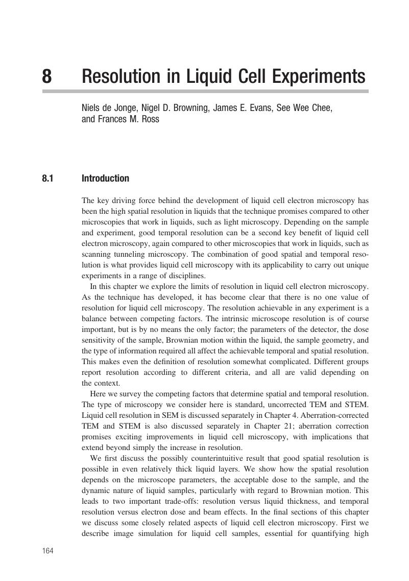
Image of the first page of this content. For PDF version, please use the ‘Save PDF’ preceeding this image.'
Type
Chapter
Information
Publisher: Cambridge University Press
Print publication year: 2016

Access options

Get access to the full version of this content by using one of the access options below. (Log in options will check for institutional or personal access. Content may require purchase if you do not have access.)

Book purchase

Temporarily unavailable

References

Hell, S. W., Far-field optical nanoscopy. Science, 316 (2007), 11531158.CrossRefGoogle ScholarPubMed
Reimer, L. and Kohl, H., Transmission Electron Microscopy: Physics of Image Formation (New York: Springer, 2008).Google Scholar
de Jonge, N. and Ross, F. M., Electron microscopy of specimens in liquid. Nat. Nanotechnol., 6 (2011), 695704.CrossRefGoogle ScholarPubMed
Peckys, D. B., Veith, G. M., Joy, D. C. and de Jonge, N., Nanoscale imaging of whole cells using a liquid enclosure and a scanning transmission electron microscope. PLoS One, 4 (2009), e8214.CrossRefGoogle Scholar
Klein, K. L., Anderson, I. M. and de Jonge, N., Transmission electron microscopy with a liquid flow cell. J. Microsc., 242 (2011), 117123.CrossRefGoogle ScholarPubMed
Woehl, T. J., Jungjohann, K. L., Evans, J. E. et al., Experimental procedures to mitigate electron beam induced artifacts during in situ fluid imaging of nanomaterials. Ultramicroscopy, 127 (2013), 5363.CrossRefGoogle ScholarPubMed
Ring, E. A. and de Jonge, N., Video-frequency scanning transmission electron microscopy of moving gold nanoparticles in liquid. Micron, 43 (2012), 10781084.CrossRefGoogle ScholarPubMed
Zaluzek, N. J., The influence of Cs/Cc correction in analytical imaging and spectroscopy in scanning and transmission electron microscopy. Ultramicroscopy, 151 (2015), 240249.CrossRefGoogle Scholar
Parsons, D. F., Matricardi, V. R., Moretz, R. C. and Turner, J. N., Electron microscopy and diffraction of wet unstained and unfixed biological objects. Adv. Biol. Med. Phys., 15 (1974), 161270.CrossRefGoogle ScholarPubMed
Egerton, R. F., Control of radiation damage in the TEM, Ultramicroscopy, 127 (2012), 100108.CrossRefGoogle ScholarPubMed
Rose, A., The sensitivity performance of the human eye on an absolute scale. J. Opt Soc Am., 38 (1948), 196208.CrossRefGoogle Scholar
Pierson, J., Sani, M., Tomova, C., Godsave, S. and Peters, P. J., Toward visualization of nanomachines in their native cellular environment. Histochem. Cell Biol., 132 (2009), 253262.CrossRefGoogle ScholarPubMed
Hoenger, A. and McIntosh, J. R., Probing the macromolecular organization of cells by electron tomography. Curr. Opin. Cell Biol., 21 (2009), 8996.CrossRefGoogle ScholarPubMed
Howells, M. R., Beetz, T., Chapman, H. N. et al., An assessment of the resolution limitation due to radiation-damage in X-ray diffraction microscopy. J Electron Spectrosc. Relat. Phenomena, 170 (2009), 412.CrossRefGoogle ScholarPubMed
Bammes, B. E., Jakana, J., Schmid, M. F. and Chiu, W., Radiation damage effects at four specimen temperatures from 4 to 100 K. J. Struct. Biol., 169 (2010), 331341.CrossRefGoogle ScholarPubMed
Stahlberg, H. and Walz, T., Molecular electron microscopy: state of the art and current challenges. ACS Chem. Biol., 3 (2008), 268281.CrossRefGoogle ScholarPubMed
Matricardi, V. R., Moretz, R. C. and Parsons, D. F., Electron diffraction of wet proteins: catalase. Science, 177 (1972), 268270.CrossRefGoogle ScholarPubMed
Schneider, N. M., Norton, M. M., Mendel, B. J. et al., Electron–water interactions and implications for liquid cell electron microscopy. J. Phys. Chem. C, 118 (2014), 2237322382.CrossRefGoogle Scholar
Hermannsdörfer, J., de Jonge, N. and Verch, A., Electron beam induced chemistry of gold nanoparticles in saline solution. Chem. Commun., 51 (2015), 1639316396.CrossRefGoogle ScholarPubMed
Peckys, D. B. and de Jonge, N., Liquid scanning transmission electron microscopy: imaging protein complexes in their native environment in whole eukaryotic cells. Microsc. Microanal., 20 (2014), 346365.CrossRefGoogle ScholarPubMed
Peckys, D. B., Mazur, P., Gould, K. L. and de Jonge, N., Fully hydrated yeast cells imaged with electron microscopy. Biophys. J., 100 (2011), 25222529.CrossRefGoogle ScholarPubMed
Chee, S. W., Loh, D., Mirsaidov, U. and Matsudaira, P., Probing nanoparticle dynamics in 200 nm thick liquid layers at millisecond time resolution. Microsc. Microanal., 21 (Suppl 3) (2015), 267268.CrossRefGoogle Scholar
de Jonge, N., Peckys, D. B., Kremers, G. J. and Piston, D. W., Electron microscopy of whole cells in liquid with nanometer resolution. Proc. Natl. Acad. Sci. USA, 106 (2009), 21592164.CrossRefGoogle ScholarPubMed
de Jonge, N., Poirier-Demers, N., Demers, H., Peckys, D. B. and Drouin, D., Nanometer-resolution electron microscopy through micrometers-thick water layers. Ultramicroscopy, 110 (2010), 11141119.CrossRefGoogle ScholarPubMed
Schuh, T. and de Jonge, N., Liquid scanning transmission electron microscopy: nanoscale imaging in micrometers-thick liquids. C. R. Phys., 15 (2014), 214223.CrossRefGoogle Scholar
Demers, H., Poirier-Demers, N., Drouin, D. and de Jonge, N., Simulating STEM imaging of nanoparticles in micrometers-thick substrates. Microsc. Microanal., 16 (2010), 795804.CrossRefGoogle ScholarPubMed
Zheng, H., Claridge, S. A., Minor, A. M., Alivisatos, A. P. and Dahmen, U., Nanocrystal diffusion in a liquid thin film observed by in situ transmission electron microscopy. Nano Lett., 9 (2009), 24602465.CrossRefGoogle Scholar
Zheng, H., Smith, R. K., Jun, Y. W. et al., Observation of single colloidal platinum nanocrystal growth trajectories. Science, 324, (2009), 13091312.CrossRefGoogle ScholarPubMed
Chen, X. and Wen, J., In situ wet-cell TEM observation of gold nanoparticle motion in an aqueous solution. Nano. Res. Lett., 7 (2012), 598.CrossRefGoogle Scholar
White, E. R., Mecklenburg, M., Shevitski, B., Singer, S. B. and Regan, B. C., Charged nanoparticle dynamics in water induced by scanning transmission electron microscopy. Langmuir, 28 (2012), 36953698.CrossRefGoogle ScholarPubMed
Liu, Y., Lin, X.-M., Sun, Y. and Rajh, T., In situ visualization of self-assembly of charged gold nanoparticles. J. Am. Chem. Soc., 135 (2013), 37643767.CrossRefGoogle ScholarPubMed
Verch, A., Pfaff, M. and De Jonge, N., Exceptionally slow movement of gold nanoparticles at a solid:liquid interface investigated by scanning transmission electron microscopy. Langmuir, 31 (2015), 69566964.CrossRefGoogle Scholar
Contarato, D., Denes, P., Doering, D., Joseph, J. and Krieger, B., High speed, radiation hard CMOS pixel sensors for transmission electron microscopy. Phys. Procedia, 37 (2013), 15041510.CrossRefGoogle Scholar
Drouin, D., Couture, A. R., Gauvin, R. et al., CASINO V2.42: a fast and easy-to-use modeling tool for scanning electron microscopy and microanalysis users. Scanning, 29 (2007), 92101.CrossRefGoogle ScholarPubMed
LeBeau, J. M., D’Alfonso, A. J., Findlay, S. D., Stemmer, S. and Allen, L. J., Quantitative comparisons of contrast in experimental and simulated bright-field scanning transmission electron microscopy images. Phys. Rev. B, 80 (2009), 174106.CrossRefGoogle Scholar
Kirkland, E. J., Loane, R. F. and Silcox, J., Simulation of annular dark field STEM images using a modified multislice method. Ultramicroscopy, 23 (1987), 7796.CrossRefGoogle Scholar
Ishizuka, K., A practical approach for STEM image simulation based on the FFT multi-slice method. Ultramicroscopy, 90 (2002), 7183.CrossRefGoogle Scholar
Welch, D. A., Faller, R., Evans, J. E. and Browning, N. D., Simulating realistic imaging conditions for in-situ liquid microscopy. Ultramicroscopy, 135 (2013), 3642.CrossRefGoogle ScholarPubMed
de Jonge, N., Pfaff, M. and Peckys, D. B., Practical aspects of transmission electron microscopy in liquid. Adv. Imag. Electron Phys., 186 (2014), 137.CrossRefGoogle Scholar
Abellan, P., Woehl, T. J., Parent, L. R. et al., Factors controlling quantitative liquid (scanning) transmission electron microscopy. Chem. Commun., 50 (2014), 48734880.CrossRefGoogle Scholar
Grogan, J. M., Park, J. H., Ye, X. et al., Liquid cell in-situ electron microscopy: interfacial phenomena and electrochemical deposition. Microsc. Microanal., 18 (2012), 11601161.CrossRefGoogle Scholar
Park, J. H., Schneider, N. M., Grogan, J. M. et al., Control of electron beam-induced Au nanocrystal growth kinetics through solution chemistry. Nano Lett., 15 (2015), 53145320.CrossRefGoogle ScholarPubMed
Patterson, J. P., Abellan, P., Denny, M. S. Jr. et al., Observing the growth of metal–organic frameworks by in situ liquid cell transmission electron microscopy, J. Am. Chem. Soc., 137 (2015), 73227328.CrossRefGoogle ScholarPubMed
Jungjohann, K. L., Evans, J. E., Aguiar, J. A., Arslan, I. and Browning, N. D., Atomic scale imaging and spectroscopy for in situ liquid scanning transmission electron microscopy. Microsc. Microanal., 18 (2012), 621627.CrossRefGoogle ScholarPubMed
Grand, D., Bernas, A. and Amouyal, E., Photo-ionization of aqueous indole – conduction band edge and energy gap in liquid water. Chem. Phys., 44 (1979), 7379.CrossRefGoogle Scholar
Egerton, R. F., Electron Energy Loss Spectroscopy (New York: Plenum, 1996).CrossRefGoogle Scholar
Malis, T., Cheng, S. C. and Egerton, R. F., EELS log-ratio technique for specimen thickness measurement in the TEM. J. Electron Microsc. Tech., 8 (1988), 193200.CrossRefGoogle ScholarPubMed
Iakoubovskii, K., Mitsubishi, K., Nakayama, Y. and Furuya, K., Mean free path of inelastic electron scattering in elemental solids and oxides using transmission electron microscopy: atomic number dependent oscillatory behavior. Phys. Rev. B, 77 (2008), 104102.CrossRefGoogle Scholar
Hahn, M., Seredynski, J. and Baumeister, W., Inactivation of catalase monolayers by irradiation with 100kV electrons. Proc. Natl. Acad. Sci. USA, 73 (1976), 823827.CrossRefGoogle Scholar
Woehl, T. J., Evans, J. E., Arslan, I., Ristenpart, W. D. and Browning, N. D., Direct in-situ determination of the mechanisms controlling nanoparticle nucleation and growth, ACS Nano, 6 (2012), 85998610.CrossRefGoogle ScholarPubMed
Nishiyama, H., Suga, M., Ogura, T. et al., Atmospheric scanning electron microscope observes cells and tissues in open medium through silicon nitride film. J. Struct. Biol., 169 (2010), 438449.CrossRefGoogle ScholarPubMed
Evans, J. E., Jungjohann, K. L., Browning, N. D. and Arslan, I., Controlled growth of nanoparticles from solution with in situ liquid transmission electron microscopy. Nano Lett., 11 (2011), 28092813.CrossRefGoogle ScholarPubMed

Save book to Kindle

To save this book to your Kindle, first ensure no-reply@cambridge.org is added to your Approved Personal Document E-mail List under your Personal Document Settings on the Manage Your Content and Devices page of your Amazon account. Then enter the ‘name’ part of your Kindle email address below. Find out more about saving to your Kindle.

Note you can select to save to either the @free.kindle.com or @kindle.com variations. ‘@free.kindle.com’ emails are free but can only be saved to your device when it is connected to wi-fi. ‘@kindle.com’ emails can be delivered even when you are not connected to wi-fi, but note that service fees apply.

Find out more about the Kindle Personal Document Service.

Available formats
×

Save book to Dropbox

To save content items to your account, please confirm that you agree to abide by our usage policies. If this is the first time you use this feature, you will be asked to authorise Cambridge Core to connect with your account. Find out more about saving content to Dropbox.

Available formats
×

Save book to Google Drive

To save content items to your account, please confirm that you agree to abide by our usage policies. If this is the first time you use this feature, you will be asked to authorise Cambridge Core to connect with your account. Find out more about saving content to Google Drive.

Available formats
×