Skip to main content Accessibility help
×
Hostname: page-component-745bb68f8f-kw2vx Total loading time: 0 Render date: 2025-01-27T17:16:20.345Z Has data issue: false hasContentIssue false

8 - Toward Real-Space Crystallography

from Core Section

Published online by Cambridge University Press:  03 March 2022

Thomas F. Kelly
Affiliation:
Steam Instruments, Inc.
Brian P. Gorman
Affiliation:
Colorado School of Mines
Simon P. Ringer
Affiliation:
University of Sydney
Get access

Summary

We discuss how ASAT has the potential to make important advances on critical frontiers in crystallography. These key frontiers include unequivocal quantification of the nearest-neighbour relationships in materials, compositional information, and details of the degree of both short-range order and long-range order. Interfaces represent a particular opportunity. We discuss the present challenges in experimental microscopy-based methods to incorporate both the structural crystallographic information at crystal interfaces with the local chemical compositional information. We anticipate that ASAT will drive forward the field of interface science and interface engineering.

Type
Chapter
Information
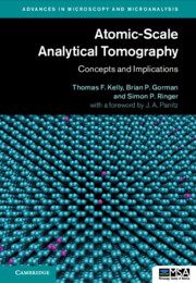
Atomic-Scale Analytical Tomography
Concepts and Implications
, pp. 145 - 159
Publisher: Cambridge University Press
Print publication year: 2022

Access options

Get access to the full version of this content by using one of the access options below. (Log in options will check for institutional or personal access. Content may require purchase if you do not have access.)

References

Gault, B., Moody, M. P., Cairney, J. M., and Ringer, S. P., “Atom Probe Crystallography,” Mater. Today, vol. 15, no. 9, pp. 378386, 2012.Google Scholar
Committee on Integrated Computational Materials Engineering, National Materials Advisory Board, Division on Engineering and Physical Sciences, National Research Council, Integrated Computational Materials Engineering: A Transformational Discipline for Improved Competitiveness and National Security. National Academies Press, 2008.Google Scholar
Eades, J. A. and Steeds, J. W., “Real Space Crystallography,” Phys. Bull., vol. 26, no. 3, pp. 108109, Mar. 1975, doi: https://doi.org/10.1088/0031-9112/26/3/024.CrossRefGoogle Scholar
De Rosier, D. J. and Klug, A., “Reconstruction of Three Dimensional Structures from Electron Micrographs,” Nature, vol. 217, no. 5124, pp. 130134, Jan. 1968, doi: https://doi.org/10.1038/217130a0.Google Scholar
Henderson, R. and Unwin, P. N. T., “Three-Dimensional Model of Purple Membrane Obtained by Electron Microscopy,” Nature, vol. 257, no. 5521, pp. 2832, Sep. 1975, doi: https://doi.org/10.1038/257028a0.CrossRefGoogle ScholarPubMed
Tsong, T. T., Atom-Probe Field Ion Microscopy: Field Ion Emission and Surfaces and Interfaces at Atomic Resolution. Cambridge, UK: Cambridge University Press, 1990.CrossRefGoogle Scholar
Walko, R. J. and Muller, E. W., “Self-Imaging of a Surface by Field Desorption,” Phys. Stat. Sol. A, vol. 9, pp. K9K10, 1972.CrossRefGoogle Scholar
Yu, Suchorski, Ernst, N, Schmidt, W. A. et al., “Field Desorption and Field Evaporation of Metals,” Prog. Surf. Sci., vol. 53, no. 2–4, pp. 135153, 1996.Google Scholar
Moore, A. J. W. and Spink, J. A., “Field Evaporation from Tungsten and the Bonding of Surface Atoms,” Surf. Sci., vol. 12, pp. 479496, 1968.CrossRefGoogle Scholar
Moore, A. J. W. and Spink, J. A., “Field Evaporation of Tungsten Atoms,” Surf. Sci., vol. 17, pp. 262266, 1969.CrossRefGoogle Scholar
Moore, A. J. W., “The Simulation of FIM Desorption Patterns,” Philos. Mag. A, vol. 43, no. 3, pp. 803814, 1981.CrossRefGoogle Scholar
Cerezo, A., Warren, P. J., and Smith, G. D. W., “Some Aspects of Image Projection in the Field-Ion Microscope,” Ultramicroscopy, vol. 79, pp. 251257, 1999.Google Scholar
Oberdorfer, C., Eich, S. M., and Schmitz, G., “A Full-Scale Simulation Approach for Atom Probe Tomography,” Ultramicroscopy, vol. 128, pp. 5567, 2013, doi: https://doi.org/10.1016/j.ultramic.2013.01.005.CrossRefGoogle ScholarPubMed
Vurpillot, F. and Oberdorfer, C., “Modeling Atom Probe Tomography: A Review,” Ultramicroscopy, vol. 159, Part 2, pp. 202216, Dec. 2015, doi: https://doi.org/10.1016/j.ultramic.2014.12.013.CrossRefGoogle ScholarPubMed
Wallace, N. D., Ceguerra, A. V., Breen, A. J., and Ringer, S. P., “On the Retrieval of Crystallographic Information from Atom Probe Microscopy Data via Signal Mapping from the Detector Coordinate Space,” Ultramicroscopy, vol. 189, pp. 6575, Jun. 2018, doi: https://doi.org/10.1016/j.ultramic.2018.02.006.CrossRefGoogle ScholarPubMed
Haley, D., Bagot, P. A. J., and Moody, M. P., “DF-Fit: A Robust Algorithm for Detection of Crystallographic Information in Atom Probe Tomography Data,” Microsc. Microanal., vol. 25, no. 2, pp. 331337, 2019, doi: https://doi.org/10.1017/S1431927618015507.CrossRefGoogle ScholarPubMed
Kühbach, M., Breen, A., Herbig, M., and Gault, B., “Building a Library of Simulated Atom Probe Data for Different Crystal Structures and Tip Orientations Using TAPSim,” Microsc. Microanal., vol. 25, pp. 320330, 2019, doi: https://doi.org/10.1017/S1431927618016252.CrossRefGoogle ScholarPubMed
Gault, B., de Geuser, F., Stephenson, L. T. et al., “Estimation of the Reconstruction Parameters for Atom Probe Tomography,” Microsc. Microanal., vol. 14, no. 4, pp. 296305, 2008.CrossRefGoogle Scholar
Gault, B. et al., “Dynamic Reconstruction for Atom Probe Tomography,” Ultramicroscopy, vol. 111, pp. 16191624, 2011.CrossRefGoogle ScholarPubMed
Day, A. C., Ceguerra, A. V., and Ringer, S. P., “Introducing a Crystallography-Mediated Reconstruction (CMR) Approach to Atom Probe Tomography,” Microsc. Microanal., vol. 25 no. 2, pp. 288300, 2019, doi: https://doi.org/10.1017/S1431927618015593.CrossRefGoogle ScholarPubMed
Breen, A. J. et al., “Correlating Atom Probe Crystallographic Measurements with Transmission Kikuchi Diffraction Data,” Microsc. Microanal., vol. 23, no. 2, pp. 279290, 2017, doi: https://doi.org/10.1017/S1431927616012605.Google Scholar
Howe, J. M., Physical Metallurgy: 3-Volume Set. Amsterdam: Elsevier, 2014.Google Scholar
Padmanabhan, K. A. and Gleiter, H., “On the Structure of Grain/interphase Boundaries and Interfaces,” Beilstein J. Nanotechnol., vol. 5, no. 1, pp. 16031615, Sep. 2014, doi: https://doi.org/10.3762/bjnano.5.172.Google Scholar
Dillon, S. J., Tang, M., Carter, W. C., and Harmer, M. P., “Complexion: A New Concept for Kinetic Engineering in Materials Science,” Acta Mater., vol. 55, no. 18, pp. 62086218, Oct. 2007, doi: https://doi.org/10.1016/j.actamat.2007.07.029.CrossRefGoogle Scholar
Patala, S., “Understanding Grain Boundaries – the Role of Crystallography, Structural Descriptors and Machine Learning,” Comput. Mater. Sci., vol. 162, pp. 281294, May 2019, doi: https://doi.org/10.1016/j.commatsci.2019.02.047.Google Scholar
Keller, R. R. and Geiss, R. H., “Transmission EBSD from 10 nm Domains in a Scanning Electron Microscope,” J. Microsc., vol. 245, no. 3, pp. 245251, Mar. 2012, doi: https://doi.org/10.1111/j.1365-2818.2011.03566.x.CrossRefGoogle Scholar
Trimby, P. W., “Orientation Mapping of Nanostructured Materials Using Transmission Kikuchi Diffraction in the Scanning Electron Microscope,” Ultramicroscopy, vol. 120, pp. 1624, Sep. 2012, doi: https://doi.org/10.1016/j.ultramic.2012.06.004.Google Scholar
Tugcu, K. et al., “Enhanced Grain Refinement of an Al–Mg–Si Alloy by High-Pressure Torsion Processing at 100°C,” Mater. Sci. Eng. A, vol. 552, pp. 415418, Aug. 2012, doi: https://doi.org/10.1016/j.msea.2012.05.063.Google Scholar
Farabi, E., Hodgson, P. D., Rohrer, G. S., and Beladi, H., “Five-Parameter Intervariant Boundary Characterization of Martensite in Commercially Pure Titanium,” Acta Mater., vol. 154, pp. 147160, Aug. 2018, doi: https://doi.org/10.1016/j.actamat.2018.05.023.CrossRefGoogle Scholar
Farabi, E., Tari, V., Hodgson, P. D., Rohrer, G. S., and Beladi, H., “On the Grain Boundary Network Characteristics in a Martensitic Ti–6Al–4 V Alloy,” J. Mater. Sci., vol. 55, no. 31, pp. 1529915321, Nov. 2020, doi: https://doi.org/10.1007/s10853-020-05075-7.CrossRefGoogle Scholar
DeMott, R., Collins, P., Kong, C. et al., “3D Electron Backscatter Diffraction Study of α Lath Morphology in Additively Manufactured Ti-6Al-4 V,” Ultramicroscopy, vol. 218, p. 113073, Nov. 2020, doi: https://doi.org/10.1016/j.ultramic.2020.113073.CrossRefGoogle Scholar
Ganesh, K. J., Kawasaki, M., Zhou, J. P., and Ferreira, P. J., “D-STEM: A Parallel Electron Diffraction Technique Applied to Nanomaterials,” Microsc. Microanal., vol. 16, no. 5, pp. 614621, Oct. 2010, doi: https://doi.org/10.1017/S1431927610000334.Google Scholar
Vincent, R. and Midgley, P. A., “Double Conical Beam-Rocking System for Measurement of Integrated Electron Diffraction Intensities,” Ultramicroscopy, vol. 53, no. 3, pp. 271282, 1994, doi: https://doi.org/10.1016/0304-3991(94)90039-6.Google Scholar
Rauch, E. F. and Véron, M., “Automated Crystal Orientation and Phase Mapping in TEM,” Mater. Charact., vol. 98, pp. 19, Dec. 2014, doi: https://doi.org/10.1016/j.matchar.2014.08.010.CrossRefGoogle Scholar
Ruiz-Zepeda, F., Arizpe-Zapata, J. A., Bahema, D., Ponce, A., and Garcia-Gutierrez, D. I., “Electron Diffraction and Crystal Orientation Phase Mapping Under Scanning Transmission Electron Microscopy,” in Advanced Transmission Electron Microscopy: Applications to Nanomaterials, Deepak, F. L., Mayoral, A., and Arenal, R., eds. Springer International Publishing AG, 2015.Google Scholar
Zuo, J.-M., “Electron Nanodiffraction,” in Springer Handbook of Microscopy, Hawkes, P. W. and Spence, J. C. H., eds. Cham: Springer International Publishing, 2019, pp. 905969.Google Scholar
Savitzky, B. H. et al., “py4DSTEM: A Software Package for Multimodal Analysis of Four-Dimensional Scanning Transmission Electron Microscopy Datasets,” ArXiv200309523 Cond-Mat Physics, Mar. 2020, accessed: May 19, 2020. [Online]. Available: http://arxiv.org/abs/2003.09523.Google Scholar
Yen, H.-W. et al., “Role of Stress-Assisted Martensite in the Design of Strong Ultrafine-Grained Duplex Steels,” Acta Mater., vol. 82, pp. 100114, Jan. 2015, doi: https://doi.org/10.1016/j.actamat.2014.09.017.CrossRefGoogle Scholar
Liddicoat, P. V. et al., “Nanostructural Hierarchy Increases the Strength of Aluminium Alloys,” Nat. Commun., vol. 1, p. 63, Sep. 2010, doi: https://doi.org/10.1038/ncomms1062.CrossRefGoogle ScholarPubMed
Yao, L., Ringer, S. P., Cairney, J. M., and Miller, M. K., “The Anatomy of Grain Boundaries: Their Structure and Atomic-Level Solute Distribution,” Scr. Mater., vol. 69, no. 8, pp. 622625, Oct. 2013, doi: https://doi.org/10.1016/j.scriptamat.2013.07.013.CrossRefGoogle Scholar
Kirova, E. M. and Pisarev, V. V., “Morphological Aspect of Crystal Nucleation in Wall-Confined Supercooled Metallic Film,” J. Phys. Condens. Matter, vol. 33, no. 3, p. 034003, Oct. 2020, doi: https://doi.org/10.1088/1361-648X/abba6b.CrossRefGoogle ScholarPubMed
Ojovan, M. I. and Louzguine-Luzgin, D. V., “Revealing Structural Changes at Glass Transition via Radial Distribution Functions,” J. Phys. Chem. B, vol. 124, no. 15, pp. 31863194, Apr. 2020, doi: https://doi.org/10.1021/acs.jpcb.0c00214.Google Scholar
Thompson, K., Geiser, B., Gerstl, S. A., and Sebastian, J., “Investigations of Dopant Clustering in Si via Radial Distribution Function,” Microsc. Microanal., vol. 12, no. Supplement S02, pp. 17341735, 2006, doi: https://doi.org/10.1017/S1431927606065391.Google Scholar
Haley, D., Petersen, T., Barton, G., and Ringer, S. P., “Influence of Field Evaporation on Radial Distribution Functions in Atom Probe Tomography,” Philos. Mag., vol. 89, no. 11, pp. 925943, 2009, doi: https://doi.org/10.1080/14786430902821610.CrossRefGoogle Scholar
Zhou, J., Odqvist, J., Thuvander, M., and Hedström, P., “Quantitative Evaluation of Spinodal Decomposition in Fe-Cr by Atom Probe Tomography and Radial Distribution Function Analysis,” Microsc. Microanal., vol. 19, no. 3, pp. 665675, 2013, doi: https://doi.org/10.1017/S1431927613000470.Google Scholar
Ceguerra, A. V, Powles, R. C, Moody, M. P, and Ringer, S. P, “Quantitative Description of Atomic Architecture in Solid Solutions: A Generalized Theory for Multicomponent Short-Range Order,” Phys. Rev. B, vol. 82, no. 13, p. 132201, 2010.Google Scholar
Ceguerra, A. V, Moody, M. P, Powles, R. C, Petersen, T. C, Marceau, R. K. W, and Ringer, S. P, “Short-Range Order in Multicomponent Materials,” Acta Crystallogr. A, vol. 68, no. 5, pp. 547560, Sep. 2012, doi: https://doi.org/10.1107/S0108767312025706.Google Scholar

Save book to Kindle

To save this book to your Kindle, first ensure no-reply@cambridge.org is added to your Approved Personal Document E-mail List under your Personal Document Settings on the Manage Your Content and Devices page of your Amazon account. Then enter the ‘name’ part of your Kindle email address below. Find out more about saving to your Kindle.

Note you can select to save to either the @free.kindle.com or @kindle.com variations. ‘@free.kindle.com’ emails are free but can only be saved to your device when it is connected to wi-fi. ‘@kindle.com’ emails can be delivered even when you are not connected to wi-fi, but note that service fees apply.

Find out more about the Kindle Personal Document Service.

Available formats
×

Save book to Dropbox

To save content items to your account, please confirm that you agree to abide by our usage policies. If this is the first time you use this feature, you will be asked to authorise Cambridge Core to connect with your account. Find out more about saving content to Dropbox.

Available formats
×

Save book to Google Drive

To save content items to your account, please confirm that you agree to abide by our usage policies. If this is the first time you use this feature, you will be asked to authorise Cambridge Core to connect with your account. Find out more about saving content to Google Drive.

Available formats
×