Skip to main content Accessibility help
×
Hostname: page-component-78c5997874-j824f Total loading time: 0 Render date: 2024-11-13T05:23:49.478Z Has data issue: false hasContentIssue false

Chapter 5 - Ultrasound assessment of normal fetal anatomy

from Section 2 - Embryology, fetal physiology and fetal anatomy

Published online by Cambridge University Press:  05 April 2016

Bidyut Kumar
Affiliation:
Wrexham Maelor Hospital
Zarko Alfirevic
Affiliation:
University of Liverpool
Get access

Summary

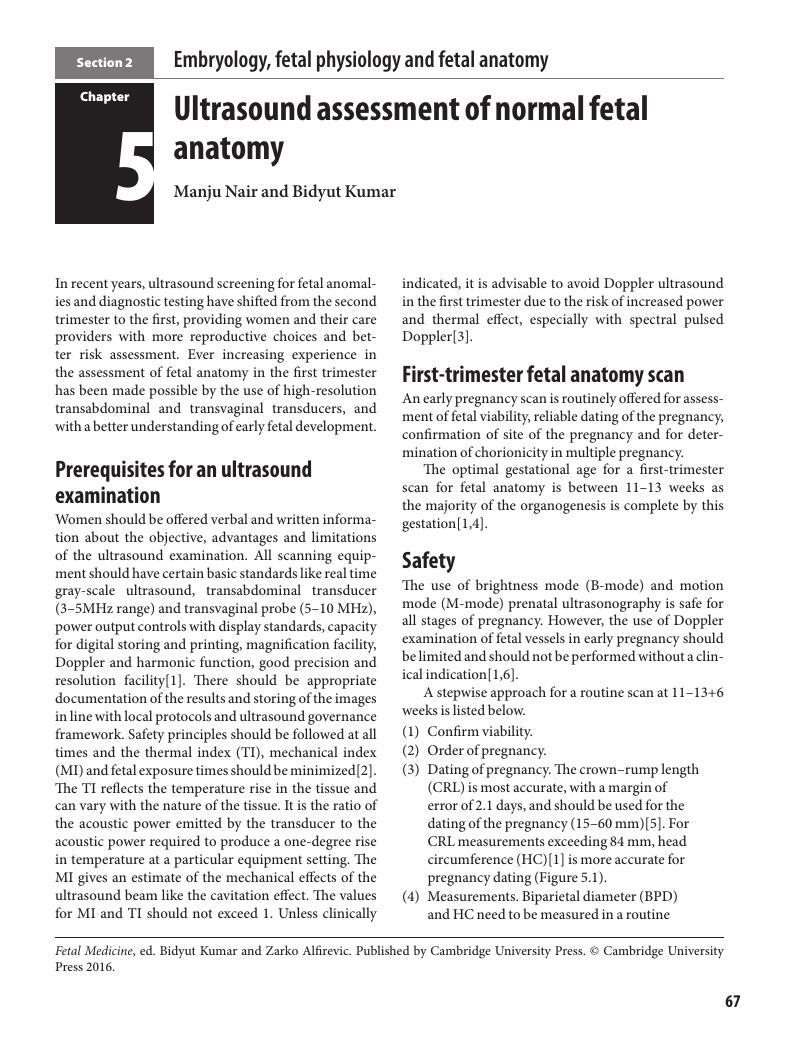
Image of the first page of this content. For PDF version, please use the ‘Save PDF’ preceeding this image.'
Type
Chapter
Information
Fetal Medicine , pp. 67 - 80
Publisher: Cambridge University Press
Print publication year: 2016

Access options

Get access to the full version of this content by using one of the access options below. (Log in options will check for institutional or personal access. Content may require purchase if you do not have access.)

References

Salomon, LJ, Alfirevic, Z, Bilardo, CM, et al. ISUOG Practice Guidelines: performance of first-trimester fetal ultrasound scan. Ultrasound Obstet Gynecol 2013; 41: 102–13.Google Scholar
British Medical Ultrasound Society. Guidelines for the safe use of diagnostic ultrasound equipment. London: BMUS; 2009.Google Scholar
Salvesen, K, Lees, C, Abramowicz, J, et al. on behalf of the Board of the International Society of Ultrasound in Obstetrics and Gynaecology (ISUOG). ISUOG statement on the safe use of Doppler in the11 to 13 + 6-week fetal ultrasound examination.Ultrasound Obstet Gynecol 2011; 37(6): 628.CrossRefGoogle Scholar
Souka, AP, Nicolaides, KH. Diagnosis of fetal abnormalities at the 10–14-week scan. Ultrasound Obstet Gynecol 1997; 10: 429–42.Google Scholar
Bogota-Ángel, SP, Bega, G. Ultrasound in pregnancy: if, when, what. In: Obstetric Evidence-Based Guidelines. Vincenzo Bergh, E (ed). London: Informa Health Care; 2007: 23–9.Google Scholar
Duck, FA. Is it safe to use diagnostic ultrasound during the first trimester? Ultrasound Obstet Gynecol 1999; 13: 385–88.Google Scholar
Khalil, A, Coates, A, Papageorghiou, A, et al. Biparietal diameter at 11–13 weeks gestation in foetuses with open spina bifida. Ultrasound Obstet Gynaecol 2013; 42(4): 409–15.Google Scholar
Twining, P, McHugo, JM, Pilling, DW.Textbook of Fetal Abnormalities. London: Churchill Livingstone; 2000.Google Scholar
Sepulveda, W, Wong, AE, Fauchon, DE. Fetal spinal anomalies in a first-trimester sonographic screening program for aneuploidy. Prenat Diagn 2011; 31(1): 107–14.CrossRefGoogle Scholar
National Collaborating Centre for Women’s and Children’s Health. NICE Clinical Guideline: Antenatal care – routine care for the healthy pregnant woman. London: RCOG Press; 2008: 13479.Google Scholar
Chudleigh, P, Thilaganathan, B. Obstetric Ultrasound: How, Why and When, 3rd edn. London: Churchill Livingstone/Elsevier; 2004.Google Scholar
Salomon, J, Alfirevic, Z, Berghella, V, et al. ISUOG Clinical Standards Committee. Practice guidelines for performance of the routine mid-trimester fetal ultrasound scan. Ultrasound Obstet Gynecol 2011; 37: 116–26.Google Scholar
Maarse, W, Bergé, SJ, Pistorius, L, et al. Diagnostic accuracy of transabdominal ultrasound in detecting prenatal cleft lip and palate: a systematic review. Ultrasound Obstet Gynecol 2010; 35(4): 495502.Google Scholar
Rotten, D, Levaillant, JM. Two- and three-dimensional sonographic assessment of the fetal face. 2. Analysis of cleft lip, alveolus and palate. Ultrasound Obstet Gynecol 2004; 24(4): 402–11.Google Scholar
Carvalho, JS, Allan, LD, et al. ISUOG practice guidelines (updated): sonographic screening examination of the fetal heart. Ultrasound Obstet Gynecol 2013; 41: 348–59.Google Scholar
ISUOG. Sonographic examination of the fetal central nervous system: guidelines for performing the ‘basic examination’ and the ‘fetal neurosonogram’. Ultrasound Obstet Gynecol 2007; 29: 109–16.Google Scholar
D’Alton, M, Mercer, B, Riddick, E, et al. Serial thoracic versus abdominal circumference ratios for the prediction of pulmonary hypoplasia in premature rupture of the membranes remote from term. Am J Obstet Gynecol 1992; 166: 658–63.Google Scholar
Chitty, LS, Altman, DG. Appendix: Charts of fetal measurements. In: Rodeck, CH, Whittle, MJ (eds). Fetal Medicine Basic Science and Clinical Practice, 2nd edn. London: Churchill Livingstone/Elsevier; 2009: 738–44.Google Scholar
Lap, C, Manten, G, Mulder, E, et al. A prospective longitudinal ultrasound study of the fetal small bowel and colon diameters. Ultrasound Obstet Gynecol 2012; 40: 294.Google Scholar

Save book to Kindle

To save this book to your Kindle, first ensure coreplatform@cambridge.org is added to your Approved Personal Document E-mail List under your Personal Document Settings on the Manage Your Content and Devices page of your Amazon account. Then enter the ‘name’ part of your Kindle email address below. Find out more about saving to your Kindle.

Note you can select to save to either the @free.kindle.com or @kindle.com variations. ‘@free.kindle.com’ emails are free but can only be saved to your device when it is connected to wi-fi. ‘@kindle.com’ emails can be delivered even when you are not connected to wi-fi, but note that service fees apply.

Find out more about the Kindle Personal Document Service.

Available formats
×

Save book to Dropbox

To save content items to your account, please confirm that you agree to abide by our usage policies. If this is the first time you use this feature, you will be asked to authorise Cambridge Core to connect with your account. Find out more about saving content to Dropbox.

Available formats
×

Save book to Google Drive

To save content items to your account, please confirm that you agree to abide by our usage policies. If this is the first time you use this feature, you will be asked to authorise Cambridge Core to connect with your account. Find out more about saving content to Google Drive.

Available formats
×