Skip to main content Accessibility help
×
Hostname: page-component-78c5997874-xbtfd Total loading time: 0 Render date: 2024-11-14T09:52:55.714Z Has data issue: false hasContentIssue false

3 - Flow Cytometry of Normal Blood, Bone Marrow and Lymphatic Tissue

Published online by Cambridge University Press:  01 February 2018

Anna Porwit
Affiliation:
Lunds Universitet, Sweden
Marie Christine Béné
Affiliation:
Université de Nantes, France
Get access

Summary

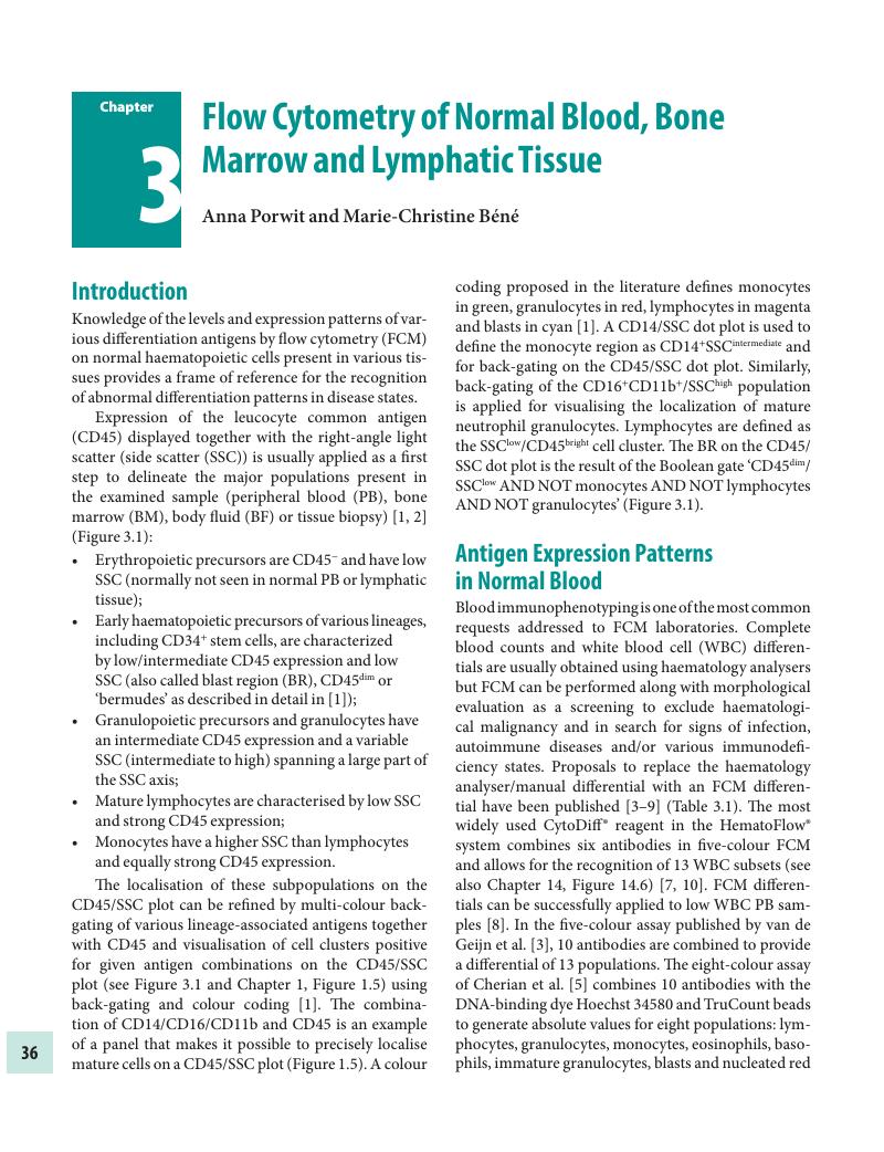
Image of the first page of this content. For PDF version, please use the ‘Save PDF’ preceeding this image.'
Type
Chapter
Information
Publisher: Cambridge University Press
Print publication year: 2018

Access options

Get access to the full version of this content by using one of the access options below. (Log in options will check for institutional or personal access. Content may require purchase if you do not have access.)

References

Arnoulet, C, Bene, MC, Durrieu, F, et al. Four- and five-color flow cytometry analysis of leukocyte differentiation pathways in normal bone marrow: a reference document based on a systematic approach by the GTLLF and GEIL. Cytometry B Clin Cytom; 78 (2010):410.CrossRefGoogle ScholarPubMed
Borowitz, MJ, Guenther, KL, Shults, KE, and Stelzer, GT. Immunophenotyping of acute leukemia by flow cytometric analysis. Use of CD45 and right-angle light scatter to gate on leukemic blasts in three-color analysis. Am J Clin Pathol; 100 (1993):534–40.CrossRefGoogle ScholarPubMed
van de Geijn, GJ, van Rees, V, van Pul-Bom, N, et al. Leukoflow: multiparameter extended white blood cell differentiation for routine analysis by flow cytometry. Cytometry A; 79 (2011):694706.CrossRefGoogle ScholarPubMed
Roussel, M, Davis, BH, Fest, T, and Wood, BL, H International Council for Standardization in. Toward a reference method for leukocyte differential counts in blood: comparison of three flow cytometric candidate methods. Cytometry A; 81 (2012):973–82.Google Scholar
Cherian, S, Levin, G, Lo, WY, et al. Evaluation of an 8-color flow cytometric reference method for white blood cell differential enumeration. Cytometry B Clin Cytom; 78 (2010):319–28.Google ScholarPubMed
Bjornsson, S, Wahlstrom, S, Norstrom, E, et al. Total nucleated cell differential for blood and bone marrow using a single tube in a five-color flow cytometer. Cytometry B Clin Cytom; 74 (2008):91103.CrossRefGoogle Scholar
Faucher, JL, Lacronique-Gazaille, C, Frebet, E, et al. ‘6 markers/5 colors’ extended white blood cell differential by flow cytometry. Cytometry A; 71 (2007):934–44.Google Scholar
Kim, AH, Lee, W, Kim, M, Kim, Y, and Han, K. White blood cell differential counts in severely leukopenic samples: a comparative analysis of different solutions available in modern laboratory hematology. Blood Res; 49 (2014):120–6.CrossRefGoogle ScholarPubMed
Park, SH, Park, BG, Park, CJ, et al. An extended leukocyte differential count (16 types of circulating leukocytes) using the CytoDiff flow cytometric system can provide information for the discrimination of sepsis severity and prediction of outcome in sepsis patients. Cytometry B Clin Cytom; 86 (2014):244–56.CrossRefGoogle Scholar
Allou, K, Vial, JP, Bene, MC, and Lacombe, F. The routine leukocyte differential flow cytometry HematoFlow method: a new flagging system for automatic validation. Cytometry B Clin Cytom; 88 (2015):375–84.CrossRefGoogle Scholar
Melzer, S, Zachariae, S, Bocsi, J, et al. Reference intervals for leukocyte subsets in adults: results from a population-based study using 10-color flow cytometry. Cytometry B Clin Cytom; 88 (2015):270–81.CrossRefGoogle Scholar
Mahdi, T, Rajab, A, Padmore, R, and Porwit, A. Characteristics of lymphoproliferative disorders with more than one aberrant cell population as detected by 10-color flow cytometry. Cytometry B Clin Cytom (2016).Google Scholar
van Dongen, JJ, Lhermitte, L, Bottcher, S, et al. EuroFlow antibody panels for standardized n-dimensional flow cytometric immunophenotyping of normal, reactive and malignant leukocytes. Leukemia; 26 (2012):1908–75.CrossRefGoogle ScholarPubMed
Bogaert, DJ, De Bruyne, M, Debacker, V, et al. The immunophenotypic fingerprint of patients with primary antibody deficiencies is partially present in their asymptomatic first-degree relatives. Haematologica; 102 (2017):192202.CrossRefGoogle ScholarPubMed
Brady, KA, Atwater, SK, and Lowell, CA. Flow cytometric detection of CD10 (cALLA) on peripheral blood B lymphocytes of neonates. Br J Haematol; 107 (1999):712–15.CrossRefGoogle Scholar
Mahnke, YD, Brodie, TM, Sallusto, F, Roederer, M, and Lugli, E. The who's who of T-cell differentiation: human memory T-cell subsets. Eur J Immunol; 43 (2013):2797–809.CrossRefGoogle ScholarPubMed
Rovati, B, Mariucci, S, Poma, R, et al. An eight-colour flow cytometric method for the detection of reference values of lymphocyte subsets in selected healthy donors. Clin Exp Med; 14 (2014):249–59.CrossRefGoogle ScholarPubMed
Hedley, BD, Keeney, M, Popma, J, and Chin-Yee, I. Novel lymphocyte screening tube using dried monoclonal antibody reagents. Cytometry B Clin Cytom; 88 (2015):361–70.CrossRefGoogle ScholarPubMed
Rajab, A, editor. Ten-color 15 antibody flow cytometry panel for immunophenotyping of lymphocyte population. The 30th Annual Meeting of the International Clinical Cytometry Society; 2015; Denver, CO, USA: ICCS.Google Scholar
Tosato, F, Bucciol, G, Pantano, G, et al. Lymphocytes subsets reference values in childhood. Cytometry A; 87 (2015):81–5.CrossRefGoogle ScholarPubMed
Szczepanski, T, van der Velden, VH, and van Dongen, JJ. Flow-cytometric immunophenotyping of normal and malignant lymphocytes. Clin Chem Lab Med; 44 (2006):775–96.CrossRefGoogle ScholarPubMed
Mandala, WL, Ananworanich, J, Apornpong, T, et al. Control lymphocyte subsets: can one country's values serve for another's? J Allergy Clin Immunol; 134 (2014):759761 e758.CrossRefGoogle ScholarPubMed
Zhang, K, Wang, F, Zhang, M, et al. Reference ranges of lymphocyte subsets balanced for age and gender from a population of healthy adults in Chongqing district of China. Cytometry B Clin Cytom; 90 (2016):538–42.CrossRefGoogle ScholarPubMed
Sagnia, B, Ateba Ndongo, F, Ndiang Moyo Tetang, S, et al. Reference values of lymphocyte subsets in healthy, HIV-negative children in Cameroon. Clin Vaccine Immunol; 18 (2011):790–95.CrossRefGoogle ScholarPubMed
Valiathan, R, Deeb, K, Diamante, M, et al. Reference ranges of lymphocyte subsets in healthy adults and adolescents with special mention of T cell maturation subsets in adults of South Florida. Immunobiology; 219 (2014):487–96.CrossRefGoogle ScholarPubMed
Nieto, WG, Almeida, J, Romero, A, et al. MBL Primary Health Care Group of Salamanca for the Study of. Increased frequency (12%) of circulating chronic lymphocytic leukemia-like B-cell clones in healthy subjects using a highly sensitive multicolor flow cytometry approach. Blood; 114 (2009):33–7.CrossRefGoogle Scholar
Durrieu, F, Genevieve, F, Arnoulet, C, et al. Normal levels of peripheral CD19(+) CD5(+) CLL-like cells: toward a defined threshold for CLL follow-up -- a GEIL-GOELAMS study. Cytometry B Clin Cytom; 80 (2011):346–53.Google Scholar
Lundell, AC, Johansen, S, Adlerberth, I, et al. High proportion of CD5+ B cells in infants predicts development of allergic disease. J Immunol; 193 (2014):510–18.CrossRefGoogle ScholarPubMed
Clavarino, G, Delouche, N, Vettier, C, et al. Novel strategy for phenotypic characterization of human B lymphocytes from precursors to effector cells by flow cytometry. PLoS One; 11 (2016):e0162209.CrossRefGoogle Scholar
Boldt, A, Borte, S, Fricke, S, et al. Eight-color immunophenotyping of T-, B-, and NK-cell subpopulations for characterization of chronic immunodeficiencies. Cytometry B Clin Cytom; 86 (2014):191206.CrossRefGoogle Scholar
Aggarwal, N, Fischer, J, Swerdlow, SH, and Craig, FE. Splenic lymphoid subsets with less well-recognized phenotypes mimic aberrant antigen expression. Am J Clin Pathol; 140 (2013):787–94.CrossRefGoogle ScholarPubMed
Bjorkstrom, NK, Beziat, V, Cichocki, F, et al. CD8 T cells express randomly selected KIRs with distinct specificities compared with NK cells. Blood; 120 (2012):3455–65.CrossRefGoogle ScholarPubMed
Roden, AC, Morice, WG, and Hanson, CA. Immunophenotypic attributes of benign peripheral blood gammadelta T cells and conditions associated with their increase. Arch Pathol Lab Med; 132 (2008):1774–80.CrossRefGoogle ScholarPubMed
Vasudev, A, Ying, CT, Ayyadhury, S, et al. Gamma/delta T cell subsets in human aging using the classical alpha/beta T cell model. J Leukoc Biol; 96 (2014):647–55.CrossRefGoogle ScholarPubMed
Cooper, MA, Fehniger, TA, and Caligiuri, MA. The biology of human natural killer-cell subsets. Trends Immunol; 22 (2001):633–40.CrossRefGoogle ScholarPubMed
Moretta, L, Montaldo, E, Vacca, P, et al. Human natural killer cells: origin, receptors, function, and clinical applications. Int Arch Allergy Immunol; 164 (2014):253–64.CrossRefGoogle ScholarPubMed
Montaldo, E, Vacca, P, Moretta, L, and Mingari, MC. Development of human natural killer cells and other innate lymphoid cells. Semin Immunol; 26 (2014):107–13.CrossRefGoogle ScholarPubMed
Zambello, R, Teramo, A, Barila, G, Gattazzo, C, and Semenzato, G. Activating KIRs in Chronic Lymphoproliferative Disorder of NK Cells: protection from Viruses and Disease Induction? Front Immunol; 5 (2014):72.CrossRefGoogle ScholarPubMed
Wood, B. Multicolor immunophenotyping: human immune system hematopoiesis. Methods Cell Biol; 75 (2004):559–76.CrossRefGoogle ScholarPubMed
van Lochem, EG, van der Velden, VH, Wind, HK, et al. Immunophenotypic differentiation patterns of normal hematopoiesis in human bone marrow: reference patterns for age-related changes and disease-induced shifts. Cytometry B Clin Cytom; 60 (2004):113.CrossRefGoogle ScholarPubMed
Malik, M, Chiles, J, 3rd, Xi, HS, et al. Genetics of CD33 in Alzheimer's disease and acute myeloid leukemia. Hum Mol Genet; 24 (2015):3557–70.CrossRefGoogle Scholar
Ziegler-Heitbrock, L. Blood monocytes and their subsets: established features and open questions. Front Immunol; 6 (2015):423.CrossRefGoogle ScholarPubMed
Hudig, D, Hunter, KW, Diamond, WJ, and Redelman, D. Properties of human blood monocytes. I. CD91 expression and log orthogonal light scatter provide a robust method to identify monocytes that is more accurate than CD14 expression. Cytometry B Clin Cytom; 86 (2014):111–20.CrossRefGoogle ScholarPubMed
Hudig, D, Hunter, KW, Diamond, WJ, and Redelman, D. Properties of human blood monocytes. II. Monocytes from healthy adults are highly heterogeneous within and among individuals. Cytometry B Clin Cytom; 86 (2014):121–34.CrossRefGoogle ScholarPubMed
Wong, L, Hill, BL, Hunsberger, BC, et al. Automated analysis of flow cytometric data for measuring neutrophil CD64 expression using a multi-instrument compatible probability state model. Cytometry B Clin Cytom; 88 (2015):227–35.CrossRefGoogle ScholarPubMed
Hoffmann, JJ. Neutrophil CD64: a diagnostic marker for infection and sepsis. Clin Chem Lab Med; 47 (2009):903–16.CrossRefGoogle ScholarPubMed
Cimato, TR, Furlage, RL, Conway, A, and Wallace, PK. Simultaneous measurement of human hematopoietic stem and progenitor cells in blood using multicolor flow cytometry. Cytometry B Clin Cytom; 90 (2016):415–23.CrossRefGoogle ScholarPubMed
Han, X, Jorgensen, JL, Brahmandam, A, et al. Immunophenotypic study of basophils by multiparameter flow cytometry. Arch Pathol Lab Med; 132 (2008):813–19.CrossRefGoogle Scholar
Yu, YR, Hotten, DF, Malakhau, Y, et al. Flow cytometric analysis of myeloid cells in human blood, bronchoalveolar lavage, and lung tissues. Am J Respir Cell Mol Biol; 54 (2016):1324.CrossRefGoogle Scholar
Dzionek, A, Fuchs, A, Schmidt, P, et al. BDCA-2, BDCA-3, and BDCA-4: three markers for distinct subsets of dendritic cells in human peripheral blood. J Immunol; 165 (2000):6037–46.CrossRefGoogle ScholarPubMed
Gabrilovich, DI. Myeloid-derived suppressor cells. Cancer Immunol Res; 5 (2017):38.CrossRefGoogle Scholar
Terstappen, LW, Safford, M, and Loken, MR. Flow cytometric analysis of human bone marrow. III. Neutrophil maturation. Leukemia; 4 (1990):657–63.Google Scholar
Terstappen, LW and Loken, MR. Myeloid cell differentiation in normal bone marrow and acute myeloid leukemia assessed by multi-dimensional flow cytometry. Anal Cell Pathol; 2 (1990):229–40.Google ScholarPubMed
Terstappen, LW and Levin, J. Bone marrow cell differential counts obtained by multidimensional flow cytometry. Blood Cells; 18 (1992):311–30; discussion 331–12.Google ScholarPubMed
Terstappen, LW, Huang, S, and Picker, LJ. Flow cytometric assessment of human T-cell differentiation in thymus and bone marrow. Blood; 79 (1992):666–77.CrossRefGoogle ScholarPubMed
Lucio, P, Parreira, A, van den Beemd, MW, et al. Flow cytometric analysis of normal B cell differentiation: a frame of reference for the detection of minimal residual disease in precursor-B-ALL. Leukemia; 13 (1999):419–27.CrossRefGoogle ScholarPubMed
Porwit-MacDonald, A, Bjorklund, E, Lucio, P, et al. BIOMED-1 concerted action report: flow cytometric characterization of CD7+ cell subsets in normal bone marrow as a basis for the diagnosis and follow-up of T cell acute lymphoblastic leukemia (T-ALL). Leukemia; 14 (2000):816–25.CrossRefGoogle Scholar
Macedo, A, Orfao, A, Ciudad, J, et al. Phenotypic analysis of CD34 subpopulations in normal human bone marrow and its application for the detection of minimal residual disease. Leukemia; 9 (1995):1896–901.Google Scholar
Matarraz, S, Lopez, A, Barrena, S, et al. The immunophenotype of different immature, myeloid and B-cell lineage-committed CD34+ hematopoietic cells allows discrimination between normal/reactive and myelodysplastic syndrome precursors. Leukemia; 22 (2008):1175–83.CrossRefGoogle ScholarPubMed
Kussick, SJ, and Wood, BL. Using 4-color flow cytometry to identify abnormal myeloid populations. Arch Pathol Lab Med; 127 (2003):1140–47.CrossRefGoogle ScholarPubMed
Della Porta, MG, Malcovati, L, Invernizzi, R, et al. Flow cytometry evaluation of erythroid dysplasia in patients with myelodysplastic syndrome. Leukemia; 20 (2006):549–55.Google ScholarPubMed
Huang, S, and Terstappen, LW. Lymphoid and myeloid differentiation of single human CD34+, HLA-DR+, CD38- hematopoietic stem cells. Blood; 83 (1994):1515–26.CrossRefGoogle ScholarPubMed
Yahata, T, Muguruma, Y, Yumino, S, et al. Quiescent human hematopoietic stem cells in the bone marrow niches organize the hierarchical structure of hematopoiesis. Stem Cells; 26 (2008):3228–36.CrossRefGoogle ScholarPubMed
Bjorklund, E, Gruber, A, Mazur, J, et al. CD34+ cell subpopulations detected by 8-color flow cytometry in bone marrow and in peripheral blood stem cell collections: application for MRD detection in leukemia patients. Int J Hematol; 90 (2009):292302.CrossRefGoogle ScholarPubMed
van Rhenen, A, Feller, N, Kelder, A, et al. High stem cell frequency in acute myeloid leukemia at diagnosis predicts high minimal residual disease and poor survival. Clin Cancer Res; 11 (2005):6520–7.CrossRefGoogle Scholar
Goardon, N, Nikolousis, E, Sternberg, A, et al. Reduced CD38 expression on CD34+ cells as a diagnostic test in myelodysplastic syndromes. Haematologica; 94 (2009):1160–3.CrossRefGoogle Scholar
Notta, F, Doulatov, S, Laurenti, E, et al. Isolation of single human hematopoietic stem cells capable of long-term multilineage engraftment. Science; 333 (2011):218–21.CrossRefGoogle ScholarPubMed
Mirabelli, P, Di Noto, R, Lo Pardo, C, et al. Extended flow cytometry characterization of normal bone marrow progenitor cells by simultaneous detection of aldehyde dehydrogenase and early hematopoietic antigens: implication for erythroid differentiation studies. BMC Physiol; 8 (2008):13.CrossRefGoogle ScholarPubMed
Jafari, K, Tierens, A, Rajab, A, et al. Visualization of cell composition and maturation in the bone marrow using 10-color flow cytometry and radar plots. Cytometry B Clin Cytom (2017).Google ScholarPubMed
Stetler-Stevenson, M, Arthur, DC, Jabbour, N, et al. Diagnostic utility of flow cytometric immunophenotyping in myelodysplastic syndrome. Blood; 98 (2001):979–87.CrossRefGoogle ScholarPubMed
Loken, MR, Chu, SC, Fritschle, W, Kalnoski, M, and Wells, DA. Normalization of bone marrow aspirates for hemodilution in flow cytometric analyses. Cytometry B Clin Cytom; 76 (2009):2736.CrossRefGoogle ScholarPubMed
Brooimans, RA, Kraan, J, van Putten, W, et al. Flow cytometric differential of leukocyte populations in normal bone marrow: influence of peripheral blood contamination. Cytometry B Clin Cytom; 76 (2009):1826.CrossRefGoogle ScholarPubMed
Elghetany, MT, Ge, Y, Patel, J, Martinez, J, and Uhrova, H. Flow cytometric study of neutrophilic granulopoiesis in normal bone marrow using an expanded panel of antibodies: correlation with morphologic assessments. J Clin Lab Anal; 18 (2004):3641.CrossRefGoogle ScholarPubMed
Elghetany, MT. Surface antigen changes during normal neutrophilic development: a critical review. Blood Cells Mol Dis; 28 (2002):260–74.CrossRefGoogle ScholarPubMed
Edvardsson, L, Dykes, J, Olsson, ML, and Olofsson, T. Clonogenicity, gene expression and phenotype during neutrophil versus erythroid differentiation of cytokine-stimulated CD34+ human marrow cells in vitro. Br J Haematol; 127 (2004):451–63.CrossRefGoogle ScholarPubMed
Matarraz, S, Almeida, J, Flores-Montero, J, et al. Introduction to the diagnosis and classification of monocytic-lineage leukemias by flow cytometry. Cytometry B Clin Cytom; 92 (2017):218–27.CrossRefGoogle Scholar
Loken, MR, Shah, VO, Dattilio, KL, and Civin, CI. Flow cytometric analysis of human bone marrow: I. Normal erythroid development. Blood; 69 (1987):255–63.CrossRefGoogle Scholar
Eidenschink Brodersen, L, Menssen, AJ, Wangen, JR, et al. Assessment of erythroid dysplasia by ‘difference from normal’ in routine clinical flow cytometry workup. Cytometry B Clin Cytom; 88 (2015):125–35.CrossRefGoogle ScholarPubMed
Laranjeira, P, Rodrigues, R, Carvalheiro, T, et al. Expression of CD44 and CD35 during normal and myelodysplastic erythropoiesis. Leuk Res; 39 (2015):361–70.CrossRefGoogle ScholarPubMed
Seegmiller, AC, Kroft, SH, Karandikar, NJ, and McKenna, RW. Characterization of immunophenotypic aberrancies in 200 cases of B acute lymphoblastic leukemia. Am J Clin Pathol; 132 (2009):940–9.CrossRefGoogle Scholar
Campana, D, Janossy, G, Bofill, M, et al. Human B cell development. I. Phenotypic differences of B lymphocytes in the bone marrow and peripheral lymphoid tissue. J Immunol; 134 (1985):1524–30.CrossRefGoogle Scholar
Campana, D and Coustan-Smith, E. Minimal residual disease studies by flow cytometry in acute leukemia. Acta Haematol; 112 (2004):815.CrossRefGoogle ScholarPubMed
Loken, MR, Shah, VO, Dattilio, KL, and Civin, CI. Flow cytometric analysis of human bone marrow. II. Normal B lymphocyte development. Blood; 70 (1987):1316–24.CrossRefGoogle ScholarPubMed
McKenna, RW, Washington, LT, Aquino, DB, Picker, LJ, and Kroft, SH. Immunophenotypic analysis of hematogones (B-lymphocyte precursors) in 662 consecutive bone marrow specimens by 4-color flow cytometry. Blood; 98 (2001):2498–507.CrossRefGoogle Scholar
Janossy, G, Bofill, M, and Schuurman, HJ. Human B-lymphoid differentiation: normal versus malignant. Neth J Med; 39 (1991):232–43.Google ScholarPubMed
Blom, B and Spits, H. Development of human lymphoid cells. Annu Rev Immunol; 24 (2006):287320.CrossRefGoogle ScholarPubMed
Fuda, FS, Karandikar, NJ, and Chen, W. Significant CD5 expression on normal stage 3 hematogones and mature B Lymphocytes in bone marrow. Am J Clin Pathol; 132 (2009):733–7.CrossRefGoogle ScholarPubMed
Flores-Montero, J, de Tute, R, Paiva, B, et al. Immunophenotype of normal vs. myeloma plasma cells: toward antibody panel specifications for MRD detection in multiple myeloma. Cytometry B Clin Cytom; 90 (2016):6172.CrossRefGoogle ScholarPubMed
Klein, F, Feldhahn, N, Lee, S, et al. T lymphoid differentiation in human bone marrow. Proc Natl Acad Sci U S A; 100 (2003):6747–52.CrossRefGoogle ScholarPubMed
Six, EM, Bonhomme, D, Monteiro, M, et al. A human postnatal lymphoid progenitor capable of circulating and seeding the thymus. J Exp Med; 204 (2007):3085–93.CrossRefGoogle Scholar
Freud, AG, Yokohama, A, Becknell, B, et al. Evidence for discrete stages of human natural killer cell differentiation in vivo. J Exp Med; 203 (2006):1033–43.CrossRefGoogle ScholarPubMed
Escribano, L, Orfao, A, Diaz-Agustin, B, et al. Indolent systemic mast cell disease in adults: immunophenotypic characterization of bone marrow mast cells and its diagnostic implications. Blood; 91 (1998):2731–6.CrossRefGoogle ScholarPubMed
Orfao, A, Escribano, L, Villarrubia, J, et al. Flow cytometric analysis of mast cells from normal and pathological human bone marrow samples: identification and enumeration. Am J Pathol; 149 (1996):1493–9.Google ScholarPubMed
Escribano, L, Diaz-Agustin, B, Bellas, C, et al. Utility of flow cytometric analysis of mast cells in the diagnosis and classification of adult mastocytosis. Leuk Res; 25 (2001):563–70.CrossRefGoogle ScholarPubMed
Diao, J, Zhao, J, Winter, E, and Cattral, MS. Recruitment and differentiation of conventional dendritic cell precursors in tumors. J Immunol; 184 (2010):1261–7.CrossRefGoogle ScholarPubMed
Laane, E, Tani, E, Bjorklund, E, et al. Flow cytometric immunophenotyping including Bcl-2 detection on fine needle aspirates in the diagnosis of reactive lymphadenopathy and non-Hodgkin's lymphoma. Cytometry B Clin Cytom; 64 (2005):3442.CrossRefGoogle ScholarPubMed
Pascual, V, Liu, YJ, Magalski, A, et al. Analysis of somatic mutation in five B cell subsets of human tonsil. J Exp Med; 180 (1994):329–39.CrossRefGoogle ScholarPubMed
Caron, G, Le Gallou, S, Lamy, T, Tarte, K, and Fest, T. CXCR4 expression functionally discriminates centroblasts versus centrocytes within human germinal center B cells. J Immunol; 182 (2009):7595–602.CrossRefGoogle ScholarPubMed
Hogerkorp, CM and Borrebaeck, CA. The human CD77- B cell population represents a heterogeneous subset of cells comprising centroblasts, centrocytes, and plasmablasts, prompting phenotypical revision. J Immunol; 177 (2006):4341–9.CrossRefGoogle ScholarPubMed
Mantei, K and Wood, BL. Flow cytometric evaluation of CD38 expression assists in distinguishing follicular hyperplasia from follicular lymphoma. Cytometry B Clin Cytom; 76 (2009):315–20.Google Scholar
Kussick, SJ, Kalnoski, M, Braziel, RM, and Wood, BL. Prominent clonal B-cell populations identified by flow cytometry in histologically reactive lymphoid proliferations. Am J Clin Pathol; 121 (2004):464–72.CrossRefGoogle ScholarPubMed
Santegoets, SJ, Dijkgraaf, EM, Battaglia, A, et al. Monitoring regulatory T cells in clinical samples: consensus on an essential marker set and gating strategy for regulatory T cell analysis by flow cytometry. Cancer Immunol Immunother; 64 (2015):1271–86.CrossRefGoogle Scholar
Weinstein, JS, Lezon-Geyda, K, Maksimova, Y, et al. Global transcriptome analysis and enhancer landscape of human primary T follicular helper and T effector lymphocytes. Blood; 124 (2014):3719–29.CrossRefGoogle ScholarPubMed
Laurent, C, Fazilleau, N, and Brousset, P. A novel subset of T-helper cells: follicular T-helper cells and their markers. Haematologica; 95 (2010):356–8.CrossRefGoogle Scholar
Colovai, AI, Giatzikis, C, Ho, EK, et al. Flow cytometric analysis of normal and reactive spleen. Mod Pathol; 17 (2004):918–27.CrossRefGoogle ScholarPubMed
Li, S, Juco, J, Mann, KP, and Holden, JT. Flow cytometry in the differential diagnosis of lymphocyte-rich thymoma from precursor T-cell acute lymphoblastic leukemia/lymphoblastic lymphoma. Am J Clin Pathol; 121 (2004):268–74.CrossRefGoogle ScholarPubMed
Yuan, J, Gali, VL, Perry, DA, et al. Flow cytometric characteristics of extrathymic thymocytes in adenoid tissue: a case report and comparison to normal thymus and thymoma. Cytometry B Clin Cytom (2017).CrossRefGoogle Scholar
Ohgami, RS, Zhao, S, Ohgami, JK, et al. TdT+ T-lymphoblastic populations are increased in Castleman disease, in Castleman disease in association with follicular dendritic cell tumors, and in angioimmunoblastic T-cell lymphoma. Am J Surg Pathol; 36 (2012):1619–28.CrossRefGoogle Scholar
Yu, GH, Vergara, N, Moore, EM, and King, RL. Use of flow cytometry in the diagnosis of lymphoproliferative disorders in fluid specimens. Diagn Cytopathol; 42 (2014):664–70.CrossRefGoogle ScholarPubMed
van de Geijn, GM, van Gent, M, van Pul-Bom, N, et al. A new flow cytometric method for differential cell counting in ascitic fluid. Cytometry B Clin Cytom; 90 (2016):506–11.CrossRefGoogle ScholarPubMed
Song, JY, Filie, AC, Venzon, D, Stetler-Stevenson, M, and Yuan, CM. Flow cytometry increases the sensitivity of detection of leukemia and lymphoma cells in bronchoalveolar lavage specimens. Cytometry B Clin Cytom; 82 (2012):305–12.Google ScholarPubMed

Save book to Kindle

To save this book to your Kindle, first ensure coreplatform@cambridge.org is added to your Approved Personal Document E-mail List under your Personal Document Settings on the Manage Your Content and Devices page of your Amazon account. Then enter the ‘name’ part of your Kindle email address below. Find out more about saving to your Kindle.

Note you can select to save to either the @free.kindle.com or @kindle.com variations. ‘@free.kindle.com’ emails are free but can only be saved to your device when it is connected to wi-fi. ‘@kindle.com’ emails can be delivered even when you are not connected to wi-fi, but note that service fees apply.

Find out more about the Kindle Personal Document Service.

Available formats
×

Save book to Dropbox

To save content items to your account, please confirm that you agree to abide by our usage policies. If this is the first time you use this feature, you will be asked to authorise Cambridge Core to connect with your account. Find out more about saving content to Dropbox.

Available formats
×

Save book to Google Drive

To save content items to your account, please confirm that you agree to abide by our usage policies. If this is the first time you use this feature, you will be asked to authorise Cambridge Core to connect with your account. Find out more about saving content to Google Drive.

Available formats
×