Skip to main content Accessibility help
×
  • Cited by 5
Publisher:
Cambridge University Press
Online publication date:
November 2012
Print publication year:
2012
Online ISBN:
9780511842153

Book description

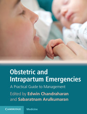
Every day, approximately 1000 women die from preventable causes related to pregnancy and childbirth, most of which result from common treatable complications, such as haemorrhage, infections, pre-eclampsia and obstructed labour, which have not been recognized in time or treated properly. Every unborn child also faces risk of stillbirth, birth trauma, oxygen deprivation and neonatal death or long-term brain damage during birthing. Obstetric and Intrapartum Emergencies: A Practical Guide to Management is written by a wide variety of obstetric experts in developing and developed countries and provides an easy-to-use guide to recognize and treat perinatal emergencies before it is too late. The text includes learning tools such as 'Key Pearls' and 'Key Pitfalls', a section on managing emergencies in a low-resource setting and contains detailed illustrations throughout. This book is a practical and invaluable guide for obstetricians, neonatologists, midwives, medical students and the wider perinatal team.

Refine List

Actions for selected content:

Select all | Deselect all
  • View selected items
  • Export citations
  • Download PDF (zip)
  • Save to Kindle
  • Save to Dropbox
  • Save to Google Drive

Save Search

You can save your searches here and later view and run them again in "My saved searches".

Please provide a title, maximum of 40 characters.
×

Contents


Page 2 of 2


  • 21 - Palpitations during pregnancy
    pp 145-149
  • View abstract

    Summary

    This chapter discusses the maternal and fetal implications, diagnostic signs, and management strategies for amniotic fluid embolus (AFE). Pulmonary oedema, acute respiratory distress syndrome, disseminated intravascular coagulopathy (DIC), pulmonary embolus, haemorrhage, right then let cardiac failure, cerebrovascular events, cardiorespiratory arrest, death are maternal implications of AFE. The first-line management involves resuscitation strategies. The main aim of early delivery is to facilitate and improve outcome of maternal resuscitation. The second-line management includes diagnosis and supportive care. The purpose of ICU is to monitor observations, maintain haemodynamic instability and reduce iatrogenic and disease complications. Options of treatment include diuretics, inotropes and steroids. Plasma exchange, haemofiltration and extracorporeal membrane oxygenation have been used in treatment. Regular fire drills involving maternal collapse on the labour ward can ensure that a robust system is in place for the acute management of AFE.
  • 22 - Breathlessness
    pp 150-153
  • View abstract

    Summary

    This chapter discusses the implications, diagnostic signs and management strategies for uterine rupture. Upper-segment caesarean section scar has a higher risk of uterine rupture compared with lower-segment caesarean section (LSCS) scar. The diagnostic signs depend on the site, extent and timing of the uterine rupture. Assessment and resuscitation involves assessing the vital signs and providing initial supportive treatment following management of haemorrhagic shock and resuscitation of a collapsed woman. Recognition of cephalopelvic disproportion or malposition is essential prior to augmentation of labour in all women, especially with secondary inertia or prolongation of the second stage of labour. Continuous electronic fetal heart monitoring is indicated for woman undergoing vaginal birth after caesarean (VBACS) or trial of labour with a scarred uterus. Primary precautions to prevent uterine rupture are most important. Increased motivation and encouraging early prenatal care enables the detection of risk factors which could be managed appropriately.
  • 25 - Psychiatric emergencies
    pp 170-175
  • View abstract

    Summary

    This chapter discusses the types, implications and management strategies of breech delivery. Breech presentation at time of delivery is associated with increased perinatal mortality and morbidity. Any factor that affects the uterine shape and tone, passenger (fetal size, maturity, structure and number) and passage (both bony pelvis and sot tissues) can predispose to breech presentation. Before allowing vaginal breech delivery it is important to confirm the presenting part by performing a vaginal examination. An episiotomy may be performed as a prophylactic measure when the breech delivery is imminent, even in multiparous women. It has been advocated to prevent possibility of soft tissue dystocia. For simplicity, conduct of assisted vaginal breech delivery will be considered in three parts: Delivery of the legs and buttocks; Delivery of the trunk and shoulders and Delivery of the 'after-coming' head.
  • 27 - Diabeticketoacidosis in pregnancy
    pp 185-190
  • View abstract

    Summary

    Cord prolapse is an obstetric emergency with a high risk of perinatal mortality. Selective transvaginal scanning in women with high-risk factors such as a transverse lie, malpresentation (e.g. footling or lexed breech) or high presenting part of fetus, may be useful. Amniotomy is contraindicated if the cord is palpable below or by the side of the presenting part during vaginal examination. Women with prelabour (prolonged) rupture of membranes (PROM) should be offered a speculum examination irrespective of the period of gestation. A digital vaginal examination indicates the presence of PROM or preterm PROM (PPROM) with cardiotocograph (CTG) abnormalities such as variable decelerations, prolonged decelerations and bradycardia and a suspicion of cord prolapse. Emergency caesarean section (CS) is frequently needed but assisted or operative vaginal delivery may be possible if the cervix is fully dilated. Post-delivery debriefing and counselling of parents and risk-management discussions are needed.
  • 29 - Musculoskeletalconsiderations in pregnancy
    pp 198-203
  • View abstract

    Summary

    The main aim of fetal monitoring is to timely identify and hence to salvage fetuses that are at risk of intrapartum hypoxic injury, whilst avoiding unnecessary operative intervention to fetuses that are normoxic or those who are mounting a good compensatory response. Cardiotocography (CTG) interpretation based on pattern recognition leads to unnecessary interventions as well as lack of action as all the CTG patterns of fetal neurological injury are not currently known and the specific CTG patterns do not correlate with poor neonatal outcomes. Intrapartum hypoxia should be suspected when there are changes in the baseline heart rate (i.e. below 110 beats per minute (bpm) or above 160 bpm) and/or presence of decelerations (on auscultation for 1 min after a uterine contraction) on intermittent auscultation. The decelerations are classified as early, late and variable in relation to the uterine contractions.
  • 31 - General anaesthesia and failed intubation
    pp 214-220
  • View abstract

    Summary

    Shoulder dystocia occurs when the baby's head has been born but a shoulder becomes stuck behind the mother's pelvic bone, resulting in a delivery that requires additional obstetric manoeuvres to release the shoulder after gentle downward traction has failed. Failure of external rotation of the fetal head and turtle sign, the retraction of the fetal head into the vagina from the perineum, are the key diagnostic signs. First line manoeuvres (SPR) and second line manoeuvres are carried out to manage shoulder dystocia. If facilities for safe and immediate emergency caesarean sections are not available, then clinicians should be trained on symphysiotomy as the main second-line measure. A metal catheter, scalpel handle and blade and suitable local anaesthetic should be made available in birth settings. All staff providing intrapartum care should undergo annual skills and drills training on the management of shoulder dystocia.
  • 33 - Transfusion,anaphylactic and drug reactions
    pp 227-232
  • View abstract

    Summary

    Dizygotic twin rate has increased by about 50% over the past two decades due to the wide use of ovarian stimulation regimes in the treatment of subfertility. Diagnosis can be made by ultrasound in the first trimester and the chorionicity has to be assessed before 14 weeks. Continuous cardiotocograph (CTG) monitoring of both twins is mandatory. There is no ideal time interval between delivery of the first and second twin. Continuous electronic fetal monitoring of the second twin is mandatory, after the birth of the first twin. Cord prolapse is more common after the delivery of the first twin and should be anticipated. Vaginal delivery is always preferable to caesarean delivery in low-resource settings when the first twin is vertex. When the second twin is non-vertex internal podalic version and breech extraction should be the aim over emergency caesarean delivery, if there are no other contraindications for vaginal birth.
  • 35 - Neonatal resuscitation and the management of immediate neonatalproblems
    pp 241-249
  • View abstract

    Summary

    Instrumental vaginal deliveries can be hazardous in inexperienced hands and should be undertaken with due care and supervision. In cases of fetal distress it is essential that the instrumental delivery be straight forward as the combination of trauma and hypoxia is potentially damaging to the fetus. In general, ventouse delivery is preferred when the position is occipitotransverse or occipito-posterior to allow for autorotation of the fetal head during traction unless the accoucher is experienced in Kielland's rotational forceps delivery. Where maternal expulsive efforts may be compromised, forceps may be better than ventouse delivery. Maternal complications are higher with forceps whilst neonatal complications are more common with the use of ventouse. Although instrumental delivery is a service provided in both basic and comprehensive essential obstetric care, it is under-used in low-resource settings. After delivery, an adequate review of overall conduct of the delivery, perineal repair and postpartum care should follow.
  • 37 - Peri-and postmortem caesarean section
    pp 256-259
  • View abstract

    Summary

    Depending on the urgency, caesarean sections (CS) are categorised as: grade 1, grade 2, grade 3 and grade 4. Grades 1-3 are all types of emergency CS with differing degrees of urgency. Regional anaesthesia is appropriate for the majority of cases but general anaesthesia may be needed in a grade 1 CS. Lower segment transverse incision is the commonest incision used and is associated with the lowest intra-operative haemorrhage as the lower segment is thin at this level. Classical incision is a rarely used vertical incision in the upper segment of the uterus. Vicryl no.1 on a blunt needle for uterine closure and Vicryl no.1 on a sharp needle for rectus sheath are commonly used sutures. Manual separation of the placenta before spontaneous separation increases the blood loss during CS and also the risk of postoperative endometritis.
  • 39 - Simulationtraining
    pp 264-268
  • View abstract

    Summary

    Unintended trauma and complications during caesarean section (CS) include: (i) difficult delivery of the fetus (ii) fetal injuries (iii) bleeding (iv) bladder and/or ureteric injuries (v) small or large bowel injuries. The main causes of bleeding are: tone, tissue, trauma, and thrombin. Care should be taken to avoid inadvertent injury to adjacent structures (ureters and bladder) and these extensions should be secured with haemostatic sutures. Inferior epigastric vessels are notorious for retraction and hence prompt recognition of injury and immediate repair is mandatory to avoid primary surgical haemorrhage and return to theatre. Care should be taken to clamp, cut and ligate omental vessels whilst performing adhesiolysis. It is good practice to always check for bladder injuries after closure of the uterine incision and before closing the peritoneal cavity. Obstetricians should be able to recognise organ damage and repair the injury in low-resource settings.
  • 40 - Risk management (emergency obstetric and intrapartum care)
    pp 269-274
  • View abstract

    Summary

    Acute puerperal uterine inversion is a rare, potentially life-threatening complication of pregnancy. A dilated cervix with a relaxed uterus and simultaneous downward traction on the fundus are the possible factors leading to inversion of the uterus. The best way to manage the neurogenic component would be to reposition the uterus. The first-line procedure which is commonly used for manual replacement of uterus is referred to as Johnson's manoeuvre. Use of tocolytics in a situation where postpartum haemorrhage is a common accompaniment is fraught with danger. There are a few techniques which have been used during surgery to reduce the inverted uterus: Huntingdon's operation, Haultain's operation and hysterectomy. It is recommended to use an uterotonic drug in the initial phase of management after repositioning. Oxytocin infusion, misoprostol per rectum or prostaglandins can be used for this purpose.

Page 2 of 2


Metrics

Full text views

Total number of HTML views: 0
Total number of PDF views: 0 *
Loading metrics...

Book summary page views

Total views: 0 *
Loading metrics...

* Views captured on Cambridge Core between #date#. This data will be updated every 24 hours.

Usage data cannot currently be displayed.