Skip to main content Accessibility help
×
  • Cited by 2
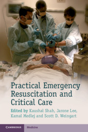
  • Edited by Kaushal Shah, Department of Emergency Medicine, Mount Sinai School of Medicine, New York, Jarone Lee, Department of Emergency Medicine, Massachusetts General Hospital, Boston, Kamal Medlej, American University of Beirut, Scott D. Weingart, Department of Emergency Medicine, Mount Sinai School of Medicine, New York
Publisher:
Cambridge University Press
Online publication date:
November 2013
Print publication year:
2013
Online ISBN:
9781139523936

Book description

Acute resuscitation and care of unstable and critically ill patients can be a daunting experience for all trainees in the emergency department or the intensive care unit. The practical, easy-to-read and evidence-based information in Practical Emergency Resuscitation and Critical Care will help all physicians understand and begin management of these patients. This book offers the collaborative expertise of dozens of critical care physicians from different specialities, including but not limited to: emergency medicine, surgery, medicine and anaesthesia. Divided into sections by medical entities, it covers essential topics that are likely to be encountered in the emergency department where critical care often begins. The portable format and bullet point style content allows all practitioners instant access to the principle information that is necessary for the diagnosis and management of critical care patients.

Refine List

Actions for selected content:

Select all | Deselect all
  • View selected items
  • Export citations
  • Download PDF (zip)
  • Save to Kindle
  • Save to Dropbox
  • Save to Google Drive

Save Search

You can save your searches here and later view and run them again in "My saved searches".

Please provide a title, maximum of 40 characters.
×

Contents


Page 1 of 3


  • 2 - Monitoring
    pp 10-16
  • 3 - Airway management
    pp 17-27
  • 4 - Mechanical ventilation
    pp 28-34
  • 10 - Thoracic trauma
    pp 71-81
  • View abstract

    Summary

    This chapter discusses the diagnosis, evaluation and management of shock. It presents special circumstances which make diagnosis and management of shock difficult in pediatric and pregnant patients. Shock should be suspected when patients present with a constellation of signs including ill-appearance, tachycardia, tachypnea, hypotension, and oliguria. The principles of shock management include specific therapy for treating the underlying cause, and general therapy to manage the shock syndrome. Recognition of shock is difficult due to variations in age-dependent vital signs, difficulty in assessing mental status, and the non-specificity of early manifestations of shock such as irritability and poor feeding. Elderly patients experience significantly more morbidity and mortality from all causes of shock due to their limited ability to augment cardiac output and maintain vascular tone. Elderly patients often have multiple comorbidities or use multiple medications that distort the diagnosis and management of shock.
  • 12 - Severe pelvic trauma
    pp 90-95
  • View abstract

    Summary

    This chapter discusses the basic methods and principles of monitoring for proper management of the critically ill patient in the emergency setting. Pulse oximetry provides continuous measurement of a patient's oxygenation status in the case of respiratory monitoring. Capnography measures the partial pressure or concentration of expired carbon dioxide (CO2), the end-tidal carbon dioxide (EtCO2). Ultrasonography of the inferior vena cava (IVC) can be useful in determining fluid responsiveness during non-invasive hemodynamic monitoring. Invasive hemodynamic monitoring provides data via catheters inserted in central veins or arteries. Central venous pressure is obtained by placing a central venous catheter (CVC) in the internal jugular or subclavian vein. The CVP should be interpreted with caution in critically ill patients that have known heart disease or structural cardiac anomalies. Pulse pressure variation (PPV) and systolic pressure variation (SPV) can be used to determine fluid responsiveness in a mechanically ventilated patient.
  • 13 - Compartment syndrome
    pp 96-99
  • View abstract

    Summary

    This chapter discusses the management of airway. Oxygenation is the primary concern in airway management. As hemoglobin and oxygen bind cooperatively, desaturation is slow above SpO2 90%. Below 90%, hemoglobin molecules quickly lose bound oxygen, and critical hypoxia can occur in seconds. Due to the technical aspects of pulse oximetry, there is a lag of up to 2 minutes in the measured SpO2. Therefore, reading in the 80-90% range may indicate that the actual SpO2 is much lower. Laryngoscopy should be abandoned when SpO2 reads 90% in order for the patient to be reoxygenated. The goal of preoxygenation is not merely to achieve a SpO2 of 100%, but also to de-nitrogenate the lungs, completely filling the lungs with oxygen to act as an oxygen reservoir during laryngoscopy. Principles of laryngoscopy are identical for direct and video laryngoscopy, with the exception of different positioning.
  • 14 - Soft tissue injury: crush injury, arterial injury, and open fractures
    pp 100-107
  • View abstract

    Summary

    This chapter discusses the management of mechanical ventilation. The different modes of ventilation are controlled mandatory ventilation (CMV), assist volume control, synchronized intermittent mandatory ventilation (SIMV), pressure control (PC), and pressure support (PS). The two ventilation strategies that can be used in critically ill patients in the emergency department are lung protective strategy and obstructive strategy. Both of these strategies utilize the assist control (AC) volume cycled mode of ventilation. The lung protective strategy is designed for patients with acute lung injury (ALI) or acute respiratory distress syndrome (ARDS), or who are at risk for lung injury. Obstructive strategy is designed for patients with obstructive lung disease (i.e., asthma or COPD) whose airways are constricted and therefore require a longer time to fully exhale. There are two basic pressures that should be monitored in mechanically ventilated patients: peak inspiratory pressure and plateau pressure.
  • 15 - Burns
    pp 108-114
  • View abstract

    Summary

    This chapter discusses the management of intensive care unit (ICU) patient in the emergency department. Frequent reassessment of ICU boarders is essential. Since mortality starts to increase at about 6 hours, it is reasonable to completely reassess an ICU boarder every 2-4 hours. Assessments should focus on fundamentals of care and disease-specific goals. Volume status is a critical component of resuscitation and management. Accurate "Ins and Outs" are frequently poorly recorded in ICU boarders. This can be remedied by asking the nurses to never take down an empty IV bag, and to number each IV bag with a permanent marker prior to administration. The ICU is completely responsible for patient care, orders, and management. The emergency physician is available for emergent interventions and acute deterioration while the patient is in the emergency department (ED). Shared responsibility of care is often dictated by a consensus interdepartmental policy.
  • Section 3 - Neurological emergencies
    pp 115-138
  • View abstract

    Summary

    This chapter discusses the management of trauma. The primary survey for a trauma patient is performed with regard to airway, breathing and circulation. Several airway adjuncts are available to assist in endotracheal intubation, including the gum elastic bougie, supraglottic airway devices, videolaryngoscopy, or fiberoptic scopes. The breathing evaluation include visualization of chest rise, auscultation of breath sounds, palpation of the chest wall feeling for crepitus or flail segments, and assuring that the trachea is midline. The patient is examined for signs of hemorrhage, including all compartments that can hold life-threatening amounts of blood loss. Evaluation for disability in the primary survey should include Glasgow Coma Scale (GCS), neurological examination to rule out neurological deficit, and pupil examination for signs of intracranial injury. Emergency department thoracotomy (EDT) is a resuscitative procedure that has low survival rate and should be performed in unique circumstances.
  • 18 - Status epilepticus
    pp 130-133
  • View abstract

    Summary

    This chapter discusses the diagnosis, evaluation and management of severe traumatic brain injury (TBI). Early intubation is indicated in all severe TBI patients, and pretreatment may help minimize increase in intracranial pressure (ICP) during intubation. All severe TBI patients should have a neurosurgical evaluation early in their ED course. If the patient has a seizure, treatment and loading with 1g of phenytoin or fosphenytoin is indicated. The decision to place invasive ICP monitoring should generally be made in conjunction with neurosurgery. The indications for invasive ICP monitoring (external ventricular drain or intraparenchymal monitor) include severe TBI and a computed tomography (CT) showing hematomas, contusions, swelling, herniation, or compressed basal cisterns. Propofol or pentobarbital coma may be induced for severe, but nonsurgical TBI. The goal is to reduce cerebral activity and oxygen demand. These agents cause hypotension and decreased cerebral perfusion pressure (CPP) and should only be initiated with neurosurgical input.
  • 19 - Acute spinal cord compression
    pp 134-138
  • View abstract

    Summary

    This chapter discusses the pathophysiology and critical management of spinal cord trauma. It describes the most common and significant injuries to the spinal cord. Penetrating injuries can result in a complete or partial spinal cord transection. Following the immediate trauma, secondary injury can occur to the spinal cord within minutes to hours. The mechanisms of secondary injury to the spinal cord include hypoxia, ischemia, inflammation, edema, necrosis, electrolyte and ion disturbances, excitotoxicity and apoptosis. Early intubation is considered for all patients with cervical spinal cord injuries who demonstrate any signs of inadequate ventilation or oxygenation in order to minimize secondary spinal cord injuries. Care should be taken during the intubation of patients with cervical spine injuries to minimize any movement of the neck that may cause worsening of the injury. The use of airway adjuncts, such as video laryngoscopy or fiberoptic techniques, may be preferable to direct laryngoscopy.
  • Section 4 - Cardiovascular emergencies
    pp 139-196
  • View abstract

    Summary

    This chapter discusses the diagnosis, evaluation and management of neck trauma. It presents special considerations with regard to immobilization and the safety of removing the cervical collar for penetrating neck trauma. High-resolution computed tomography angiography (CTA) is the initial diagnostic study of choice in the stable patient with penetrating neck trauma or blunt neck trauma when blunt cerebrovascular injury is suspected. CTA can be the initial diagnostic study of choice regardless of zone of injury. CTA is particularly useful for zone I and III penetrating injuries, which are more difficult to evaluate by physical examination. Unstable patients with penetrating injuries require immediate surgical consultation and exploration in the OR. Unstable patients include those patients with hard signs: clear airway injury (air bubbling through wound), hemodynamic instability despite resuscitation, uncontrolled bleeding (including expanding hematoma), or evolving neurological deficit.

Page 1 of 3


Metrics

Altmetric attention score

Full text views

Total number of HTML views: 0
Total number of PDF views: 0 *
Loading metrics...

Book summary page views

Total views: 0 *
Loading metrics...

* Views captured on Cambridge Core between #date#. This data will be updated every 24 hours.

Usage data cannot currently be displayed.