Skip to main content Accessibility help
×
  • Cited by 2
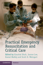
  • Edited by Kaushal Shah, Department of Emergency Medicine, Mount Sinai School of Medicine, New York, Jarone Lee, Department of Emergency Medicine, Massachusetts General Hospital, Boston, Kamal Medlej, American University of Beirut, Scott D. Weingart, Department of Emergency Medicine, Mount Sinai School of Medicine, New York
Publisher:
Cambridge University Press
Online publication date:
November 2013
Print publication year:
2013
Online ISBN:
9781139523936

Book description

Acute resuscitation and care of unstable and critically ill patients can be a daunting experience for all trainees in the emergency department or the intensive care unit. The practical, easy-to-read and evidence-based information in Practical Emergency Resuscitation and Critical Care will help all physicians understand and begin management of these patients. This book offers the collaborative expertise of dozens of critical care physicians from different specialities, including but not limited to: emergency medicine, surgery, medicine and anaesthesia. Divided into sections by medical entities, it covers essential topics that are likely to be encountered in the emergency department where critical care often begins. The portable format and bullet point style content allows all practitioners instant access to the principle information that is necessary for the diagnosis and management of critical care patients.

Refine List

Actions for selected content:

Select all | Deselect all
  • View selected items
  • Export citations
  • Download PDF (zip)
  • Save to Kindle
  • Save to Dropbox
  • Save to Google Drive

Save Search

You can save your searches here and later view and run them again in "My saved searches".

Please provide a title, maximum of 40 characters.
×

Contents


Page 2 of 3


  • 21 - Tachyarrhythmias
    pp 146-152
  • View abstract

    Summary

    This chapter discusses the diagnosis, evaluation and management of thoracic trauma including pneumothorax, hemothorax, cardiac tamponade, aortic injury, trachebronchial injury and flail chest. Tension pneumothorax presents with hypotension, tachypnea, tachycardia, distended neck veins, diminished or absent breath sounds on the affected side, and tracheal deviation away from the side of injury. Retained hemothorax following tube thoracostomy is a risk factor for infection, and should generally prompt early video assisted thoracic surgery (VATS). Traumatic pericardial tamponade must be treated with immediate surgical thoracotomy to address the cause of the bleeding into the pericardium. The critical patient with aortic injury who survives transport to the emergency department has a high probability of aortic rupture resulting in complete hemodynamic collapse and death if not immediately diagnosed and treated. Tracheal transection is associated with multiple other severe injuries. Endotracheal intubation and mechanical ventilation are indicated for the decompensating patient with flail chest.
  • 22 - Bradyarrhythmias
    pp 153-157
  • View abstract

    Summary

    This chapter discusses the diagnosis, evaluation and management of solid organ abdominal trauma. It presents special circumstances which make diagnosis and management of solid organ abdominal trauma difficult in pediatric patients. The primary survey for solid organ abdominal trauma should be aimed at determining which patients need immediate laparotomy versus those that are stable for further diagnostic workup. Vital signs provide a key to hemodynamic stability. Unstable patients with blunt or penetrating trauma to the abdomen require immediate laparotomy. All patients after significant trauma, both blunt and penetrating, should receive screening AP chest radiography. Given small anteroposterior diameter and developing abdominal musculature, children are more vulnerable to blunt forces. The most likely reason for sudden deterioration in a trauma patient with solid organ injury is hemorrhagic shock; therefore more aggressive resuscitation has to be considered and the process to get patient to the OR for laparotomy is facilitated.
  • 23 - Post–cardiac arrest care
    pp 158-163
  • View abstract

    Summary

    This chapter discusses the diagnosis, evaluation and management of pelvic trauma. The most commonly used classification system for pelvic fractures is the Young-Burgess system. This system categorizes injuries on the basis of mechanism of injury and can be used to predict the risk of blood loss. Severely injured patients are often hypotensive and require early intubation and fluid resuscitation. It is important to keep pelvic fractures high in the differential diagnosis when evaluating any patient with multiple injuries. Tachycardia is usually the first abnormal vital sign that may lead to the diagnosis of acute blood loss in severe pelvic trauma. The primary goal is early reduction of pelvic volume, which decreases venous hemorrhage through tamponade and clot formation, thereby improving mortality. Circumferential wrapping of the pelvis with a sheet is an easy and inexpensive option for pelvic binding. Commercially available pelvic binders are also an excellent option.
  • 24 - Acute decompensated heart failure
    pp 164-170
  • View abstract

    Summary

    This chapter discusses the diagnosis, evaluation and management of compartment syndrome. In trauma, the anterior compartment of the leg is the most common location of compartment syndrome; however, it is possible for it to occur in any extremity compartment. Symptoms commonly arise within 2 hours of injury but can also present up to 6 days later. The additional findings of paresthesias, anesthesia, paralysis, poikilothermia, and pulselessness are very late findings and should not be relied upon in the initial evaluation of compartment syndrome. The treatment of compartment syndrome is immediate relief of the pressure and this starts with removing any constricting devices, bandages or casts. Surgical intervention is the definitive treatment in which complete fasciotomy is performed in the operating room (OR) by trauma, vascular, or orthopedic surgeons. Neutral elevation is the preferred position. Raising the limb above the heart decreases perfusion without decreasing compartment pressures.
  • 25 - Aortic dissection
    pp 171-176
  • View abstract

    Summary

    This chapter discusses the diagnosis, evaluation and management of soft tissue injury: crush injury, arterial injury, and open fractures. The crush syndrome comprises the systemic manifestations that arise as a result of a crush injury once the external force is removed. Arterial injury can lead to hypotension if the hemorrhage is not addressed aggressively with source control and resuscitation with fluids. The mangled extremity severity score (MESS) is the most widely validated classification system of the lower extremity when evaluating the severity of open fractures. Limb viability is related to vascular status, patient age, duration of ischemia, and absorbed energy. Hypovolemic shock is the leading cause of death after soft tissue injury. Placement of two large-bore IVs and aggressive fluid resuscitation is necessary in the hypotensive individual. Cardiac arrhythmias and cardiac arrest contribute to a large percentage of early deaths in crush injury patients.
  • 26 - Pericarditis and myocarditis
    pp 177-184
  • View abstract

    Summary

    This chapter discusses the diagnosis, evaluation and management of burns. Respiratory insufficiency or failure may result from mechanical or physiological mechanisms following a thermal injury. Thermal injuries can result in a significant impact to the cardiovascular system. The cardiovascular insufficiency observed following thermal injury may result from direct cardiac suppression via inflammatory mediators, alterations in preload, cardiac contractility, or peripheral vascular tone. The secondary assessment of a thermal injured patient should follow a systematic approach similar to that of a trauma patient. The thermal injuries should be evaluated and classified based on degree and extent of injury. A Lund and Browder chart can assist in the establishment of the extent and depth of thermal injury based on the body part affected. Partial thickness burns greater than 10% total body surface area (TBSA) is one of the criteria for patients who would benefit from transfer to a burn center.
  • 27 - Hypertensive emergencies
    pp 185-190
  • View abstract

    Summary

    This chapter discusses the diagnosis, evaluation and management of ischemic strokes. Patients with ischemic strokes may present with altered mental status or airway compromise requiring immediate treatment. If the patient is not protecting his/her airway due to neurological deficits or level of consciousness, intubation will be required. If possible, the neurological examination prior to intubation has to be assessed. Blood pressure monitoring is essential for maintaining brain perfusion and decreasing the risk of conversion to hemorrhagic stroke. The most likely cause of sudden decompensation is intracranial hemorrhage during tissue plasminogen activator (tPA) administration or increased intracranial pressure due to cerebral edema. The goal should be to maintain normocarbia while minimizing positive endexpiratory pressure (PEEP). Increased PEEP can lower venous return to the heart, leading to reduced cardiac output and worsening cerebral perfusion. Consultant neurosurgery can be pivotal during cerebellar infarcts or massive hemispheric infarcts.
  • Section 5 - Respiratory emergencies
    pp 197-228
  • View abstract

    Summary

    This chapter discusses the diagnosis, evaluation and management of intracranial hemorrhage. It describes types of intracranial hemorrhage, including subarachnoid hemorrhage (SAH), subdural hemorrhage (SDH), epidural hemorrhage (EDH) and intracerebral hemorrhage (ICH). Emergent non-contrast head CT is the cornerstone for detection of ICH. MRI is equally effective in identifying ICH and better at detecting predisposing underlying parenchymal or vascular anomalies. Emergency department management focuses on protecting cerebral perfusion by balancing the forces of mean arterial pressure (MAP) and intracranial pressure (ICP). Patients should be positioned with the head of the bed elevated to 30 degrees to support cerebral venous drainage to reduce ICP. The main goal of emergency management is to temporize ICP changes, avoid secondary insults (e.g., hypoxia and hypotension) and protect cerebral perfusion pressure (CPP) while expediting neurosurgical evaluation for possible life-saving surgical intervention.
  • 30 - Asthma
    pp 203-207
  • View abstract

    Summary

    This chapter discusses the diagnosis, evaluation and management of status epilepticus. Seizures, which may be the result of central nervous system (CNS) infection, require early and empiric antibiotics, antivirals, and possibly steroids, ideally before lumbar puncture is performed. Seizures may require additional treatment and can be refractory to first-line agents (i.e., benzodiazepines) and second-line agents (i.e., phenytoin, phenobarbital, and valproate). If seizures are refractory to first- and second-line agents, levetiracetam or lacosamide, or induction with general anesthesia by inhalational anesthetics has to be considered. The most likely causes for sudden decompensation are airway compromise/respiratory failure, sepsis/septic shock, and recurrent seizure activity. Patients requiring multiple boluses of medications or continuous infusions should be considered for intubation for airway protection. Patients with an infectious etiology may rapidly progress to sepsis and require additional hemodynamic support. Prolonged seizure activity with or without overt muscle twitching is associated with increased mortality.
  • 31 - Chronic obstructive pulmonary disease
    pp 208-212
  • View abstract

    Summary

    This chapter discusses the diagnosis, evaluation and management of acute spinal cord compression. It presents special circumstances which make diagnosis and management of Cauda equina syndrome difficult. Spinal shock is characterized by a loss of spinal cord function below the level of the lesion. Cervical and thoracic level lesions may be associated with respiratory compromise. The spinal shock results in a disruption of sympathetic innervation causing unopposed parasympathetic tone, which may also cause hypotension and bradyarrhythmias (neurogenic shock). The spinal shock is characterized by flaccid paralysis and loss of bladder/bowel control. The diagnosis of acute spinal cord compression is suggested by history and physical examination, and confirmed by radiography or surgical intervention. Clinical presentations may vary depending on the level of neurological injury. The most likely causes for sudden decompensation in spinal cord include expansion of the ending lesion causing worsening neurological compromise or a high cervical/thoracic lesion.
  • 32 - Acute respiratory distress syndrome
    pp 213-217
  • View abstract

    Summary

    This chapter discusses the diagnosis, evaluation and management of acute coronary syndrome (ACS). ACS is a spectrum of disease and can present as acute myocardial infarction (AMI) or unstable angina (UA). There are four main elements in the diagnosis of ACS: clinical history, physical examination, electrocardiogram findings and cardiac biomarkers. Any patient with ST-segment elevation myocardial infarction (STEMI) should undergo reperfusion with percutaneous coronary intervention (PCI) within 90 minutes of presentation. Fibrinolytics should be used for patients unable to undergo PCI within the recommended timeframe. Beta-antagonists have been shown to benefit post-MI patients within 24 hours of the initial event when administered orally. ACE inhibitors are also recommended within 24 hours post event, but not in the immediate treatment of ACS. The three most common reasons for decompensation of the ACS patient include cardiac arrhythmias, cardiogenic shock with congestive heart failure, and mechanical complications.
  • 34 - Pulmonary embolism
    pp 223-228
  • View abstract

    Summary

    This chapter discusses the diagnosis, evaluation and management of tachyarrhythmias. Ironically, the classic presentation for tachyarrhythmias mostly consists of non-specific symptoms. Patients may complain of palpitations, chest pain, lightheadedness, dyspnea, or non-specific weakness. Further evaluation will reveal a rapid heart rate on physical examination or on the electrocardiogram (ECG). Patients with unstable tachyarrhythmias present with signs and symptoms of hypoperfusion and hemodynamic compromise while still maintaining a palpable pulse. Patients who do not have a palpable pulse are deemed to be in cardiac arrest and are treated according to Advanced Cardiovascular Life Support (ACLS) guidelines. Once tachyarrhythmia is confirmed, consideration should be given to whether the arrhythmia has an underlying noncardiac etiology such as a toxic ingestion or a metabolic disturbance. The primary goal with a patient in sinus tachycardia (ST) is to treat the underlying condition rather than the tachycardia itself.
  • Section 6 - Gastrointestinal emergencies
    pp 229-288
  • View abstract

    Summary

    This chapter discusses the diagnosis, evaluation and management of bradyarrhythmias. Sinus bradycardia occurs in 15-20% of patients with acute myocardial infarction secondary to ischemia of the sinoatrial (SA) node. Syncope may result from primary dysrhythmia or from reduced cardiac output. A 12-lead electrocardiogram (ECG) is essential for the diagnosis of bradycardia and to differentiate between the different types of bradyarrhythmias. History should focus particularly on symptoms of ischemic heart disease, and on medications such as nodal blockers. As the bradycardia worsens, cardiac output decreases as well. This results in hypotension and hypoperfusion that need to be corrected using medications or a pacemaker. In addition, a decrease in cardiac output can result in pulmonary edema. Even with the development of pulmonary edema, the bradycardia is the first thing that needs to be addressed. Treatment of the cardiogenic pulmonary edema can be instituted afterward.
  • 36 - Abdominal aortic aneurysms
    pp 237-244
  • View abstract

    Summary

    This chapter talks about the management of post-cardiac arrest care. It discusses the special circumstances in which adequate hemodynamic stability cannot be achieved during post-cardiac arrest care. An immediate assessment of a patient after the return of spontaneous circulation should include a focused history (usually obtained from bystanders or emergency medical services personnel), physical examination, diagnostic testing, and imaging studies. The physical examination should follow the ABCs, checking the airway for appropriate endotracheal tube (ETT) placement, the presence of bilateral breath sounds, circulatory status and blood pressure, heart rate and rhythm, disability with neurological response and Glasgow coma scale, and exposure to fully expose the patient and complete the examination. The extent of brain injury and cardiovascular instability are the major determinants of mortality after cardiac arrest. Brain injury is responsible for mortality in 68% of out-of-hospital arrests and 23% of in-hospital arrests.
  • 37 - Acute pancreatitis
    pp 245-252
  • View abstract

    Summary

    This chapter discusses the diagnosis, evaluation and management of acute decompensated heart failure (ADHF). Left-sided heart failure classically presents with dyspnea, usually related to pulmonary vascular congestion. Patients with severe ADHF present with respiratory distress and impending respiratory failure. The associated symptoms may include frothy oral secretions, diaphoresis, and hypoxia. Patients may also have other symptoms related to poor cardiac output and poor perfusion such as chest pain and altered mental status. Patients may be hypertensive or hypotensive depending on the etiology of symptoms and hypotension can be indicative of cardiogenic shock and is particularly concerning. Important elements of history include past history of cardiac dysfunction and potential causes of new cardiac dysfunction. When approaching a patient with ADHF, one must be sure to address any underlying cause while simultaneously managing the physiological derangements. Patients who become hypoxic, lethargic, or more confused despite noninvasive positive-pressure ventilation (NPPV) should be intubated.
  • 38 - Fulminant hepatic failure
    pp 253-260
  • View abstract

    Summary

    This chapter discusses the diagnosis, evaluation and management of aortic dissection. In addition to chest pain, patients may present with focal neurological deficits secondary to the physical obstruction of either one of the carotid arteries by an intimal flap, or false lumen propagation. Vascular obstruction and ischemia may occur at any level, leading to syncope, stroke symptoms, acute myocardial infarction (frequently from right coronary artery compromise), mesenteric ischemia, paraplegia (from hypoperfusion of the spinal arteries), or limb ischemia. Cardiogenic shock may also arise as a complication of dissection into the pericardium resulting in cardiac tamponade. Beck's triad of hypotension, muffled heart sounds, and jugular venous distension can sometimes be found. Electrocardiogram (ECG) findings rarely aid in the diagnosis, though ST elevations may be present in as many as 20% of patients due to ostial coronary involvement.
  • 39 - Acute mesenteric ischemia
    pp 261-267
  • View abstract

    Summary

    This chapter discusses the diagnosis, evaluation and management of pericarditis and myocarditis. The diagnosis of myocarditis is nearly and entirely clinical but is supported by noninvasive diagnostic methods such as cardiovascular magnetic resonance imaging and cardiac biomarkers such as troponin and CK-MB may be elevated. Cardiac MRI is a sensitive and noninvasive test to diagnose acute myocarditis. Cardiac biopsy is the gold standard for diagnosis of myocarditis and provides immunohistological evidence of an inflammatory cell infiltrate with or without myocyte damage. The management of pericarditis is based on providing symptom relief and reducing the inflammation. The treatment for myocarditis is mostly symptomatic and targeted at the underlying cause. It largely parallels the treatment of pericarditis, unless ventricular function or the conduction system is impaired. Patients with acutely decreased ventricular function should be admitted to the hospital for further evaluation.
  • 40 - The surgical abdomen
    pp 268-273
  • View abstract

    Summary

    This chapter discusses the diagnosis, evaluation and management of hypertensive emergencies. It describes special considerations for aortic dissection, acute ischemic stroke, acute intracerebral hemorrhage (ICH), and preeclampsia and eclampsia. The critical management of hypertensive emergencies depends on the presence of end-organ damage. Only patients with a diagnosis of hypertensive emergency require immediate interventions in the emergency department for lowering blood pressure. Patients with chronically elevated blood pressure may suffer detrimental consequences if their blood pressure is lowered too quickly. Dramatic and rapid decreases in blood pressure can result in critical hypoperfusion of the brain, heart, and kidneys, resulting in ischemia or infarction. Patients with hypertensive urgency can be managed as outpatients as long as reliable follow-up can be arranged. They are usually started on oral antihypertensives with a goal of lowering their blood pressure to less than 160/100 mmHg over 12-48 hours.
  • 41 - Abdominal compartment syndrome
    pp 274-277
  • View abstract

    Summary

    This chapter discusses the diagnosis, evaluation and management of valvular diseases including aortic stenosis (AS), aortic regurgitation (AR), mitral stenosis (MS) and mitral regurgitation (MR). Patients with AS are particularly sensitive to changes in cardiac output due to the pressure gradient across the aortic valve. Evaluation of any valvular pathology begins with the history and physical examination, and paying attention to whether valvular defect has been previously noted. If a new acute AR is discovered, the diagnosis of aortic dissection should be ruled out with a CTA or a transthoracic or transesophageal echocardiogram. Blood culture and antibiotics may be indicated if endocarditis or a perivalvular abscess is suspected. Characterization of valve dysfunction in the emergency department is not imperative if patients are hemodynamically stable. Echocardiography should be considered in patients who are hemodynamically unstable. Acute valvular dysfunction is usually secondary to a precipitating critical problem.
  • 42 - Esophageal perforation and mediastinitis
    pp 278-283
  • View abstract

    Summary

    This chapter discusses the diagnosis, evaluation and management of upper airway emergencies. It presents special circumstances with regard to foreign bodies in the airway. The first step in the evaluation of the patient with suspected upper airway emergency is to determine the need for emergent intubation or surgical airway. If possible, a brief history should be obtained focusing on history of cancer, allergies, exposure to medications including ACE inhibitors, family history of C1 esterase inhibitor deficiency, trauma, and recent surgery. A targeted physical examination should include assessment for stridor, hoarseness, urticaria, edema of skin, lips, mouth, and throat. Given the high-risk, time-sensitive nature of these presentations, all practitioners should be familiar with their local resources, algorithms, and airway management options prior to seeing patients. In patients with a rapidly evolving upper airway obstruction, awake evaluation can provide invaluable information about potential complications before paralytics are administered.
  • Section 7 - Renal emergencies
    pp 289-324
  • View abstract

    Summary

    This chapter discusses the diagnosis, evaluation and management of asthma. Airflow restriction may be severe, leading to asthma patients presenting in an upright or tripod position, with cyanosis, altered mental status, and respiratory arrest. Asthma exacerbations initially produce tachypnea and a resultant low carbon dioxide level; a normal or elevated carbon dioxide level may indicate fatigue and impending respiratory failure. Laboratory testing and ECG should be used to differentiate asthma exacerbations from alternative etiologies or comorbid conditions. Patients should be placed on supplemental oxygen therapy as needed to maintain adequate oxygen saturations. Patients must be monitored for signs of impending respiratory failure. Constant positive airway pressure (CPAP) and bi-level positive airway pressure (Bi-PAP) may be considered for patients with severe asthma. The goal of ventilator management in asthmatic is to oxygenate and ventilate without worsening hyperinflation, which causes barotrauma and hemodynamic instability.
  • 45 - Common electrolyte disorders (sodium, potassium, calcium, magnesium)
    pp 299-313
  • View abstract

    Summary

    This chapter discusses the diagnosis, evaluation and management of chronic obstructive pulmonary disease (COPD). Airflow restriction may be severe, leading to patients presenting in an upright or tripod position, with cyanosis, altered mental status, and respiratory arrest. Patients should be placed on supplemental oxygen therapy as needed to maintain adequate oxygen saturations of 88-92%. Over-oxygenating the COPD patient can lead to worsening ventilation-perfusion mismatch and apnea. Patients must be monitored for signs of impending respiratory failure. CPAP and BiPAP may be considered for certain patients with moderate to severe COPD exacerbations. The goal of ventilator management in the COPD patient is to oxygenate and ventilate without causing barotrauma and hemodynamic instability. If patients acutely decompensate while receiving invasive or noninvasive positive pressure ventilation, the possibility of pneumothorax and intrinsic positive end-expiratory pressure (auto-PEEP) should be considered.
  • 46 - Acute kidney injury and emergent dialysis
    pp 314-319
  • View abstract

    Summary

    This chapter discusses the diagnosis, evaluation and management of acute respiratory distress syndrome (ARDS). Pulmonary edema in ARDS is heterogeneous and leads to atelectatic or consolidated areas of lung interspersed with relatively unaffected regions, creating areas of intrapulmonary shunt, which results in hypoxemia that does not improve with oxygen administration alone. As pulmonary edema accumulates in the initial exudative phase of the disease, patients become dyspneic and demonstrate increased work of breathing. Due to worsening lung compliance, tidal volumes decrease and respiratory rate increases. Patients become progressively hypoxemic due to both worsening V/Q mismatch and shunt physiology. The respiratory failure is not fully explained by cardiac failure or volume overload. If a known risk factor for ARDS is not present, objective assessment such as echocardiography should be obtained to rule out hydrostatic edema. On a chest radiograph, ARDS looks essentially the same as cardiogenic pulmonary edema.

Page 2 of 3


Metrics

Altmetric attention score

Full text views

Total number of HTML views: 0
Total number of PDF views: 0 *
Loading metrics...

Book summary page views

Total views: 0 *
Loading metrics...

* Views captured on Cambridge Core between #date#. This data will be updated every 24 hours.

Usage data cannot currently be displayed.