- NG
nasogastric
- PEG
percutaneous endoscopic gastrostomy
Indications
Enteral feeding is used for patients in situations where they are unable to meet their nutritional requirements through the oral route. In practice this is usually because eating and drinking are unsafe due to failure of adequate airway protection during swallowing or inaccessible due to obstruction of the upper gastrointestinal tract, for example, by head and neck cancers. Less commonly enteral feeding is necessary where nutritional requirements are particularly high, to supplement oral feeding (e.g. cystic fibrosis).
The 2010 British Artificial Nutrition Support survey found that the most common group of reasons for home enteral tube feeding in 2009 was ‘central nervous system and mental health’ disorders; accounting for 47% of all new registrations(1). Forty-two percent of these central nervous system registrations were for vascular disease, predominantly cerebrovascular accident. Twenty-five to 40% of all patients who suffer a cerebrovascular accident develop dysphagia. Degenerative central nervous system conditions such as multiple sclerosis and motor neurone disease are also a frequent indication for enteral feeding accounting for 14% of all new registrations. The fastest-growing indication for gastrostomy insertion, however, is cancer (accounting for 37% of new registrations in 2009), with head and neck cancers actually being a more frequent indication of enteral feeding than cerebrovascular accident in this period. The current indications are summarised in Table 1.
Table 1. Diagnosis at registration for home enteral nutrition (from British Artificial Nutrition Survey 2010)

MND, motor neurone disease; MS, multiple sclerosis; GI, gastrointestinal; CF, cystic fibrosis; CNS, central nervous system.
The British Artificial Nutrition Support survey also indicates the types of tube being used for home enteral nutrition with a gradual increase in the proportion of jejunostomy rather than gastrostomy tubes being placed over the past 10 years (up to 8% in 2009) but no significant increase in the use of nasogastric (NG) tubes in the community (between 13 and 18%).
Technique of insertion
Endoscopic insertion of gastrostomy tube (‘pull technique’)
Patients are brought to the endoscopy unit and are made to lie on their left side. They are lightly sedated usually using midazolam. A routine gastroscopy is performed and then the stomach is inflated with air. The patient is turned on their back and the epigastric area cleaned with an antiseptic solution. The best site for tube insertion is determined by transillumination of the abdominal wall from the gastroscope light. Indentation of the abdominal wall by the assistant enables the endoscopist to ensure that the internal position of the tube will be satisfactory. The abdominal wall is then infiltrated with local anaesthetic and a cannula passed into the gastric lumen. A suture is passed through the cannula and caught with a snare passed by the endoscopist down the working channel of the endoscope. The endoscope and snare are then withdrawn through the patient's mouth. The percutaneous endoscopic gastrostomy (PEG) tube is attached to the oral end of the suture and the assistant then pulls the suture and PEG tube back through the mouth and up through the anterior abdominal wall until the internal bumper sits snugly against the anterior wall of the stomach (see Fig. 1). The external part of the gastrostomy tube can then be cut to a convenient length for use.

Fig. 1. Schematic diagram of a gastrostomy tube in situ.
Radiological insertion of gastrostomy tube
Radiological insertion of gastrostomy tubes requires an NG tube to inflate the stomach with air and to approximate it to the anterior abdominal wall. The gastrostomy site is then infiltrated with local anaesthetic and the gastric wall is fixed to the anterior abdominal wall either with a suturing kit or T-fasteners. This procedure is known as ‘gastropexy’. The gastrostomy tube is then inserted directly through the abdominal wall into the stomach under screening, usually using a guidewire and a series of dilators. The internal fixation device can either be a pigtail catheter or a ‘pop-out’ bolster.
Jejunostomy and per oral image-guided gastrostomy tubes
Feeding tubes can also be placed in the jejunum using similar endoscopic and radiological methods. A new technique of per oral image-guided gastrostomy has also been developed whereby the stomach is inflated via an NG tube and directly punctured through the skin. A guidewire and then catheter is then passed into the stomach and manipulated up the oesophagus and out of the mouth. The gastrostomy tube is mounted over a guidewire and pulled down into the stomach in a way similar to an endoscopically placed gastrostomy. This technique avoids the need to perform a gastropexy.
Complications
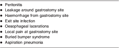
There are a number of potential complications of the procedure of gastrostomy tube insertion and these are, in the main, similar for both endoscopically and radiologically placed tubes. In addition, for endoscopically placed tubes there are all the usual risks associated with a standard upper gastrointestinal endoscopy (i.e. oversedation, aspiration pneumonia during the procedure, etc.). The most common complications are summarised in Table 2.
Table 2. Potential complications of gastrostomy insertion

Peritonitis is one of the most serious complications and can occur if the gastric wall is not properly pulled up to the anterior abdominal wall or if colon or small bowel become interposed between the two. The risk of bleeding at the time of the procedure should be minimised by a routine check of blood clotting and platelet count. Infection around the exit site of the gastrostomy tube is the most common complication of the procedure and is usually satisfactorily treated with antiseptic measures, daily change of dressing and antibiotics if necessary. Some units have successfully introduced screening and treatment for methicillin-resistant Staphylococcus aureus prior to gastrostomy insertion in addition to routine intravenous antibiotics given at the time of the procedure to minimise this risk(Reference Thomas, Cantrill and Waghorn2). The passage of the gastrostomy tube through the oesophagus can occasionally produce lacerations. Some patients complain of local pain at the site of gastrostomy insertion for a few days after the procedure. This occurs most often with more aware patients with more muscular abdominal walls and usually settles with simple analgesia. Once the stoma track has become established, patients are encouraged to push their tubes in by a few centimetres and rotate them on a daily basis. This is to prevent the gastric mucosa growing over the internal bumper of the gastrostomy tube (the ‘Buried Bumper Syndrome’ see Fig. 2). This may manifest as a gradual blocking of the tube. Although the bumper can usually be freed endoscopically using a needle–knife spincterotome, a formal laparotomy is sometimes required to remove the gastrostomy tube from the anterior abdominal wall.

Fig. 2. Series of endoscopic photographs showing the development of buried bumper syndrome. (a) Internal bumper of gastrostomy becoming retracted into gastric wall. (b) Gastric mucosa growing over internal bumper. (c) Internal bumper has become buried in the gastric mucosa so that the site of the bumper is now difficult to identify endoscopically.
The overall complication rate of gastrostomy tube insertion has been estimated at 8–30% with major complications occurring at a rate of 1–4%(Reference Mathus-Vliegen and Koning3–Reference Rabeneck, Wray and Petersen7). Mortality rates in the 30 d post-gastrostomy insertion depend in part on the initial indication and so vary considerably. Sanders et al. published Kaplan–Meier survival curves for a range of indications and showed considerable variation (see Fig. 3)(Reference Sanders, Carter and D'Silva8). More recent data (presented at BAPEN 2010) have shown similar survival curves.

Fig. 3. Survival of patients undergoing gastrostomy insertion divided by diagnosis(Reference Sanders, Carter and D'Silva8). Reprinted by permission from Macmillan Publishers Ltd.
Predictors of higher post-procedure mortality include age, co-morbidity (particularly diabetes), low BMI, current or recent pneumonia, low serum albumin level and high C-reactive protein(Reference Zopf, Maiss and Konturek9, Reference Janes, Price and Khan10). Some studies have suggested that the risks associated with gastrostomy placement are rising and this may be due to a willingness to place tubes in a higher-risk population. The 2004 Report of the National Confidential Enquiry into Patient Outcome and Death found worryingly high rates of early death after PEG insertion(11). Of 692 deaths associated with PEG placement, fourteen patients (2%) died on the day of the procedure, and a further 18% (126/692) died within the first 3 d following the procedure. A total of 43% (236/692) of deaths occurred within 1 week and a further 26% (183/692) in the second week. The most common cause of death after a PEG placement was respiratory failure. One in five PEG procedures were felt to be futile by the expert advisors and processes for patient selection for the procedure were felt to be inadequate.
Patient selection
Clearly choosing the right patients for gastrostomy insertion is crucial. There is no need for gastrostomy in situations where feeding is likely to be short term, an NG tube is well tolerated and the tube is being looked after safely. Dilemmas usually occur when a patient finds the NG tube uncomfortable or is agitated and repeatedly pulls out or displaces the tube. A decision to place a gastrostomy tube must take into consideration the risks and benefits for a particular patient, including, where possible, the patient's preference, and with a clear plan for what is to be achieved by feeding.
Patients judged to be mentally competent have the right to accept or refuse treatment once they are in full possession of all the information regarding the risks and benefits. Judging whether a particular patient has the capacity to make a particular decision requires a careful assessment. The criteria for this judgement (in England and Wales) are laid out in the Mental Capacity Act 2005(12). This states that patients must have an understanding of the decision that they need to make, why they need to make it and the likely consequences, in addition to the ability to understand, retain and weigh up the information relevant to the decision. The patient also needs to have a method to communicate this. If a patient is judged to be lacking in capacity, then the decisions made on the patient's behalf must be in his/her best interests and made by the healthcare professional responsible for carrying out the treatment. Deciding on whether a gastrostomy tube should be inserted usually involves discussions with a patient's family, close friends or carers to try to determine what the patient would have wanted for themselves and what the consequences of the procedure would be for the patient. In cases where there is no-one else other than paid staff to represent a patient, an Independent Mental Capacity Advisor may be appointed to support and represent the patient.
Placing feeding tubes near the end of life is inappropriate in most cases but this decision needs a multidisciplinary approach, as recommended by the Royal College of Physicians Working Party Report on oral feeding difficulties and dilemmas(13). This report suggests a clinical approach based on determining the underlying diagnosis, the severity of the disease, the prognosis of the patient, the mechanism of the feeding problem and the risks to the patient of eating and drinking.
Dementia
Artificial feeding of patients suffering with dementia is a very controversial area. The British Artificial Nutrition Support survey found in 2009 that the proportion of patients with dementia recorded as being the reason for home enteral feeding was falling, with only forty-nine new cases registered in 2009 (compared with 109 in 2007). Oral feeding problems usually occur late in the disease process and at a stage where it is not possible to determine the patient's wishes regarding their care. Although, practically speaking, the insertion of a gastrostomy tube is usually uncomplicated, most studies suggest that survival post-gastrostomy insertion is very poor in dementia compared with other indications for gastrostomy insertion. Some patients deemed to have an ‘unsafe swallow’ can still manage oral feeding with minimum risk if sufficient assistance is given, consistencies are altered and optimum positioning is carefully maintained. There have been no randomised controlled trials of gastrostomy feeding compared with careful hand feeding or other methods of nutritional support, and no clear benefits of gastrostomy insertion in terms of preventing pressure sores, improving functional status, improving nutritional status or extending life have been demonstrated in clinical trials(Reference Cervo, Bryan and Farber14–Reference Anderson, O'Connor and Mayer18). Use of a gastrostomy tube may also impact on quality of life by removing the patient from social interaction at mealtimes or the close attention they receive during assisted oral feeding. The pleasures of oral feeding may also be denied them if a gastrostomy feed is relied upon to provide all nutritional needs.
The decision regarding gastrostomy feeding in dementia must, however, be based on each individual case, and there are instances where it is appropriate, particularly where life expectancy seems reasonable, there is little other co-morbidity and where oral feeding is distressing for the patient with coughing or choking, yet the patient still seems hungry and keen to eat. In some circumstances the patient is able to eat but takes such a long time that they are unable to participate in other pleasurable activities. In these cases, a combined approach of assisted oral and enteral feeding is sometimes necessary.
Alternatives to gastrostomy placement
A safe oral intake is sometimes possible to maintain even in patients deemed to have an ‘unsafe swallow’ if they are managed with very careful hand feeding, with close attention to the patient's positioning and the consistencies of the foods and fluids offered. Coughing and spluttering during feeding are sometimes useful ways of clearing the airway and, unless they are distressing for the patient, are not necessarily a reason to avoid oral intake, particularly in patients who enjoy feeding and are nearing the end of their lives. Such feeding, however, is very time consuming for the carer. The Royal College of Physicians report on end-of-life care specifically states than the insertion of an artificial feeding tube to save resources or in order to transfer the patient to another facility is unethical(13).
The usual alternative to gastrostomy feeding is an NG tube. These have the advantage of being minimally invasive, and can be placed easily and safely at the bedside even in very sick patients with multiple co-morbidities. Although suitable for the provision of short-term nutrition, patients can find the tubes uncomfortable, and they are easily displaced by mistake or intentionally by an agitated or confused patient. Patients with an NG tube may complain of a sore throat or sinusitis, have difficulty in swallowing any supplementary oral nutrition and are prone to oropharyngeal candidiasis. Frequent displacement of NG tubes is a real practical problem and interferes with NG feeding, causing delays in feeding and poorer nutritional intake. Confirmation of the tip position can be difficult, and if an aspirate of fluid with a pH lower than 5·5 cannot be obtained, then radiology is required to confirm that the tip of the tube is correctly placed in the gastric lumen. Inadvertant feeding into the lungs is a recognised complication despite the widespread use of protocols for confirming the position of the tube.
Some of these disadvantages can be overcome by the use of a ‘nasal loop’ or ‘bridle’. This consists of a tape that passes around the back of the nasal septum and then clips onto the NG tube. The tape stabilises the NG tube position and effectively prevents its inadvertant removal. It also limits the movement of the tube during swallowing which some patients report as being more comfortable. The nasal loop can be placed quickly, safely and easily by the bedside using an introducer system with medical magnets mounted on flexible plastic tubes which meet at the back of the nasal septum, allowing a tape attached to one of the introducer tubes to be pulled around the septum and down the other nostril. Now that this system has become more generally available in hospitals, gastrostomy tubes should be required less often for short-term nutritional support(Reference Anderson, O'Connor and Mayer18).
Choosing the best method of nutritional support for an individual patient is often challenging. A thorough understanding of the patient's needs, home circumstances and support alongside knowledge of the risks and benefits of each route of administration of enteral nutrition is paramount in making a good decision. This is usually best achieved by a multidisciplinary team including doctors, nurses, dietitians and, where possible, the patient and their carers. For patients at the end of their lives and particularly patients with dementia, the over-riding principle should be to preserve good quality of life for the patient and avoid any intervention which merely prolongs their death.
Acknowledgement
The author and Buckinghamshire Healthcare NHS Trust declare no conflict of interest in relation to this article.


